MLLT1 Antibody (3H2) - Azide and BSA Free
Novus Biologicals | Catalog # H00004298-M01
![Immunocytochemistry/ Immunofluorescence: MLLT1 Antibody (3H2) [H00004298-M01]](https://resources.rndsystems.com/images/products/MLLT1-Antibody-3H2-Immunocytochemistry-Immunofluorescence-H00004298-M01-img0004.jpg "Immunocytochemistry/ Immunofluorescence: MLLT1 Antibody (3H2) [H00004298-M01]")
Key Product Details
Species Reactivity
Validated:
Cited:
Applications
Validated:
Cited:
Label
Antibody Source
Format
Product Specifications
Immunogen
Reactivity Notes
Specificity
Clonality
Host
Isotype
Description
Scientific Data Images for MLLT1 Antibody (3H2) - Azide and BSA Free
Immunocytochemistry/ Immunofluorescence: MLLT1 Antibody (3H2) [H00004298-M01]
Immunocytochemistry/Immunofluorescence: MLLT1 Antibody (3H2) [H00004298-M01] - Analysis of monoclonal antibody to MLLT1 on HeLa cell. Antibody concentration 10 ug/ml.
 [H00004298-M01]")
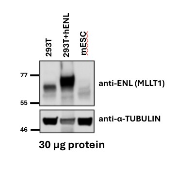
Western Blot: Mouse Monoclonal MLLT1 Antibody (3H2) [H00004298-M01]
Western Blot: Mouse Monoclonal MLLT1 Antibody (3H2) [H00004298-M01] - Western Blot showing amount of protein, cell lines tested (293T, over expression line 293T+hENL, and mESCs) and loading control blot using alpha-tubulin. Image from a verified customer review.Applications for MLLT1 Antibody (3H2) - Azide and BSA Free
Immunocytochemistry/ Immunofluorescence
Immunohistochemistry
Immunohistochemistry-Paraffin
Sandwich ELISA
Western Blot
Reviewed Applications
Read 1 review rated 3 using H00004298-M01 in the following applications:
Formulation, Preparation, and Storage
Purification
Formulation
Format
Preservative
Concentration
Shipping
Stability & Storage
Background: MLLT1
Alternate Names
Entrez Gene IDs
Gene Symbol
OMIM
UniProt
Additional MLLT1 Products
Product Documents for MLLT1 Antibody (3H2) - Azide and BSA Free
Certificate of Analysis
To download a Certificate of Analysis, please enter a lot or batch number in the search box below.
Product Specific Notices for MLLT1 Antibody (3H2) - Azide and BSA Free
This product is produced by and distributed for Abnova, a company based in Taiwan.
This product is for research use only and is not approved for use in humans or in clinical diagnosis. Primary Antibodies are guaranteed for 1 year from date of receipt.
Citations for MLLT1 Antibody (3H2) - Azide and BSA Free
 Powered by Bioz
Customer Reviews for MLLT1 Antibody (3H2) - Azide and BSA Free (1)
Have you used MLLT1 Antibody (3H2) - Azide and BSA Free?
Submit a review and receive an Amazon gift card!
$25/€18/£15/$25CAN/¥2500 Yen for a review with an image
$10/€7/£6/$10CAN/¥1110 Yen for a review without an image
Submit a review
Customer Images
-
Application: Western BlotSample Tested: mESCs and HEK293 human embryonic kidney cell lineSpecies: Human and MouseVerified Customer | Posted 07/01/2024Annnotated WB image showing amount of protein, cell lines tested (293T, over expression line 293T+hENL, and mESCs), loading control blot using alpha-tubulinWestern blot with 30µg of protein per sample, antibody at a 1:500 dilution. Used wild type HEK293T cells and HEK293Ts with over expression of hENL. We also used mouse embryonic stem cells. Used alpha-tubulin as a loading control for each well.

There are no reviews that match your criteria.
Protocols
Find general support by application which include: protocols, troubleshooting, illustrated assays, videos and webinars.
- Antigen Retrieval Protocol (PIER)
- Antigen Retrieval for Frozen Sections Protocol
- Appropriate Fixation of IHC/ICC Samples
- Cellular Response to Hypoxia Protocols
- Chromogenic IHC Staining of Formalin-Fixed Paraffin-Embedded (FFPE) Tissue Protocol
- Chromogenic Immunohistochemistry Staining of Frozen Tissue
- ClariTSA™ Fluorophore Kits
- Detection & Visualization of Antibody Binding
- ELISA Sample Preparation & Collection Guide
- ELISA Troubleshooting Guide
- Fluorescent IHC Staining of Frozen Tissue Protocol
- Graphic Protocol for Heat-induced Epitope Retrieval
- Graphic Protocol for the Preparation and Fluorescent IHC Staining of Frozen Tissue Sections
- Graphic Protocol for the Preparation and Fluorescent IHC Staining of Paraffin-embedded Tissue Sections
- Graphic Protocol for the Preparation of Gelatin-coated Slides for Histological Tissue Sections
- How to Run an R&D Systems DuoSet ELISA
- How to Run an R&D Systems Quantikine ELISA
- How to Run an R&D Systems Quantikine™ QuicKit™ ELISA
- ICC Cell Smear Protocol for Suspension Cells
- ICC Immunocytochemistry Protocol Videos
- ICC for Adherent Cells
- IHC Sample Preparation (Frozen sections vs Paraffin)
- Immunocytochemistry (ICC) Protocol
- Immunocytochemistry Troubleshooting
- Immunofluorescence of Organoids Embedded in Cultrex Basement Membrane Extract
- Immunofluorescent IHC Staining of Formalin-Fixed Paraffin-Embedded (FFPE) Tissue Protocol
- Immunohistochemistry (IHC) and Immunocytochemistry (ICC) Protocols
- Immunohistochemistry Frozen Troubleshooting
- Immunohistochemistry Paraffin Troubleshooting
- Preparing Samples for IHC/ICC Experiments
- Preventing Non-Specific Staining (Non-Specific Binding)
- Primary Antibody Selection & Optimization
- Protocol for Heat-Induced Epitope Retrieval (HIER)
- Protocol for Making a 4% Formaldehyde Solution in PBS
- Protocol for VisUCyte™ HRP Polymer Detection Reagent
- Protocol for the Fluorescent ICC Staining of Cell Smears - Graphic
- Protocol for the Fluorescent ICC Staining of Cultured Cells on Coverslips - Graphic
- Protocol for the Preparation & Fixation of Cells on Coverslips
- Protocol for the Preparation and Chromogenic IHC Staining of Frozen Tissue Sections
- Protocol for the Preparation and Chromogenic IHC Staining of Frozen Tissue Sections - Graphic
- Protocol for the Preparation and Chromogenic IHC Staining of Paraffin-embedded Tissue Sections
- Protocol for the Preparation and Chromogenic IHC Staining of Paraffin-embedded Tissue Sections - Graphic
- Protocol for the Preparation and Fluorescent ICC Staining of Cells on Coverslips
- Protocol for the Preparation and Fluorescent ICC Staining of Non-adherent Cells
- Protocol for the Preparation and Fluorescent ICC Staining of Stem Cells on Coverslips
- Protocol for the Preparation and Fluorescent IHC Staining of Frozen Tissue Sections
- Protocol for the Preparation and Fluorescent IHC Staining of Paraffin-embedded Tissue Sections
- Protocol for the Preparation of Gelatin-coated Slides for Histological Tissue Sections
- Protocol for the Preparation of a Cell Smear for Non-adherent Cell ICC - Graphic
- Quantikine HS ELISA Kit Assay Principle, Alkaline Phosphatase
- Quantikine HS ELISA Kit Principle, Streptavidin-HRP Polymer
- R&D Systems Quality Control Western Blot Protocol
- Sandwich ELISA (Colorimetric) – Biotin/Streptavidin Detection Protocol
- Sandwich ELISA (Colorimetric) – Direct Detection Protocol
- TUNEL and Active Caspase-3 Detection by IHC/ICC Protocol
- The Importance of IHC/ICC Controls
- Troubleshooting Guide: ELISA
- Troubleshooting Guide: Immunohistochemistry
- Troubleshooting Guide: Western Blot Figures
- Western Blot Conditions
- Western Blot Protocol
- Western Blot Protocol for Cell Lysates
- Western Blot Troubleshooting
- Western Blot Troubleshooting Guide
- View all Protocols, Troubleshooting, Illustrated assays and Webinars
FAQs for MLLT1 Antibody (3H2) - Azide and BSA Free
-
Q: I'm attempting to work out conditions for IF using this antibody (H00004298-M01). However, I'm having a hard time finding publications that have published using this specific antibody and the spec sheet does not outline conditions very well. Any and all help is appreciated.
A:
This antibody happens to be one that we distribute for a Taiwanese company called Abnova. As such all of the testing is performed in their labs, please see their protocol (http://www.abnova.com/protocol_pdf/Immunofluorescence.pdf). Additionally, this antibody was used at 10ug/mL on HeLa cells.