MMP-12 Antibody - BSA Free
Novus Biologicals | Catalog # NBP1-31225
![Western Blot: MMP-12 Antibody [NBP1-31225]](https://resources.rndsystems.com/images/products/MMP-12-Antibody-Western-Blot-NBP1-31225-img0015.jpg "Western Blot: MMP-12 Antibody [NBP1-31225]")
Key Product Details
Species Reactivity
Validated:
Cited:
Applications
Validated:
Cited:
Label
Antibody Source
Format
Product Specifications
Immunogen
Reactivity Notes
Localization
Clonality
Host
Isotype
Theoretical MW
Disclaimer note: The observed molecular weight of the protein may vary from the listed predicted molecular weight due to post translational modifications, post translation cleavages, relative charges, and other experimental factors.
Scientific Data Images for MMP-12 Antibody - BSA Free
Western Blot: MMP-12 Antibody [NBP1-31225]
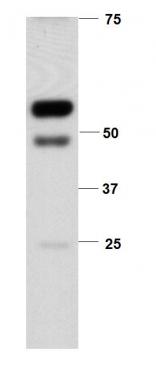
Western Blot: MMP-12 Antibody [NBP1-31225] - Whole cell extract (30 ug) was separated by 10% SDS-PAGE, and the membrane was blotted with MMP12 antibody [N3C1], Internal diluted at 1:1000. The HRP-conjugated anti-rabbit IgG antibody (NBP2-19301) was used to detect the primary antibody.![Immunocytochemistry/ Immunofluorescence: MMP-12 Antibody [NBP1-31225]](https://resources.rndsystems.com/images/products/MMP-12-Antibody-Immunocytochemistry-Immunofluorescence-NBP1-31225-img0007.jpg "Immunocytochemistry/ Immunofluorescence: MMP-12 Antibody [NBP1-31225]")
Immunocytochemistry/ Immunofluorescence: MMP-12 Antibody [NBP1-31225]
Immunocytochemistry/Immunofluorescence: MMP-12 Antibody [NBP1-31225] - Analysis of methanol-fixed H1299, using antibody at 1:500 dilution.![Immunohistochemistry-Paraffin: MMP-12 Antibody [NBP1-31225]](https://resources.rndsystems.com/images/products/MMP-12-Antibody-Immunohistochemistry-Paraffin-NBP1-31225-img0013.jpg "Immunohistochemistry-Paraffin: MMP-12 Antibody [NBP1-31225]")
Immunohistochemistry-Paraffin: MMP-12 Antibody [NBP1-31225]
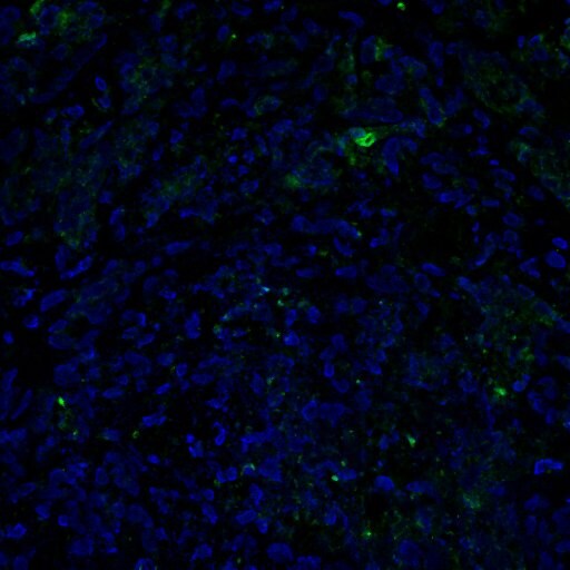
Immunohistochemistry-Paraffin: MMP-12 Antibody [NBP1-31225] - Imaging of Human breast cancer tissue. MMP12 is stained by antibody (green) and nucleus is stained with DAPI (blue). Image submitted by a verified customer review.![Immunohistochemistry-Paraffin: MMP-12 Antibody [NBP1-31225]](https://resources.rndsystems.com/images/products/MMP-12-Antibody-Immunohistochemistry-Paraffin-NBP1-31225-img0008.jpg "Immunohistochemistry-Paraffin: MMP-12 Antibody [NBP1-31225]")
Immunohistochemistry-Paraffin: MMP-12 Antibody [NBP1-31225]
Immunohistochemistry-Paraffin: MMP-12 Antibody [NBP1-31225] - DLD1 xenograft, using MMP12 antibody at 1:500 dilution. Antigen Retrieval: Trilogy™ (EDTA based, pH 8.0) buffer, 15min.![Immunohistochemistry-Paraffin: MMP-12 Antibody [NBP1-31225]](https://resources.rndsystems.com/images/products/MMP-12-Antibody-Immunohistochemistry-Paraffin-NBP1-31225-img0012.jpg "Immunohistochemistry-Paraffin: MMP-12 Antibody [NBP1-31225]")
Immunohistochemistry-Paraffin: MMP-12 Antibody [NBP1-31225]
Immunohistochemistry-Paraffin: MMP-12 Antibody [NBP1-31225] - Human lung adenocarcinoma. MMP12 antibody [N3C1], Internal diluted at 1:500. Antigen Retrieval: Trilogy™ (EDTA based, pH 8.0) buffer, 15min.
Western Blot: MMP-12 Antibody - BSA Free [NBP1-31225] -
MMP12 expression is essential for DRP1/FOXM1 regulation in HNC cells. (A) The mRNA and protein expression levels of FOXM1 in siDRP1 cells were examined. Quantification of relative FOXM1 expression is shown. (B,C) Western blotting, QPCR and luciferase activity analysis of MMP12 were determined in SAS cells transfected with FOXM1 or vector control in combination with siDRP1 or negative control. Quantification of relative DRP1, Flag‐FOXM1 and MMP12 expressions is shown. (D,E) Western blotting, QPCR and luciferase activity of MMP12 were analyzed in SAS cells transfected with FOXM1 or vector control in combination with Mdivi‐1 treatment. Quantification of relative DRP1, Flag‐FOXM1, and MMP12 expressions is shown. (F) IHC staining patterns of the HNC tumor tissues for DRP1, FOXM1 and MMP12. Scale bar: 100 um. All data presented as mean +/- SD of three independent experiments. Significance calculated using t‐test. * P < 0.05, ** P < 0.01, *** P < 0.001. Image collected and cropped by CiteAb from the following open publication (https://pubmed.ncbi.nlm.nih.gov/35313071), licensed under a CC-BY license. Not internally tested by Novus Biologicals.
Western Blot: MMP-12 Antibody - BSA Free [NBP1-31225] -
MMP12 is one of the targets of DRP1 in HNC cells. (A) Heat‐map showing relative alteration of target genes belonging to EMT molecules using QPCR array analysis of SAS cell transfected with siDRP1 compared with the negative control. Red: upregulation; green: downregulation. (B) QPCR was analyzed to validate the expressions of target genes from (A). (C) Impact of DRP1 knockdown or Mdivi‐1 on MMP12 protein expression were demonstrated in SAS and HSC‐3 cells. Quantification of relative DRP1 and MMP12 expressions are shown. (D,E) Cell growth and motility were evaluated in DRP1‐depleted cells transfected with MMP12 using MTT and Transwell assays. Quantification of relative Flag‐MMP12 expression is shown. (F) Studies from GEPIA and Oncomine datasets present the increase of MMP12 mRNA in HNC samples. (G) A positive correlation between DRP1 mRNA and MMP12 mRNA was found in the Oncomine cohort (Peng database, n = 41; Estilo database, n = 31). All data are presented as mean +/- SD of three independent experiments. Significance was calculated using t‐test. In (D) (cell growth) and (F) (GEPIA), statistical analyses were performed using one‐way ANOVA followed by Tukey’s multiple comparison’s test and Wilcoxon signed‐rank test, respectively. * P < 0.05, ** P < 0.01, *** P < 0.001. Image collected and cropped by CiteAb from the following open publication (https://pubmed.ncbi.nlm.nih.gov/35313071), licensed under a CC-BY license. Not internally tested by Novus Biologicals.
Western Blot: MMP-12 Antibody - BSA Free [NBP1-31225] -
MMP12 expression is essential for DRP1/FOXM1 regulation in HNC cells. (A) The mRNA and protein expression levels of FOXM1 in siDRP1 cells were examined. Quantification of relative FOXM1 expression is shown. (B,C) Western blotting, QPCR and luciferase activity analysis of MMP12 were determined in SAS cells transfected with FOXM1 or vector control in combination with siDRP1 or negative control. Quantification of relative DRP1, Flag‐FOXM1 and MMP12 expressions is shown. (D,E) Western blotting, QPCR and luciferase activity of MMP12 were analyzed in SAS cells transfected with FOXM1 or vector control in combination with Mdivi‐1 treatment. Quantification of relative DRP1, Flag‐FOXM1, and MMP12 expressions is shown. (F) IHC staining patterns of the HNC tumor tissues for DRP1, FOXM1 and MMP12. Scale bar: 100 um. All data presented as mean +/- SD of three independent experiments. Significance calculated using t‐test. * P < 0.05, ** P < 0.01, *** P < 0.001. Image collected and cropped by CiteAb from the following open publication (https://pubmed.ncbi.nlm.nih.gov/35313071), licensed under a CC-BY license. Not internally tested by Novus Biologicals.
Western Blot: MMP-12 Antibody - BSA Free [NBP1-31225] -
MMP12 is one of the targets of DRP1 in HNC cells. (A) Heat‐map showing relative alteration of target genes belonging to EMT molecules using QPCR array analysis of SAS cell transfected with siDRP1 compared with the negative control. Red: upregulation; green: downregulation. (B) QPCR was analyzed to validate the expressions of target genes from (A). (C) Impact of DRP1 knockdown or Mdivi‐1 on MMP12 protein expression were demonstrated in SAS and HSC‐3 cells. Quantification of relative DRP1 and MMP12 expressions are shown. (D,E) Cell growth and motility were evaluated in DRP1‐depleted cells transfected with MMP12 using MTT and Transwell assays. Quantification of relative Flag‐MMP12 expression is shown. (F) Studies from GEPIA and Oncomine datasets present the increase of MMP12 mRNA in HNC samples. (G) A positive correlation between DRP1 mRNA and MMP12 mRNA was found in the Oncomine cohort (Peng database, n = 41; Estilo database, n = 31). All data are presented as mean +/- SD of three independent experiments. Significance was calculated using t‐test. In (D) (cell growth) and (F) (GEPIA), statistical analyses were performed using one‐way ANOVA followed by Tukey’s multiple comparison’s test and Wilcoxon signed‐rank test, respectively. * P < 0.05, ** P < 0.01, *** P < 0.001. Image collected and cropped by CiteAb from the following open publication (https://pubmed.ncbi.nlm.nih.gov/35313071), licensed under a CC-BY license. Not internally tested by Novus Biologicals.Applications for MMP-12 Antibody - BSA Free
Immunocytochemistry/ Immunofluorescence
Immunohistochemistry
Immunohistochemistry-Paraffin
Western Blot
Reviewed Applications
Read 2 reviews rated 4 using NBP1-31225 in the following applications:
Formulation, Preparation, and Storage
Purification
Formulation
Format
Preservative
Concentration
Shipping
Stability & Storage
Background: MMP-12
Long Name
Alternate Names
Gene Symbol
Additional MMP-12 Products
Product Documents for MMP-12 Antibody - BSA Free
Certificate of Analysis
To download a Certificate of Analysis, please enter a lot or batch number in the search box below.
Product Specific Notices for MMP-12 Antibody - BSA Free
This product is for research use only and is not approved for use in humans or in clinical diagnosis. Primary Antibodies are guaranteed for 1 year from date of receipt.
⚠ WARNING: This product can expose you to chemicals including mercury, which is known to the State of California to cause reproductive toxicity with developmental effects. For more information go to www.P65Warnings.ca.gov.Related Research Areas
Citations for MMP-12 Antibody - BSA Free
 Powered by Bioz
Customer Reviews for MMP-12 Antibody - BSA Free (2)
Have you used MMP-12 Antibody - BSA Free?
Submit a review and receive an Amazon gift card!
$25/€18/£15/$25CAN/¥2500 Yen for a review with an image
$10/€7/£6/$10CAN/¥1110 Yen for a review without an image
Submit a review
Customer Images
-
Application: Immunohistochemistry-ParaffinSample Tested: Breast cancer tissueSpecies: HumanVerified Customer | Posted 02/22/2017MMP12-green fluorochrome, nucleus-DAPI

-
Application: Western BlotSample Tested: Mouse adipose tissue homogenate, Sample Amount: 20ugSpecies: MouseVerified Customer | Posted 06/21/2011
There are no reviews that match your criteria.
Protocols
Find general support by application which include: protocols, troubleshooting, illustrated assays, videos and webinars.
- Antigen Retrieval Protocol (PIER)
- Antigen Retrieval for Frozen Sections Protocol
- Appropriate Fixation of IHC/ICC Samples
- Cellular Response to Hypoxia Protocols
- Chromogenic IHC Staining of Formalin-Fixed Paraffin-Embedded (FFPE) Tissue Protocol
- Chromogenic Immunohistochemistry Staining of Frozen Tissue
- ClariTSA™ Fluorophore Kits
- Detection & Visualization of Antibody Binding
- Fluorescent IHC Staining of Frozen Tissue Protocol
- Graphic Protocol for Heat-induced Epitope Retrieval
- Graphic Protocol for the Preparation and Fluorescent IHC Staining of Frozen Tissue Sections
- Graphic Protocol for the Preparation and Fluorescent IHC Staining of Paraffin-embedded Tissue Sections
- Graphic Protocol for the Preparation of Gelatin-coated Slides for Histological Tissue Sections
- ICC Cell Smear Protocol for Suspension Cells
- ICC Immunocytochemistry Protocol Videos
- ICC for Adherent Cells
- IHC Sample Preparation (Frozen sections vs Paraffin)
- Immunocytochemistry (ICC) Protocol
- Immunocytochemistry Troubleshooting
- Immunofluorescence of Organoids Embedded in Cultrex Basement Membrane Extract
- Immunofluorescent IHC Staining of Formalin-Fixed Paraffin-Embedded (FFPE) Tissue Protocol
- Immunohistochemistry (IHC) and Immunocytochemistry (ICC) Protocols
- Immunohistochemistry Frozen Troubleshooting
- Immunohistochemistry Paraffin Troubleshooting
- Preparing Samples for IHC/ICC Experiments
- Preventing Non-Specific Staining (Non-Specific Binding)
- Primary Antibody Selection & Optimization
- Protocol for Heat-Induced Epitope Retrieval (HIER)
- Protocol for Making a 4% Formaldehyde Solution in PBS
- Protocol for VisUCyte™ HRP Polymer Detection Reagent
- Protocol for the Fluorescent ICC Staining of Cell Smears - Graphic
- Protocol for the Fluorescent ICC Staining of Cultured Cells on Coverslips - Graphic
- Protocol for the Preparation & Fixation of Cells on Coverslips
- Protocol for the Preparation and Chromogenic IHC Staining of Frozen Tissue Sections
- Protocol for the Preparation and Chromogenic IHC Staining of Frozen Tissue Sections - Graphic
- Protocol for the Preparation and Chromogenic IHC Staining of Paraffin-embedded Tissue Sections
- Protocol for the Preparation and Chromogenic IHC Staining of Paraffin-embedded Tissue Sections - Graphic
- Protocol for the Preparation and Fluorescent ICC Staining of Cells on Coverslips
- Protocol for the Preparation and Fluorescent ICC Staining of Non-adherent Cells
- Protocol for the Preparation and Fluorescent ICC Staining of Stem Cells on Coverslips
- Protocol for the Preparation and Fluorescent IHC Staining of Frozen Tissue Sections
- Protocol for the Preparation and Fluorescent IHC Staining of Paraffin-embedded Tissue Sections
- Protocol for the Preparation of Gelatin-coated Slides for Histological Tissue Sections
- Protocol for the Preparation of a Cell Smear for Non-adherent Cell ICC - Graphic
- R&D Systems Quality Control Western Blot Protocol
- TUNEL and Active Caspase-3 Detection by IHC/ICC Protocol
- The Importance of IHC/ICC Controls
- Troubleshooting Guide: Immunohistochemistry
- Troubleshooting Guide: Western Blot Figures
- Western Blot Conditions
- Western Blot Protocol
- Western Blot Protocol for Cell Lysates
- Western Blot Troubleshooting
- Western Blot Troubleshooting Guide
- View all Protocols, Troubleshooting, Illustrated assays and Webinars
FAQs for MMP-12 Antibody - BSA Free
-
Q: Does this antibody detect the catalytic domain alone of this protein?
A:
The immunogen for this protein is between amino acids 213 and 307 of MMP12 (UniProt ID#P39900). This corresponds to a portion of the Hemopexin-like 1 domain on UniProt and does not cover the whole catalytic domain (ZnMc_MMP) which is between amino acids 109 and 263. Please check NCBI Gene ID: 4321 (https://www.ncbi.nlm.nih.gov/gene/?term=4321) for more information.