MMP-7 Antibody (8T4R8)
Novus Biologicals | Catalog # NBP3-16067
Recombinant Monoclonal Antibody
![Western Blot: MMP-7 Antibody (8T4R8) [NBP3-16067]](https://resources.rndsystems.com/images/products/MMP-7-Antibody-9D8S10-Western-Blot-NBP3-16067-img0005.jpg "Western Blot: MMP-7 Antibody (8T4R8) [NBP3-16067]")
Loading...
Key Product Details
Species Reactivity
Human
Applications
Immunohistochemistry, Immunohistochemistry-Paraffin, Western Blot
Label
Unconjugated
Antibody Source
Recombinant Monoclonal Rabbit IgG Clone # 8T4R8 expressed in HEK293
Loading...
Product Specifications
Immunogen
Recombinant fusion protein containing a sequence corresponding to amino acids 1-267 of human MMP7 (P09237). MRLTVLCAVCLLPGSLALPLPQEAGGMSELQWEQAQDYLKRFYLYDSETKNANSLEAKLKEMQKFFGLPITGMLNSRVIEIMQKPRCGVPDVAEYSLFPNSPKWTSKVVTYRIVSYTRDLPHITVDRLVSKALNMWGKEIPLHFRKVVWGTADIMIGFARGAHGDSYPFDGPGNTLAHAFAPGTGLGGDAHFDEDERWTDGSSLGINFLYAATHELGHSLGMGHSSDPNAVMYPTYGNGDPQNFKLSQDDIKGIQKLYGKRSNSRKK
Clonality
Monoclonal
Host
Rabbit
Isotype
IgG
Scientific Data Images for MMP-7 Antibody (8T4R8)
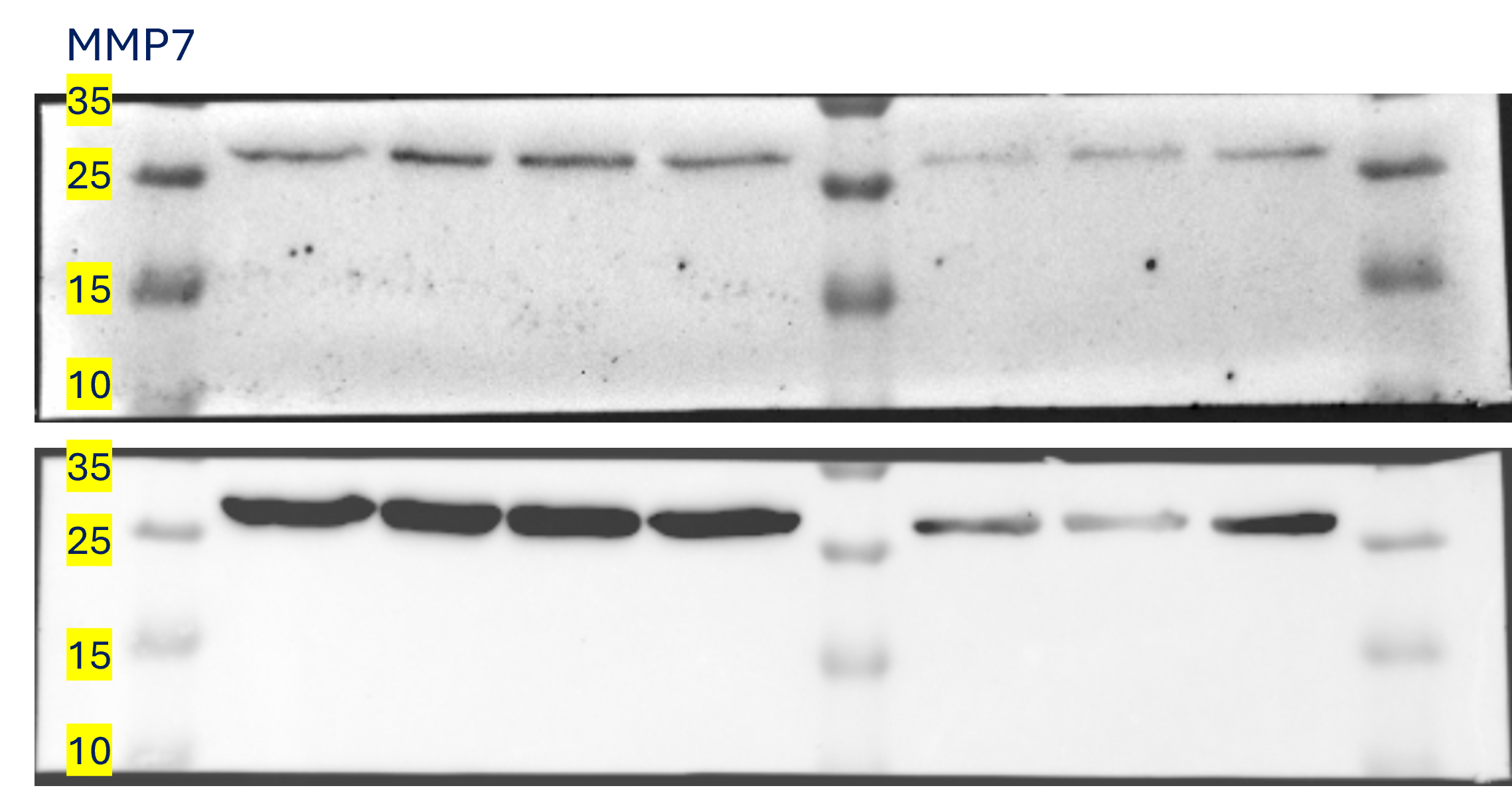
Western Blot: MMP-7 Antibody (8T4R8) [NBP3-16067]
Western Blot: MMP-7 Antibody (9D8S10) [NBP3-16067] - Analysis of extracts of various cell lines, using MMP-7 antibody (NBP3-16067 ) at 1:1000 dilution. Secondary antibody: HRP Goat Anti-Rabbit IgG (H+L) at 1:10000 dilution. Lysates/proteins: 25ug per lane. Blocking buffer: 3% nonfat dry milk in TBST. Detection: ECL Basic Kit. Exposure time: 180s. [NBP3-16067] -")
Immunohistochemistry: MMP-7 Antibody (8T4R8) [NBP3-16067] -
Immunohistochemistry: MMP-7 Antibody (8T4R8) [NBP3-16067] - Immunohistochemistry analysis of paraffin-embedded Human liver cancer tissue using MMP-7 Rabbit mAb at a dilution of 1:200 (40x lens). High pressure antigen retrieval was performed with 0.01 M citrate buffer (pH 6.0) prior to IHC staining.Applications for MMP-7 Antibody (8T4R8)
Application
Recommended Usage
Immunohistochemistry
1:50 - 1:200
Western Blot
1:500 - 1:1000
Reviewed Applications
Read 1 review rated 4 using NBP3-16067 in the following applications:
Formulation, Preparation, and Storage
Purification
Affinity purified
Formulation
PBS (pH 7.3), 50% glycerol, 0.05% BSA
Preservative
0.02% Sodium Azide
Concentration
Please see the vial label for concentration. If unlisted please contact technical services.
Shipping
The product is shipped with polar packs. Upon receipt, store it immediately at the temperature recommended below.
Stability & Storage
Store at -20C. Avoid freeze-thaw cycles.
Background: MMP-7
Long Name
Matrix Metalloproteinase 7
Alternate Names
Matrilysin, MMP7
Gene Symbol
MMP7
Additional MMP-7 Products
Product Documents for MMP-7 Antibody (8T4R8)
Certificate of Analysis
To download a Certificate of Analysis, please enter a lot or batch number in the search box below.
Product Specific Notices for MMP-7 Antibody (8T4R8)
This product is for research use only and is not approved for use in humans or in clinical diagnosis. Primary Antibodies are guaranteed for 1 year from date of receipt.
Related Research Areas
Customer Reviews for MMP-7 Antibody (8T4R8) (1)
4 out of 5
1 Customer Rating
Have you used MMP-7 Antibody (8T4R8)?
Submit a review and receive an Amazon gift card!
$25/€18/£15/$25CAN/¥2500 Yen for a review with an image
$10/€7/£6/$10CAN/¥1110 Yen for a review without an image
Submit a review
Customer Images
Showing
1
-
1 of
1 review
Showing All
Filter By:
-
Application: Western BlotSample Tested: Cancer Cell lysate , human macrophage whole cell lysate and fibroblast cellsSpecies: HumanVerified Customer | Posted 05/24/2024MMP-7 is a small molecule secreted protein in the MMP family. We have attempted to detect it in various cancer cell lines and stromal cells. It is strongly expressed in tumor-associated fibroblasts and macrophages.

There are no reviews that match your criteria.
Protocols
Find general support by application which include: protocols, troubleshooting, illustrated assays, videos and webinars.
- Antigen Retrieval Protocol (PIER)
- Antigen Retrieval for Frozen Sections Protocol
- Appropriate Fixation of IHC/ICC Samples
- Cellular Response to Hypoxia Protocols
- Chromogenic IHC Staining of Formalin-Fixed Paraffin-Embedded (FFPE) Tissue Protocol
- Chromogenic Immunohistochemistry Staining of Frozen Tissue
- ClariTSA™ Fluorophore Kits
- Detection & Visualization of Antibody Binding
- Fluorescent IHC Staining of Frozen Tissue Protocol
- Graphic Protocol for Heat-induced Epitope Retrieval
- Graphic Protocol for the Preparation and Fluorescent IHC Staining of Frozen Tissue Sections
- Graphic Protocol for the Preparation and Fluorescent IHC Staining of Paraffin-embedded Tissue Sections
- Graphic Protocol for the Preparation of Gelatin-coated Slides for Histological Tissue Sections
- IHC Sample Preparation (Frozen sections vs Paraffin)
- Immunofluorescent IHC Staining of Formalin-Fixed Paraffin-Embedded (FFPE) Tissue Protocol
- Immunohistochemistry (IHC) and Immunocytochemistry (ICC) Protocols
- Immunohistochemistry Frozen Troubleshooting
- Immunohistochemistry Paraffin Troubleshooting
- Preparing Samples for IHC/ICC Experiments
- Preventing Non-Specific Staining (Non-Specific Binding)
- Primary Antibody Selection & Optimization
- Protocol for Heat-Induced Epitope Retrieval (HIER)
- Protocol for Making a 4% Formaldehyde Solution in PBS
- Protocol for VisUCyte™ HRP Polymer Detection Reagent
- Protocol for the Preparation & Fixation of Cells on Coverslips
- Protocol for the Preparation and Chromogenic IHC Staining of Frozen Tissue Sections
- Protocol for the Preparation and Chromogenic IHC Staining of Frozen Tissue Sections - Graphic
- Protocol for the Preparation and Chromogenic IHC Staining of Paraffin-embedded Tissue Sections
- Protocol for the Preparation and Chromogenic IHC Staining of Paraffin-embedded Tissue Sections - Graphic
- Protocol for the Preparation and Fluorescent IHC Staining of Frozen Tissue Sections
- Protocol for the Preparation and Fluorescent IHC Staining of Paraffin-embedded Tissue Sections
- Protocol for the Preparation of Gelatin-coated Slides for Histological Tissue Sections
- R&D Systems Quality Control Western Blot Protocol
- TUNEL and Active Caspase-3 Detection by IHC/ICC Protocol
- The Importance of IHC/ICC Controls
- Troubleshooting Guide: Immunohistochemistry
- Troubleshooting Guide: Western Blot Figures
- Western Blot Conditions
- Western Blot Protocol
- Western Blot Protocol for Cell Lysates
- Western Blot Troubleshooting
- Western Blot Troubleshooting Guide
- View all Protocols, Troubleshooting, Illustrated assays and Webinars
Loading...