MMR/CD206/Mannose Receptor Antibody - BSA Free
Novus Biologicals | Catalog # NBP1-90020
![MMR/CD206/Mannose Receptor Antibody - BSA Free Immunohistochemistry-Paraffin: MMR/CD206/Mannose Receptor Antibody - BSA Free [NBP1-90020]](https://resources.rndsystems.com/images/products/nbp1-90020_rabbit-mmr-cd206-mannose-receptor-pab-bsa-free-15520258294298.jpg "Immunohistochemistry-Paraffin: MMR/CD206/Mannose Receptor Antibody - BSA Free [NBP1-90020]")
Key Product Details
Validated by
Species Reactivity
Validated:
Cited:
Applications
Validated:
Cited:
Label
Antibody Source
Format
Product Specifications
Immunogen
Reactivity Notes
Clonality
Host
Isotype
Scientific Data Images for MMR/CD206/Mannose Receptor Antibody - BSA Free
![MMR/CD206/Mannose Receptor Antibody - BSA Free Western Blot: MMR/CD206/Mannose Receptor Antibody - BSA Free [NBP1-90020]](https://resources.rndsystems.com/images/products/nbp1-90020_rabbit-mmr-cd206-mannose-receptor-pab-bsa-free-1552025821245.jpg "Western Blot: MMR/CD206/Mannose Receptor Antibody - BSA Free [NBP1-90020]")
Western Blot: MMR/CD206/Mannose Receptor Antibody - BSA Free [NBP1-90020]
Analysis in human liver tissue.
MMR/CD206/Mannose Receptor Antibody-NBP1-90020
Staining of human lung shows strong cytoplasmic positivity in macrophages.
MMR/CD206/Mannose Receptor Antibody-NBP1-90020
Staining of human placenta shows strong cytoplasmic positivity in Hofbauer cells.![MMR/CD206/Mannose Receptor Antibody - BSA Free Immunohistochemistry-Paraffin: MMR/CD206/Mannose Receptor Antibody - BSA Free [NBP1-90020]](https://resources.rndsystems.com/images/products/nbp1-90020_-immunohistochemistry-paraffin-639192267712343557.jpg "Immunohistochemistry-Paraffin: MMR/CD206/Mannose Receptor Antibody - BSA Free [NBP1-90020]")
Immunohistochemistry-Paraffin: MMR/CD206/Mannose Receptor Antibody - BSA Free [NBP1-90020]
Staining of human cerebral cortex shows no cytoplasmic positivity in neurons as expected.![MMR/CD206/Mannose Receptor Antibody - BSA Free Immunohistochemistry-Paraffin: MMR/CD206/Mannose Receptor Antibody - BSA Free [NBP1-90020]](https://resources.rndsystems.com/images/products/nbp1-90020_-immunohistochemistry-paraffin-639192267712649682.jpg "Immunohistochemistry-Paraffin: MMR/CD206/Mannose Receptor Antibody - BSA Free [NBP1-90020]")
![MMR/CD206/Mannose Receptor Antibody - BSA Free Immunohistochemistry-Paraffin: MMR/CD206/Mannose Receptor Antibody - BSA Free [NBP1-90020]](https://resources.rndsystems.com/images/products/nbp1-90020_-immunohistochemistry-paraffin-639192267713180927.jpg "Immunohistochemistry-Paraffin: MMR/CD206/Mannose Receptor Antibody - BSA Free [NBP1-90020]")
Immunohistochemistry-Paraffin: MMR/CD206/Mannose Receptor Antibody - BSA Free [NBP1-90020]
Staining of human liver shows moderate cytoplasmic positivity in Kupffer cells.![MMR/CD206/Mannose Receptor Antibody - BSA Free Immunohistochemistry-Paraffin: MMR/CD206/Mannose Receptor Antibody - BSA Free [NBP1-90020]](https://resources.rndsystems.com/images/products/nbp1-90020_-immunohistochemistry-paraffin-639192267712303248.jpg "Immunohistochemistry-Paraffin: MMR/CD206/Mannose Receptor Antibody - BSA Free [NBP1-90020]")
Immunohistochemistry-Paraffin: MMR/CD206/Mannose Receptor Antibody - BSA Free [NBP1-90020]
Staining of human lung shows strong cytoplasmic positivity in macrophages.![MMR/CD206/Mannose Receptor Antibody - BSA Free Immunohistochemistry-Paraffin: MMR/CD206/Mannose Receptor Antibody - BSA Free [NBP1-90020]](https://resources.rndsystems.com/images/products/nbp1-90020_-immunohistochemistry-paraffin-639192267715580522.jpg "Immunohistochemistry-Paraffin: MMR/CD206/Mannose Receptor Antibody - BSA Free [NBP1-90020]")
Immunohistochemistry-Paraffin: MMR/CD206/Mannose Receptor Antibody - BSA Free [NBP1-90020]
Staining of human placenta shows strong cytoplasmic positivity in Hofbauer cells.![MMR/CD206/Mannose Receptor Antibody - BSA Free Immunohistochemistry-Paraffin: MMR/CD206/Mannose Receptor Antibody - BSA Free [NBP1-90020]](https://resources.rndsystems.com/images/products/nbp1-90020_-immunohistochemistry-paraffin-639192267715018121.jpg "Immunohistochemistry-Paraffin: MMR/CD206/Mannose Receptor Antibody - BSA Free [NBP1-90020]")
![MMR/CD206/Mannose Receptor Antibody - BSA Free Western Blot: MMR/CD206/Mannose Receptor Antibody - BSA Free [NBP1-90020]](https://resources.rndsystems.com/images/products/nbp1-90020_-western-blot-639192267715649286.jpg "Western Blot: MMR/CD206/Mannose Receptor Antibody - BSA Free [NBP1-90020]")
Western Blot: MMR/CD206/Mannose Receptor Antibody - BSA Free [NBP1-90020]
Analysis in human liver tissue.Applications for MMR/CD206/Mannose Receptor Antibody - BSA Free
Immunohistochemistry-Paraffin
Western Blot
Reviewed Applications
Read 1 review rated 4 using NBP1-90020 in the following applications:
Formulation, Preparation, and Storage
Purification
Formulation
Format
Preservative
Concentration
Shipping
Stability & Storage
Background: MMR/CD206
Long Name
Alternate Names
Gene Symbol
Additional MMR/CD206 Products
Product Documents for MMR/CD206/Mannose Receptor Antibody - BSA Free
Certificate of Analysis
To download a Certificate of Analysis, please enter a lot or batch number in the search box below.
Product Specific Notices for MMR/CD206/Mannose Receptor Antibody - BSA Free
This product is for research use only and is not approved for use in humans or in clinical diagnosis. Primary Antibodies are guaranteed for 1 year from date of receipt.
Citations for MMR/CD206/Mannose Receptor Antibody - BSA Free
 Powered by Bioz
Customer Reviews for MMR/CD206/Mannose Receptor Antibody - BSA Free (1)
Have you used MMR/CD206/Mannose Receptor Antibody - BSA Free?
Submit a review and receive an Amazon gift card!
$25/€18/£15/$25CAN/¥2500 Yen for a review with an image
$10/€7/£6/$10CAN/¥1110 Yen for a review without an image
Submit a review
Customer Images
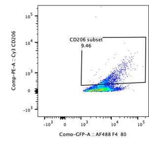
-
Application: Flow CytometrySample Tested: LiverSpecies: MouseVerified Customer | Posted 08/09/2019

There are no reviews that match your criteria.
Protocols
Find general support by application which include: protocols, troubleshooting, illustrated assays, videos and webinars.
- Antigen Retrieval Protocol (PIER)
- Antigen Retrieval for Frozen Sections Protocol
- Appropriate Fixation of IHC/ICC Samples
- Cellular Response to Hypoxia Protocols
- Chromogenic IHC Staining of Formalin-Fixed Paraffin-Embedded (FFPE) Tissue Protocol
- Chromogenic Immunohistochemistry Staining of Frozen Tissue
- ClariTSA™ Fluorophore Kits
- Detection & Visualization of Antibody Binding
- Fluorescent IHC Staining of Frozen Tissue Protocol
- Graphic Protocol for Heat-induced Epitope Retrieval
- Graphic Protocol for the Preparation and Fluorescent IHC Staining of Frozen Tissue Sections
- Graphic Protocol for the Preparation and Fluorescent IHC Staining of Paraffin-embedded Tissue Sections
- Graphic Protocol for the Preparation of Gelatin-coated Slides for Histological Tissue Sections
- IHC Sample Preparation (Frozen sections vs Paraffin)
- Immunofluorescent IHC Staining of Formalin-Fixed Paraffin-Embedded (FFPE) Tissue Protocol
- Immunohistochemistry (IHC) and Immunocytochemistry (ICC) Protocols
- Immunohistochemistry Frozen Troubleshooting
- Immunohistochemistry Paraffin Troubleshooting
- Preparing Samples for IHC/ICC Experiments
- Preventing Non-Specific Staining (Non-Specific Binding)
- Primary Antibody Selection & Optimization
- Protocol for Heat-Induced Epitope Retrieval (HIER)
- Protocol for Making a 4% Formaldehyde Solution in PBS
- Protocol for VisUCyte™ HRP Polymer Detection Reagent
- Protocol for the Preparation & Fixation of Cells on Coverslips
- Protocol for the Preparation and Chromogenic IHC Staining of Frozen Tissue Sections
- Protocol for the Preparation and Chromogenic IHC Staining of Frozen Tissue Sections - Graphic
- Protocol for the Preparation and Chromogenic IHC Staining of Paraffin-embedded Tissue Sections
- Protocol for the Preparation and Chromogenic IHC Staining of Paraffin-embedded Tissue Sections - Graphic
- Protocol for the Preparation and Fluorescent IHC Staining of Frozen Tissue Sections
- Protocol for the Preparation and Fluorescent IHC Staining of Paraffin-embedded Tissue Sections
- Protocol for the Preparation of Gelatin-coated Slides for Histological Tissue Sections
- R&D Systems Quality Control Western Blot Protocol
- TUNEL and Active Caspase-3 Detection by IHC/ICC Protocol
- The Importance of IHC/ICC Controls
- Troubleshooting Guide: Immunohistochemistry
- Troubleshooting Guide: Western Blot Figures
- Western Blot Conditions
- Western Blot Protocol
- Western Blot Protocol for Cell Lysates
- Western Blot Troubleshooting
- Western Blot Troubleshooting Guide
- View all Protocols, Troubleshooting, Illustrated assays and Webinars
FAQs for MMR/CD206/Mannose Receptor Antibody - BSA Free
-
Q: Is this product available for immunofluorescence?
A: NBP1-90020 has only been validated in western blot and IHC on paraffin-embedded tissues. If you would be using fluorescence detection on tissues, it will work for your assay. If you want to use it to detect cells, then note the alternative antibody NBP1-95964 has been validated for immunocytochemistry and may be a better choice for you.