CCL11 is a potent eosinophil chemoattractant that was originally purified from bronchoalveolar lavage fluid of guinea pigs sensitized by aerosol challenge with ovalbumin. Microsequencing of the purified protein revealed the guinea pig CCL11 to be a member of the beta (CC) chemokine family of inflammatory and immunoregulatory cytokines. cDNA clones for guinea pig, mouse and human CCL11 have been isolated. Mouse CCL11 cDNA encodes a 97 amino acid residue precursor protein from which the amino-terminal 23 amino acid residues are cleaved to generate the 74 amino acid residue mature mouse CCL11. At the protein sequence level, mature mouse CCL11 is approximately 60% identical to mature human and guinea pig CCL11. In addition, mouse CCL11 also shows high amino acid sequence identity to members of the MCP family. Mouse CCL11 is chemotactic for eosinophils, but not mononuclear cells or neutrophils. CCL11 mRNA is expressed in a variety of tissues. The expression of CCL11 mRNA is induced in cultured endothelial cells in response to IFN-gamma. In addition, CCL11 mRNA is also induced in response to the transplantation of IL-4-secreting tumor cells. The CC chemokine receptor 3 (CCR3) has now been identified to be a specific human CCL11 receptor.

Key Product Details
Species Reactivity
Validated:
Mouse
Cited:
Human, Mouse, Rat
Applications
Validated:
Immunohistochemistry, Western Blot, ELISA Capture (Matched Antibody Pair), Neutralization
Cited:
Immunohistochemistry, Immunohistochemistry-Paraffin, Immunohistochemistry-Frozen, Western Blot, Neutralization, Flow Cytometry, ELISA Development, Luminex Development
Label
Unconjugated
Antibody Source
Polyclonal Goat IgG
Loading...
Product Specifications
Immunogen
E. coli-derived recombinant mouse CCL11/Eotaxin
His24-Pro97
Accession # P48298
His24-Pro97
Accession # P48298
Specificity
Detects mouse CCL11/Eotaxin in ELISAs and Western blots. In direct ELISAs, approximately 20% cross-reactivity with recombinant human CCL11/Eotaxin is observed, and less than 5% cross-reactivity with recombinant mouse JE is observed.
Clonality
Polyclonal
Host
Goat
Isotype
IgG
Endotoxin Level
<0.10 EU per 1 μg of the antibody by the LAL method.
Scientific Data Images for Mouse CCL11/Eotaxin Antibody
Chemotaxis Induced by CCL11/Eotaxin and Neutral-ization by Mouse CCL11/ Eotaxin Antibody.
Recombinant Mouse CCL11/ Eotaxin (Catalog # 420-ME) chemoattracts the BaF3 mouse pro-B cell line transfected with mouse CCR3 in a dose-dependent manner (orange line). The amount of cells that migrated through to the lower chemotaxis chamber was measured by Resazurin (Catalog # AR002). Chemotaxis elicited by Recombinant Mouse CCL11/ Eotaxin (10 ng/mL) is neutralized (green line) by increasing concentrations of Goat Anti-Mouse CCL11/Eotaxin Antigen Affinity-purified Polyclonal Antibody (Catalog # AF-420-NA). The ND50 is typically 0.1-0.5 µg/mL.
CCL11/Eotaxin in Mouse Thymus.
CCL11/Eotaxin was detected in perfusion fixed frozen sections of mouse thymus using Goat Anti-Mouse CCL11/Eotaxin Antigen Affinity-purified Polyclonal Antibody (Catalog # AF-420-NA) at 5 µg/mL overnight at 4 °C. Tissue was stained using the NorthernLights™ 557-conjugated Anti-Goat IgG Secondary Antibody (red; Catalog # NL001) and counterstained with DAPI (blue). Specific staining was localized to cytoplasm. View our protocol for Fluorescent IHC Staining of Frozen Tissue Sections.
CCL11/Eotaxin in Mouse Colon.
CCL11/Eotaxin was detected in perfusion fixed frozen sections of mouse colon using Goat Anti-Mouse CCL11/Eotaxin Antigen Affinity-purified Polyclonal Antibody (Catalog # AF-420-NA) at 15 µg/mL overnight at 4 °C. Tissue was stained using the Anti-Goat HRP-DAB Cell & Tissue Staining Kit (brown; Catalog # CTS008) and counterstained with hematoxylin (blue). Specific staining was localized to cytoplasm. View our protocol for Chromogenic IHC Staining of Frozen Tissue Sections.
Detection of CCL11/Eotaxin by Flow Cytometry
IL4R alpha Signalling Stimulates CCL11 Production by mTEC.Analysis of TEC subsets in WT and Il4ra-/- mice at d7 post SLI (A), n=9, and (B) analysis of TEC (n=6-8) and T-cell development at d35 (n=11). (C) Representative FACS plots of thymic eosinophils in steady state WT and Il4ra-/- mice. Quantitation of thymus cellularity and thymic eosinophils at d0 (D) and d1 (E) in WT and Il4ra-/- mice, n=9 at d0, n=7-8 at d1. (F) Representative FACs plots of CCL11 expression in thymic stromal populations, showing proportions (G) and quantitation at d0 and d1 (H), with MFI of CCL11 for mTEClo, n=8. (I) qPCR analysis of Ccl11 mRNA expression E15 2-deoxyguanosine thymic organ cultures, treated for 4 days +/- IL4/IL13, with analysis conducted using a one-way ANOVA with Bonferroni post-test. (J) Representative FACS plots for expression of CCL11 by mTEClo in WT (left panel) and Il4ra-/- (right panel) with quantitation shown in (K), n=8. All data from at least 2-3 independent experiments. All bars show mean ± SEM, * p<0.05, ** p<0.01, *** p<0.001, **** p<0.0001 from an unpaired students t-test unless otherwise specified. Image collected and cropped by CiteAb from the following open publication (https://pubmed.ncbi.nlm.nih.gov/35275754), licensed under a CC-BY license. Not internally tested by R&D Systems.
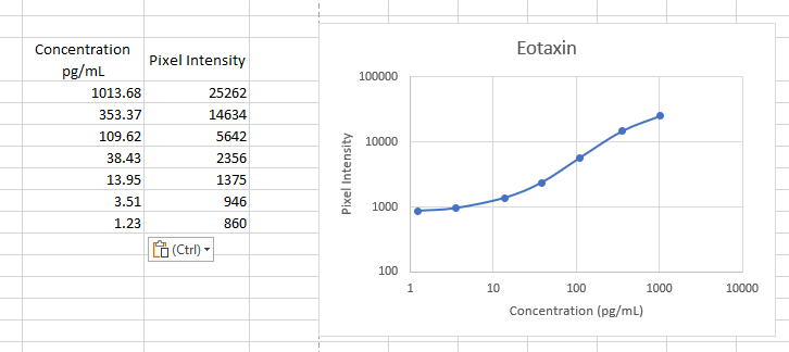
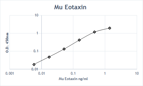
Mouse CCL11 / Eotaxin ELISA Standard Curve
Recombinant Mouse CCL11/Eotaxin (Catalog # 420-ME) was serially diluted and captured by Goat Anti-Mouse CCL11/Eotaxin Antigen Affinity-purified Polyclonal Antibody (Catalog # AF-420-NA) coated on a Clear Polystyrene Microplate (Catalog # DY990). Goat Anti-Mouse CCL11/Eotaxin Antigen Affinity-purified Polyclonal Antibody (Catalog # AF-420-NA) was biotinylated and incubated with the protein captured on the plate. Detection of the standard curve was achieved by incubating Streptavidin-HRP (Catalog # DY998)
Mouse CCL11 / Eotaxin ELISA Standard Curve
Recombinant Mouse CCL11/Eotaxin (Catalog # 420-ME) was serially diluted and captured by Goat Anti-Mouse CCL11/Eotaxin Antigen Affinity-purified Polyclonal Antibody (Catalog # AF-420-NA) coated on a Clear Polystyrene Microplate (Catalog # DY990). Goat Anti-Mouse CCL11/Eotaxin Antigen Affinity-purified Polyclonal Antibody (Catalog # AF-420-NA) was biotinylated and incubated with the protein captured on the plate. Detection of the standard curve was achieved by incubating Streptavidin-HRP (Catalog # DY998)Applications for Mouse CCL11/Eotaxin Antibody
Application
Recommended Usage
Immunohistochemistry
5-15 µg/mL
Sample: Perfusion fixed frozen sections of mouse thymus and mouse colon
Sample: Perfusion fixed frozen sections of mouse thymus and mouse colon
Western Blot
0.1 µg/mL
Sample: Recombinant Mouse CCL11/Eotaxin (Catalog # 420-ME)
Sample: Recombinant Mouse CCL11/Eotaxin (Catalog # 420-ME)
Neutralization
Measured by its ability to neutralize CCL11/Eotaxin-induced chemotaxis in the BaF3 mouse pro‑B cell line transfected with mouse CCR3. The Neutralization Dose (ND50) is typically 0.1-0.5 µg/mL in the presence of 10 ng/mL Recombinant Mouse CCL11/Eotaxin.
Mouse CCL11/Eotaxin Sandwich Immunoassay
Please Note: Optimal dilutions of this antibody should be experimentally determined.
Reviewed Applications
Read 3 reviews rated 4.7 using AF-420-NA in the following applications:
Formulation, Preparation, and Storage
Purification
Antigen Affinity-purified
Reconstitution
Reconstitute at 0.2 mg/mL in sterile PBS. For liquid material, refer to CoA for concentration.
Loading...
Formulation
Lyophilized from a 0.2 μm filtered solution in PBS with Trehalose. *Small pack size (SP) is supplied either lyophilized or as a 0.2 µm filtered solution in PBS.
Shipping
Lyophilized product is shipped at ambient temperature. Liquid small pack size (-SP) is shipped with polar packs. Upon receipt, store immediately at the temperature recommended below.
Stability & Storage
Use a manual defrost freezer and avoid repeated freeze-thaw cycles.
- 12 months from date of receipt, -20 to -70 °C as supplied.
- 1 month, 2 to 8 °C under sterile conditions after reconstitution.
- 6 months, -20 to -70 °C under sterile conditions after reconstitution.
Calculators
Background: CCL11/Eotaxin
References
- Rothenberg, M.E. et al. (1995) Proc. Natl. Acad. Sci. USA 92:8960.
- Kitaura, M. et al. (1996) J. Biol. Chem 271:7725.
- Garcia-Zepeda, E.A. et al. (1996) Nature Medicine 2:449.
- Ponath, P.D. et al. (1996) J. Clin. Invest. 97:604.
Alternate Names
Eotaxin
Gene Symbol
CCL11
UniProt
Additional CCL11/Eotaxin Products
Product Documents for Mouse CCL11/Eotaxin Antibody
Certificate of Analysis
To download a Certificate of Analysis, please enter a lot or batch number in the search box below.
Note: Certificate of Analysis not available for kit components.
Product Specific Notices for Mouse CCL11/Eotaxin Antibody
For research use only
Related Research Areas
Citations for Mouse CCL11/Eotaxin Antibody
 Powered by Bioz
Customer Reviews for Mouse CCL11/Eotaxin Antibody (3)
4.7 out of 5
3 Customer Ratings
Have you used Mouse CCL11/Eotaxin Antibody?
Submit a review and receive an Amazon gift card!
$25/€18/£15/$25CAN/¥2500 Yen for a review with an image
$10/€7/£6/$10CAN/¥1110 Yen for a review without an image
Submit a review
Customer Images
Showing
1
-
3 of
3 reviews
Showing All
Filter By:
-
Application: ELISASample Tested: SerumSpecies: MouseVerified Customer | Posted 11/28/2022Worked as capture antibody

-
Application: ELISASample Tested: SerumSpecies: MouseVerified Customer | Posted 08/07/2019
-
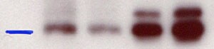
Application: Western BlotSample Tested: Rat pancreatic islet lysateVerified Customer | Posted 10/26/2015Rat pancreatic islet lysate were blotted overnight in 4 degree with polyclonal goat anti-mouse Ccl11. Serial dilution of lysate in the firs 2 lanes followed by 2 lanes of recombinant rat Ccl11. <br />Specificity: Reasonably specific<br />Sensitivity: Reasonably sensitive<br />Buffer: PBS<br />Dilution: 1/1000
There are no reviews that match your criteria.
Protocols
Find general support by application which include: protocols, troubleshooting, illustrated assays, videos and webinars.
- Antigen Retrieval Protocol (PIER)
- Antigen Retrieval for Frozen Sections Protocol
- Appropriate Fixation of IHC/ICC Samples
- Cellular Response to Hypoxia Protocols
- Chromogenic IHC Staining of Formalin-Fixed Paraffin-Embedded (FFPE) Tissue Protocol
- Chromogenic Immunohistochemistry Staining of Frozen Tissue
- ClariTSA™ Fluorophore Kits
- Detection & Visualization of Antibody Binding
- Fluorescent IHC Staining of Frozen Tissue Protocol
- Graphic Protocol for Heat-induced Epitope Retrieval
- Graphic Protocol for the Preparation and Fluorescent IHC Staining of Frozen Tissue Sections
- Graphic Protocol for the Preparation and Fluorescent IHC Staining of Paraffin-embedded Tissue Sections
- Graphic Protocol for the Preparation of Gelatin-coated Slides for Histological Tissue Sections
- IHC Sample Preparation (Frozen sections vs Paraffin)
- Immunofluorescent IHC Staining of Formalin-Fixed Paraffin-Embedded (FFPE) Tissue Protocol
- Immunohistochemistry (IHC) and Immunocytochemistry (ICC) Protocols
- Immunohistochemistry Frozen Troubleshooting
- Immunohistochemistry Paraffin Troubleshooting
- Preparing Samples for IHC/ICC Experiments
- Preventing Non-Specific Staining (Non-Specific Binding)
- Primary Antibody Selection & Optimization
- Protocol for Heat-Induced Epitope Retrieval (HIER)
- Protocol for Making a 4% Formaldehyde Solution in PBS
- Protocol for VisUCyte™ HRP Polymer Detection Reagent
- Protocol for the Preparation & Fixation of Cells on Coverslips
- Protocol for the Preparation and Chromogenic IHC Staining of Frozen Tissue Sections
- Protocol for the Preparation and Chromogenic IHC Staining of Frozen Tissue Sections - Graphic
- Protocol for the Preparation and Chromogenic IHC Staining of Paraffin-embedded Tissue Sections
- Protocol for the Preparation and Chromogenic IHC Staining of Paraffin-embedded Tissue Sections - Graphic
- Protocol for the Preparation and Fluorescent IHC Staining of Frozen Tissue Sections
- Protocol for the Preparation and Fluorescent IHC Staining of Paraffin-embedded Tissue Sections
- Protocol for the Preparation of Gelatin-coated Slides for Histological Tissue Sections
- R&D Systems Quality Control Western Blot Protocol
- TUNEL and Active Caspase-3 Detection by IHC/ICC Protocol
- The Importance of IHC/ICC Controls
- Troubleshooting Guide: Immunohistochemistry
- Troubleshooting Guide: Western Blot Figures
- Western Blot Conditions
- Western Blot Protocol
- Western Blot Protocol for Cell Lysates
- Western Blot Troubleshooting
- Western Blot Troubleshooting Guide
- View all Protocols, Troubleshooting, Illustrated assays and Webinars
Loading...
Associated Pathways

