Coagulation Factor III/Tissue Factor (TF), also known as thromboplastin and CD142, is an integral membrane protein found in a variety of cell types. It functions as a protein cofactor/receptor of Coagulation Factor VII, which is synthesized in the liver and circulated in the plasma (1). Upon binding of TF, the inactive factor VII is rapidly converted into activated VIIa. The resulting 1:1 complex of VIIa and TF initiates the coagulation pathway and has also important coagulation-independent functions such as angiognesis (2). Synthesized as a 294 amino acid precursor, mouse TF consists of a signal peptide (residues 1-28) and the mature chain (residues 29-294). As a type I membrane protein, it contains a transmembrane region (residues 252-274) and a cytoplasmic tail (residues 275-294) (3, 4). The purified rmTF corresponds to the ectodomain (residues 29-251) and is potent in activating thermolysin-processed rmCoagulation Factor VII (R&D Systems, Catalog # http://www.rndsystems.com/product_results.aspx?k=3305-SE">3305-SE) under the conditions described in the Activity Assay Protocol.
Mouse Coagulation Factor III/Tissue Factor Antibody
R&D Systems | Catalog # AF3178

Key Product Details
Validated by
Biological Validation
Species Reactivity
Validated:
Mouse
Cited:
Mouse, Transgenic Mouse
Applications
Validated:
Western Blot, Flow Cytometry, Immunoprecipitation, CyTOF-ready
Cited:
Immunohistochemistry, Immunohistochemistry-Paraffin, Western Blot, Neutralization, Flow Cytometry, Immunocytochemistry, ELISA Detection, FACS
Label
Unconjugated
Antibody Source
Polyclonal Goat IgG
Loading...
Product Specifications
Immunogen
Mouse myeloma cell line NS0-derived recombinant mouse Coagulation Factor III/Tissue Factor
Ala29-Glu251
Accession # P20352
Ala29-Glu251
Accession # P20352
Specificity
Detects mouse Coagulation Factor III in direct ELISAs and Western blots. In direct ELISAs, approximately 5% cross‑reactivity with recombinant human Coagulation Factor III is observed.
Clonality
Polyclonal
Host
Goat
Isotype
IgG
Scientific Data Images for Mouse Coagulation Factor III/Tissue Factor Antibody
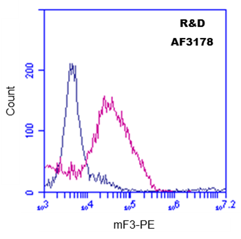
Detection of Coagulation Factor III/Tissue Factor in Raw 264.7 cells treated with 1 µg/mL LPS overnight by Flow Cytometry
Raw 264.7 cells treated with 1 µg/mL LPS overnight were stained with Goat Anti-Mouse Coagulation Factor III/Tissue Factor Antigen Affinity-purified Polyclonal Antibody (Catalog # AF3178, filled histogram) or isotype control antibody (Catalog # AB-108-C, open histogram) followed by Phycoerythrin-conjugated Anti-Goat IgG Secondary Antibody (F0107). View our protocol for Staining Membrane-associated Proteins.Applications for Mouse Coagulation Factor III/Tissue Factor Antibody
Application
Recommended Usage
CyTOF-ready
Ready to be labeled using established conjugation methods. No BSA or other carrier proteins that could interfere with conjugation.
Flow Cytometry
2.5 µg/106 cells
Sample: see below
Sample: see below
Immunoprecipitation
25 µg/mL
Sample: Conditioned cell culture medium spiked with Recombinant Mouse Coagulation Factor III/Tissue Factor (Catalog # 3178-PA), see our available Western blot detection antibodies
Sample: Conditioned cell culture medium spiked with Recombinant Mouse Coagulation Factor III/Tissue Factor (Catalog # 3178-PA), see our available Western blot detection antibodies
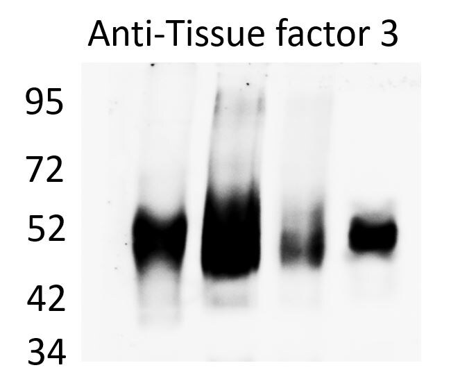
Western Blot
0.1 µg/mL
Sample: Recombinant Mouse Coagulation Factor III/Tissue Factor (Catalog # 3178-PA)
Sample: Recombinant Mouse Coagulation Factor III/Tissue Factor (Catalog # 3178-PA)
Reviewed Applications
Read 4 reviews rated 5 using AF3178 in the following applications:
Flow Cytometry Panel Builder
Bio-Techne Knows Flow Cytometry
Save time and reduce costly mistakes by quickly finding compatible reagents using the Panel Builder Tool.
Advanced Features
- Spectra Viewer - Custom analysis of spectra from multiple fluorochromes
- Spillover Popups - Visualize the spectra of individual fluorochromes
- Antigen Density Selector - Match fluorochrome brightness with antigen density

Formulation, Preparation, and Storage
Purification
Antigen Affinity-purified
Reconstitution
Reconstitute at 0.2 mg/mL in sterile PBS. For liquid material, refer to CoA for concentration.
Loading...
Formulation
Lyophilized from a 0.2 μm filtered solution in PBS with Trehalose. See Certificate of Analysis for details.
*Small pack size (-SP) is supplied either lyophilized or as a 0.2 µm filtered solution in PBS.
*Small pack size (-SP) is supplied either lyophilized or as a 0.2 µm filtered solution in PBS.
Shipping
Lyophilized product is shipped at ambient temperature. Liquid small pack size (-SP) is shipped with polar packs. Upon receipt, store immediately at the temperature recommended below.
Stability & Storage
Use a manual defrost freezer and avoid repeated freeze-thaw cycles.
- 12 months from date of receipt, -20 to -70 °C as supplied.
- 1 month, 2 to 8 °C under sterile conditions after reconstitution.
- 6 months, -20 to -70 °C under sterile conditions after reconstitution.
Calculators
Background: Coagulation Factor III/Tissue Factor
References
- Morrissey, J.H. (2004) in Handbook of Proteolytic Enzymes. Barrett, A.J. et al. (ed) Academic Press, San Diego, p. 1659.
- Versteeg, H.H. et al. (2003) Carcinogenesis 24:1009.
- Ranganathan, G. et al. (1991) J. Biol. Chem. 266:496.
- Hartzell, S. (1989) Mol. Cell. Biol. 9:2567.
Alternate Names
CD142, F3, Thromboplastin, Tissue Factor
Gene Symbol
F3
UniProt
Additional Coagulation Factor III/Tissue Factor Products
Product Documents for Mouse Coagulation Factor III/Tissue Factor Antibody
Certificate of Analysis
To download a Certificate of Analysis, please enter a lot or batch number in the search box below.
Note: Certificate of Analysis not available for kit components.
Product Specific Notices for Mouse Coagulation Factor III/Tissue Factor Antibody
For research use only
Related Research Areas
Citations for Mouse Coagulation Factor III/Tissue Factor Antibody
 Powered by Bioz
Customer Reviews for Mouse Coagulation Factor III/Tissue Factor Antibody (4)
5 out of 5
4 Customer Ratings
Have you used Mouse Coagulation Factor III/Tissue Factor Antibody?
Submit a review and receive an Amazon gift card!
$25/€18/£15/$25CAN/¥2500 Yen for a review with an image
$10/€7/£6/$10CAN/¥1110 Yen for a review without an image
Submit a review
Customer Images
Showing
1
-
4 of
4 reviews
Showing All
Filter By:
-
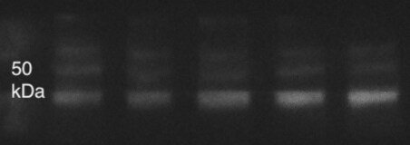
Application: Western BlotSample Tested: Brain (caudate nucleus)Species: MouseVerified Customer | Posted 10/21/2021This protein might be found in high levels in some conditions/tissues. If you calibrate a good concentration you will have a nice band around 50 KDa

-
Application: Western BlotSample Tested: Pancreatic cancer cellsSpecies: MouseVerified Customer | Posted 09/15/2021
-
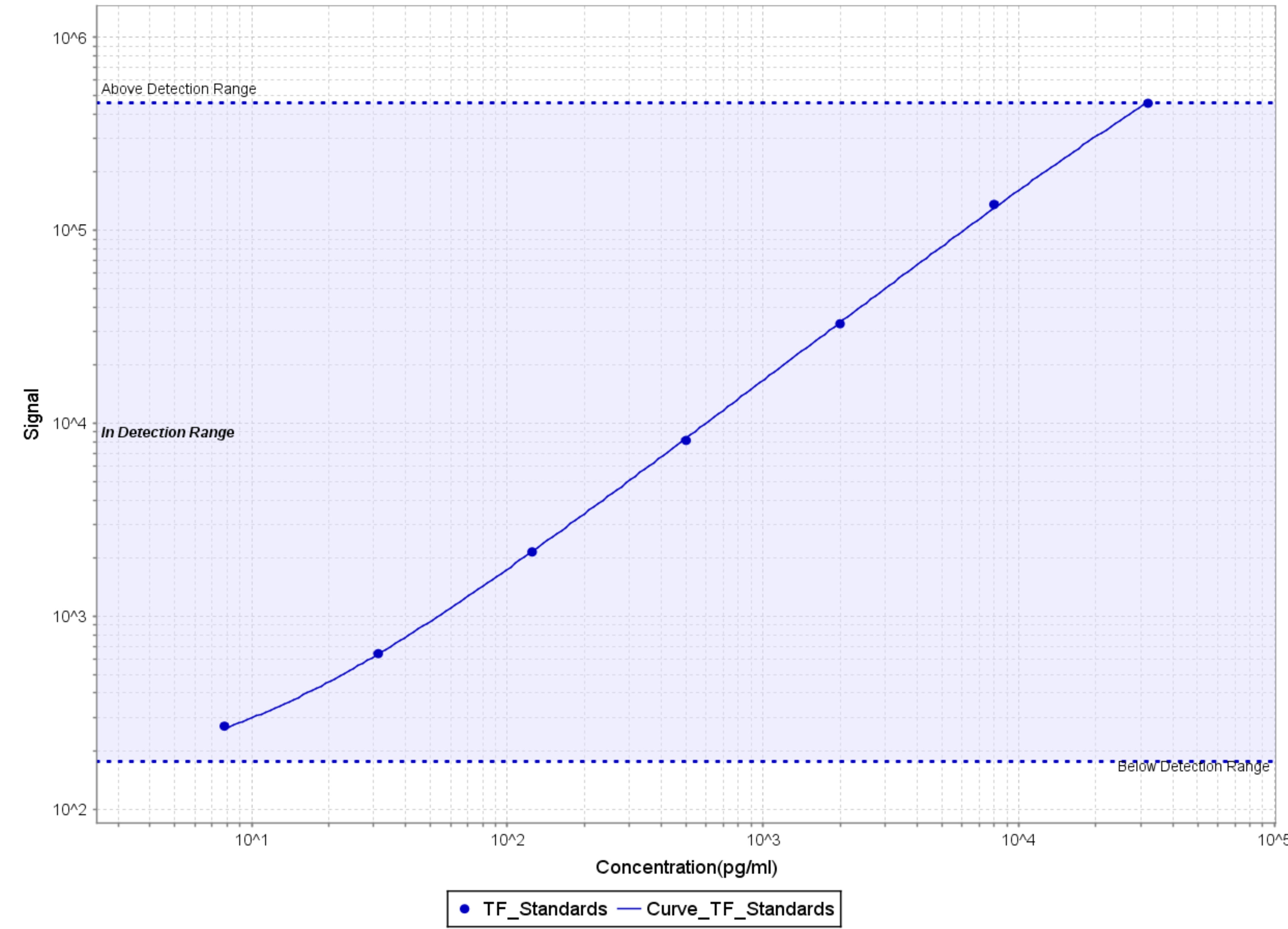
Application: MSD assaySample Tested: Serum and PlasmaSpecies: MouseVerified Customer | Posted 05/11/2018Used as detection antibody after labeling with Sulfo-Tag according to manufacturer's protocol (Meso Scale Diagnostics LLC) Detection range - 8-30,000 pg/ml
-
Application: Flow CytometrySample Tested: Blood-derived monocytesSpecies: MouseVerified Customer | Posted 04/17/2017Detection of Coagulation Factor III in Mouse mononuclear cells using Goat anti-Mouse F3 antibody (#AF3178, pink) at 2.5 ug/10^6 cells, or Isotype Control IgG (blue), followed by PE-conjugated anti-Goat secondary antibody.
There are no reviews that match your criteria.
Protocols
Find general support by application which include: protocols, troubleshooting, illustrated assays, videos and webinars.
- 7-Amino Actinomycin D (7-AAD) Cell Viability Flow Cytometry Protocol
- Cellular Response to Hypoxia Protocols
- Extracellular Membrane Flow Cytometry Protocol
- Flow Cytometry Protocol for Cell Surface Markers
- Flow Cytometry Protocol for Staining Membrane Associated Proteins
- Flow Cytometry Staining Protocols
- Flow Cytometry Troubleshooting Guide
- Immunoprecipitation Protocol
- Intracellular Flow Cytometry Protocol Using Alcohol (Methanol)
- Intracellular Flow Cytometry Protocol Using Detergents
- Intracellular Nuclear Staining Flow Cytometry Protocol Using Detergents
- Intracellular Staining Flow Cytometry Protocol Using Alcohol Permeabilization
- Intracellular Staining Flow Cytometry Protocol Using Detergents to Permeabilize Cells
- Propidium Iodide Cell Viability Flow Cytometry Protocol
- Protocol for Liperfluo
- Protocol for the Characterization of Human Th22 Cells
- Protocol for the Characterization of Human Th9 Cells
- Protocol: Annexin V and PI Staining by Flow Cytometry
- Protocol: Annexin V and PI Staining for Apoptosis by Flow Cytometry
- R&D Systems Quality Control Western Blot Protocol
- Troubleshooting Guide: Fluorokine Flow Cytometry Kits
- Troubleshooting Guide: Western Blot Figures
- Western Blot Conditions
- Western Blot Protocol
- Western Blot Protocol for Cell Lysates
- Western Blot Troubleshooting
- Western Blot Troubleshooting Guide
- View all Protocols, Troubleshooting, Illustrated assays and Webinars
Loading...
Associated Pathways
