
Key Product Details
Species Reactivity
Validated:
Mouse
Cited:
Mouse
Applications
Validated:
Immunohistochemistry, Western Blot, Neutralization
Cited:
Western Blot
Label
Unconjugated
Antibody Source
Monoclonal Rat IgG2B Clone # 840017
Loading...
Product Specifications
Immunogen
Mouse myeloma cell line NS0-derived recombinant mouse CRISP-1
Gln20-His244
Accession # Q03401
Gln20-His244
Accession # Q03401
Specificity
Detects mouse CRISP-1 in direct ELISAs.
Clonality
Monoclonal
Host
Rat
Isotype
IgG2B
Endotoxin Level
<0.10 EU per 1 μg of the antibody by the LAL method.
Scientific Data Images for Mouse CRISP-1 Antibody (840017)
Proliferation Inhibited by CRISP‑1 and Neutralization by Mouse CRISP‑1 Antibody.
Recombinant Mouse CRISP-1 inhibits proliferation in the 3A-sub E human placenta cell line in a dose-dependent manner (orange line), as measured by Resazurin (Catalog # AR002). Proliferation inhibited by Recombinant Mouse CRISP-1 (10 µg/mL) is neutralized (green line) by increasing concentrations of Rat Anti-Mouse CRISP-1 Monoclonal Antibody (Catalog # MAB4675). The ND50 is typically 10-20 µg/mL.
Detection of Mouse CRISP‑1 by Western Blot.
Western blot shows lysates of mouse epididymis tissue. PVDF membrane was probed with 1 µg/mL of Rat Anti-Mouse CRISP-1 Monoclonal Antibody (Catalog # MAB4675) followed by HRP-conjugated Anti-Rat IgG Secondary Antibody (Catalog # HAF005). A specific band was detected for CRISP-1 at approximately 27-30 kDa (as indicated). This experiment was conducted under reducing conditions and using Immunoblot Buffer Group 1.
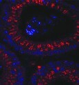
CRISP‑1 in Mouse Epididymis.
CRISP-1 was detected in perfusion fixed frozen sections of mouse epididymis using Rat Anti-Mouse CRISP-1 Monoclonal Antibody (Catalog # MAB4675) at 10 µg/mL overnight at 4 °C. Tissue was stained using the NorthernLights™ 557-conjugated Anti-Rat IgG Secondary Antibody (red; Catalog # NL013) and counterstained with DAPI (blue). Specific staining was localized to cytoplasm. View our protocol for Fluorescent IHC Staining of Frozen Tissue Sections.Applications for Mouse CRISP-1 Antibody (840017)
Application
Recommended Usage
Immunohistochemistry
8-25 µg/mL
Sample: Perfusion fixed frozen sections of mouse epididymis
Sample: Perfusion fixed frozen sections of mouse epididymis
Western Blot
1 µg/mL
Sample: Mouse epididymis tissue
Sample: Mouse epididymis tissue
Neutralization
Measured by its ability to neutralize CRISP‑1-inhibited proliferation in the 3A‑sub E human placenta cell line. The Neutralization Dose (ND50) is typically 10-20 µg/mL in the presence of 10 µg/mL Recombinant Mouse CRISP‑1.
Reviewed Applications
Read 1 review rated 5 using MAB4675 in the following applications:
Formulation, Preparation, and Storage
Purification
Protein A or G purified from hybridoma culture supernatant
Reconstitution
Reconstitute at 0.5 mg/mL in sterile PBS. For liquid material, refer to CoA for concentration.
Loading...
Formulation
Lyophilized from a 0.2 μm filtered solution in PBS with Trehalose. *Small pack size (SP) is supplied either lyophilized or as a 0.2 µm filtered solution in PBS.
Shipping
Lyophilized product is shipped at ambient temperature. Liquid small pack size (-SP) is shipped with polar packs. Upon receipt, store immediately at the temperature recommended below.
Stability & Storage
Use a manual defrost freezer and avoid repeated freeze-thaw cycles.
- 12 months from date of receipt, -20 to -70 °C as supplied.
- 1 month, 2 to 8 °C under sterile conditions after reconstitution.
- 6 months, -20 to -70 °C under sterile conditions after reconstitution.
Calculators
Background: CRISP-1
References
- Koppers, A.J. et al. (2011) Asian J. Androl. 13:111.
- Gibbs, G.M. et al. (2008) Endocr. Rev. 29:865.
- Yamazaki, Y. and T. Morita (2004) Toxicon 44:227.
- Guo, M. et al. (2005) J. Biol. Chem. 280:12405.
- Jalkanen, J. et al. (2005) Biol. Reprod. 72:1268.
- Nolan, M.A. et al. (2006) Biol. Reprod. 74:984.
- Charest, N.J. et al. (1989) Endocrinology 125:942.
- Maldera, J.A. et al. (2011) Biol. Reprod. 85:503.
- Nixon, B. et al. (2006) Biol. Reprod. 74:275.
- Roberts, K.P. et al. (2003) Biol. Reprod. 69:572.
- Busso, D. et al. (2007) Biol. Reprod. 77:848.
- Da Ros, V.G. et al. (2008) Dev. Biol. 320:12.
- Haendler, B. et al. (1993) Endocrinology 133:192.
- Mizuki, N. and M. Kasahara (1992) Mol. Cell. Endocrinol. 89:25.
- Peterson, R.L. et al. (2005) J. Investig. Dermatol. Symp. Proc. 10:238.
Long Name
Cysteine-rich Secretory Protein 1
Alternate Names
AEGL1, CRISP1
Gene Symbol
CRISP1
UniProt
Additional CRISP-1 Products
Product Documents for Mouse CRISP-1 Antibody (840017)
Certificate of Analysis
To download a Certificate of Analysis, please enter a lot or batch number in the search box below.
Note: Certificate of Analysis not available for kit components.
Product Specific Notices for Mouse CRISP-1 Antibody (840017)
For research use only
Citations for Mouse CRISP-1 Antibody (840017)
 Powered by Bioz
Customer Reviews for Mouse CRISP-1 Antibody (840017) (1)
5 out of 5
1 Customer Rating
Have you used Mouse CRISP-1 Antibody (840017)?
Submit a review and receive an Amazon gift card!
$25/€18/£15/$25CAN/¥2500 Yen for a review with an image
$10/€7/£6/$10CAN/¥1110 Yen for a review without an image
Submit a review
Customer Images
Showing
1
-
1 of
1 review
Showing All
Filter By:
-
Application: Immunocytochemistry/ImmunofluorescenceSample Tested: Epididymis tissueSpecies: MouseVerified Customer | Posted 10/27/2021

There are no reviews that match your criteria.
Protocols
Find general support by application which include: protocols, troubleshooting, illustrated assays, videos and webinars.
- Antigen Retrieval Protocol (PIER)
- Antigen Retrieval for Frozen Sections Protocol
- Appropriate Fixation of IHC/ICC Samples
- Cellular Response to Hypoxia Protocols
- Chromogenic IHC Staining of Formalin-Fixed Paraffin-Embedded (FFPE) Tissue Protocol
- Chromogenic Immunohistochemistry Staining of Frozen Tissue
- ClariTSA™ Fluorophore Kits
- Detection & Visualization of Antibody Binding
- Fluorescent IHC Staining of Frozen Tissue Protocol
- Graphic Protocol for Heat-induced Epitope Retrieval
- Graphic Protocol for the Preparation and Fluorescent IHC Staining of Frozen Tissue Sections
- Graphic Protocol for the Preparation and Fluorescent IHC Staining of Paraffin-embedded Tissue Sections
- Graphic Protocol for the Preparation of Gelatin-coated Slides for Histological Tissue Sections
- IHC Sample Preparation (Frozen sections vs Paraffin)
- Immunofluorescent IHC Staining of Formalin-Fixed Paraffin-Embedded (FFPE) Tissue Protocol
- Immunohistochemistry (IHC) and Immunocytochemistry (ICC) Protocols
- Immunohistochemistry Frozen Troubleshooting
- Immunohistochemistry Paraffin Troubleshooting
- Preparing Samples for IHC/ICC Experiments
- Preventing Non-Specific Staining (Non-Specific Binding)
- Primary Antibody Selection & Optimization
- Protocol for Heat-Induced Epitope Retrieval (HIER)
- Protocol for Making a 4% Formaldehyde Solution in PBS
- Protocol for VisUCyte™ HRP Polymer Detection Reagent
- Protocol for the Preparation & Fixation of Cells on Coverslips
- Protocol for the Preparation and Chromogenic IHC Staining of Frozen Tissue Sections
- Protocol for the Preparation and Chromogenic IHC Staining of Frozen Tissue Sections - Graphic
- Protocol for the Preparation and Chromogenic IHC Staining of Paraffin-embedded Tissue Sections
- Protocol for the Preparation and Chromogenic IHC Staining of Paraffin-embedded Tissue Sections - Graphic
- Protocol for the Preparation and Fluorescent IHC Staining of Frozen Tissue Sections
- Protocol for the Preparation and Fluorescent IHC Staining of Paraffin-embedded Tissue Sections
- Protocol for the Preparation of Gelatin-coated Slides for Histological Tissue Sections
- R&D Systems Quality Control Western Blot Protocol
- TUNEL and Active Caspase-3 Detection by IHC/ICC Protocol
- The Importance of IHC/ICC Controls
- Troubleshooting Guide: Immunohistochemistry
- Troubleshooting Guide: Western Blot Figures
- Western Blot Conditions
- Western Blot Protocol
- Western Blot Protocol for Cell Lysates
- Western Blot Troubleshooting
- Western Blot Troubleshooting Guide
- View all Protocols, Troubleshooting, Illustrated assays and Webinars
Loading...