Mouse CXCL16 (CXC chemokine 16) is a non-ELR motif-containing CXC chemokine with a transmembrane domain. CX3CL1/Fractalkine and CXCL16 are the only two transmembrane chemokines within the superfamily. Mouse CXCL16 cDNA encodes a 246 amino acid (aa) precursor protein with a putative 26 aa residue signal peptide, an 88 aa residue chemokine domain, an 87 aa residue mucin-like spacer region, a 22 aa residue transmembrane domain, and a 23 aa residue cytoplasmic tail. Mouse and human CXCL16 share 49% overall aa identity and 70% similarity in the chemokine domains. Mouse CXCL16 is produced by dendritic cells in lymphoid organ T cell zones and by cells in the splenic red pulp both as membrane-bound and soluble forms. Based on northern blot analysis, CXCL16 is also expressed in some nonlymphoid tissues such as lung, small intestine and kidney. The receptor for CXCL16 has been identified as CXCR6/Bonzo (STRL33 and TYMSTR), a receptor previously shown to be a co-receptor for HIV entry. CXCR6 is expressed on naive CD8 cells, natural killer T cells and activated CD8 and CD4 T cells.

Key Product Details
Species Reactivity
Validated:
Mouse
Cited:
Mouse, Rat, Xenograft
Applications
Validated:
Western Blot, Neutralization, Flow Cytometry, CyTOF-ready
Cited:
Immunohistochemistry, Western Blot, Neutralization, Flow Cytometry, Immunocytochemistry, In vivo assay
Label
Unconjugated
Antibody Source
Polyclonal Goat IgG
Loading...
Product Specifications
Immunogen
E. coli-derived recombinant mouse CXCL16
Asn27-Pro114
Accession # Q8BSU2
Asn27-Pro114
Accession # Q8BSU2
Specificity
Detects mouse CXCL16 in direct ELISAs and Western blots. In direct ELISAs, less than 1% cross-reactivity with recombinant human (rh) CXCL16, rhFractalkine, recombinant mouse Fractalkine, and recombinant rat Fractalkine is observed.
Clonality
Polyclonal
Host
Goat
Isotype
IgG
Endotoxin Level
<0.10 EU per 1 μg of the antibody by the LAL method.
Scientific Data Images for Mouse CXCL16 Antibody
Detection of CXCL16 in Raw 264.7 cells by Flow Cytometry
Raw 264.7 cells were stained with Goat Anti-Mouse CXCL16 Antigen Affinity-purified Polyclonal Antibody (Catalog # af503, filled histogram) or isotype control antibody (Catalog # AB-108-C, open histogram) followed by Allophycocyanin-conjugated Anti-Goat IgG Secondary Antibody (Catalog # F0108). View our protocol for Staining Membrane-associated Proteins.
Chemotaxis Induced by CXCL16 and Neutralization by Mouse CXCL16 Antibody.
Recombinant Mouse CXCL16 Chemokine Domain (Catalog # 503-CX) chemoattracts the BaF3 mouse pro‑B cell line transfected with mouse CXCR6 in a dose-dependent manner (orange line). The amount of cells that migrated through to the lower chemotaxis chamber was measured by Resazurin (Catalog # AR002). Chemotaxis elicited by Recombinant Mouse CXCL16 Chemokine Domain (7.5 ng/mL) is neutralized (green line) by increasing concentrations of Goat Anti-Mouse CXCL16 Antigen Affinity-purified Polyclonal Antibody (Catalog # AF503). The ND50 is typically 0.1-0.4 µg/mL.
Mouse CXCL16 ELISA Standard Curve
Recombinant Mouse CXCL16 Chemokine Domain (Catalog # 503-CX) was serially diluted and captured by Rat Anti-Mouse CXCL16 Monoclonal Antibody (Catalog # MAB503) coated on a Clear Polystyrene Microplate (Catalog # DY990). Goat Anti-Mouse CXCL16 Antigen Affinity-purified Polyclonal Antibody (Catalog # AF503) was biotinylated and incubated with the protein captured on the plate. Detection of the standard curve was achieved by incubating Streptavidin-HRP (Catalog # DY998)Applications for Mouse CXCL16 Antibody
Application
Recommended Usage
CyTOF-ready
Ready to be labeled using established conjugation methods. No BSA or other carrier proteins that could interfere with conjugation.
Flow Cytometry
0.25 µg/106 cells
Sample: Raw264.7 mouse monocyte/macrophage cell line
Sample: Raw264.7 mouse monocyte/macrophage cell line
Western Blot
0.1 µg/mL
Sample: Recombinant Mouse CXCL16 Chemokine Domain (Catalog # 503-CX)
Sample: Recombinant Mouse CXCL16 Chemokine Domain (Catalog # 503-CX)
Neutralization
Measured by its ability to neutralize CXCL16-induced chemotaxis in the BaF3 mouse pro‑B cell line transfected with mouse CXCR6. Matloubian, M. et al. (2000) Nature Immunol. 1:298. The Neutralization Dose (ND50) is typically 0.1-0.4 µg/mL in the presence of 7.5 ng/mL Recombinant Mouse CXCL16 Chemokine Domain.
Reviewed Applications
Read 2 reviews rated 5 using AF503 in the following applications:
Flow Cytometry Panel Builder
Bio-Techne Knows Flow Cytometry
Save time and reduce costly mistakes by quickly finding compatible reagents using the Panel Builder Tool.
Advanced Features
- Spectra Viewer - Custom analysis of spectra from multiple fluorochromes
- Spillover Popups - Visualize the spectra of individual fluorochromes
- Antigen Density Selector - Match fluorochrome brightness with antigen density

Formulation, Preparation, and Storage
Purification
Antigen Affinity-purified
Reconstitution
Reconstitute at 0.2 mg/mL in sterile PBS. For liquid material, refer to CoA for concentration.
Loading...
Formulation
Lyophilized from a 0.2 μm filtered solution in PBS with Trehalose. See Certificate of Analysis for details.
*Small pack size (-SP) is supplied either lyophilized or as a 0.2 µm filtered solution in PBS.
*Small pack size (-SP) is supplied either lyophilized or as a 0.2 µm filtered solution in PBS.
Shipping
Lyophilized product is shipped at ambient temperature. Liquid small pack size (-SP) is shipped with polar packs. Upon receipt, store immediately at the temperature recommended below.
Stability & Storage
Use a manual defrost freezer and avoid repeated freeze-thaw cycles.
- 12 months from date of receipt, -20 to -70 °C as supplied.
- 1 month, 2 to 8 °C under sterile conditions after reconstitution.
- 6 months, -20 to -70 °C under sterile conditions after reconstitution.
Calculators
Background: CXCL16
Alternate Names
CXCL16
Gene Symbol
CXCL16
UniProt
Additional CXCL16 Products
Product Documents for Mouse CXCL16 Antibody
Certificate of Analysis
To download a Certificate of Analysis, please enter a lot or batch number in the search box below.
Note: Certificate of Analysis not available for kit components.
Product Specific Notices for Mouse CXCL16 Antibody
For research use only
Related Research Areas
Citations for Mouse CXCL16 Antibody
 Powered by Bioz
Customer Reviews for Mouse CXCL16 Antibody (2)
5 out of 5
2 Customer Ratings
Have you used Mouse CXCL16 Antibody?
Submit a review and receive an Amazon gift card!
$25/€18/£15/$25CAN/¥2500 Yen for a review with an image
$10/€7/£6/$10CAN/¥1110 Yen for a review without an image
Submit a review
Customer Images
Showing
1
-
2 of
2 reviews
Showing All
Filter By:
-
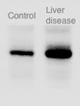
Application: Western BlotSample Tested: Liver tissueSpecies: MouseVerified Customer | Posted 07/19/2018Western blot of liver tissue from a animal model of inflammatory liver disease shows increased expression of CXCL16 protein (RIGHT), relative to control mice (LEFT).

-
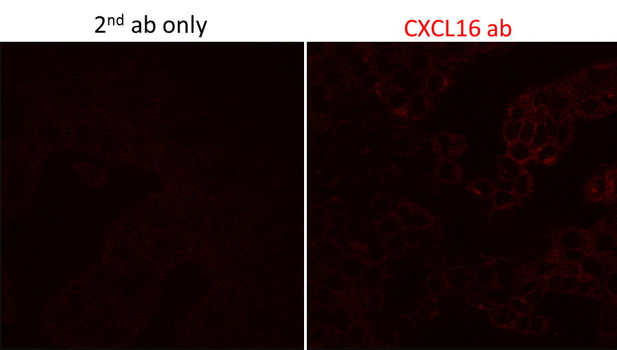
Application: Immunocytochemistry/ImmunofluorescenceSample Tested: Ovarian cell lineSpecies: MouseVerified Customer | Posted 08/30/2017
There are no reviews that match your criteria.
Protocols
Find general support by application which include: protocols, troubleshooting, illustrated assays, videos and webinars.
- 7-Amino Actinomycin D (7-AAD) Cell Viability Flow Cytometry Protocol
- Cellular Response to Hypoxia Protocols
- Extracellular Membrane Flow Cytometry Protocol
- Flow Cytometry Protocol for Cell Surface Markers
- Flow Cytometry Protocol for Staining Membrane Associated Proteins
- Flow Cytometry Staining Protocols
- Flow Cytometry Troubleshooting Guide
- Intracellular Flow Cytometry Protocol Using Alcohol (Methanol)
- Intracellular Flow Cytometry Protocol Using Detergents
- Intracellular Nuclear Staining Flow Cytometry Protocol Using Detergents
- Intracellular Staining Flow Cytometry Protocol Using Alcohol Permeabilization
- Intracellular Staining Flow Cytometry Protocol Using Detergents to Permeabilize Cells
- Propidium Iodide Cell Viability Flow Cytometry Protocol
- Protocol for Liperfluo
- Protocol for the Characterization of Human Th22 Cells
- Protocol for the Characterization of Human Th9 Cells
- Protocol: Annexin V and PI Staining by Flow Cytometry
- Protocol: Annexin V and PI Staining for Apoptosis by Flow Cytometry
- R&D Systems Quality Control Western Blot Protocol
- Troubleshooting Guide: Fluorokine Flow Cytometry Kits
- Troubleshooting Guide: Western Blot Figures
- Western Blot Conditions
- Western Blot Protocol
- Western Blot Protocol for Cell Lysates
- Western Blot Troubleshooting
- Western Blot Troubleshooting Guide
- View all Protocols, Troubleshooting, Illustrated assays and Webinars
Loading...
Associated Pathways
