Cystatin C is a member of family 2 of the cystatin superfamily (1). It is involved in processes such as tumor invasion and metastasis, inflammation and some neurological diseases. It inhibits many cysteine proteases such as papain and cathepsins B, H, K, L and S (2, 3). All mouse tissues analyzed expressed Cystatin C, with relative levels similar to those of rat and human tissues. For all three species, brain and liver had the highest and lowest levels of Cystatin C, respectively, whereas kidney, spleen and muscle had the levels in between (4). The high degree of similarity in distribution and functional properties for mouse, rat and human Cystatin C indicates that a murine model should be relevant for studies of the human disease, hereditary Cystatin C amyloid angiopathy (4).

Key Product Details
Species Reactivity
Validated:
Mouse
Cited:
Human, Mouse
Applications
Validated:
Immunohistochemistry, Western Blot, ELISA Capture (Matched Antibody Pair), Simple Western, Immunoprecipitation
Cited:
Immunohistochemistry-Paraffin, Immunohistochemistry-Frozen, Western Blot, ELISA Development
Label
Unconjugated
Antibody Source
Polyclonal Goat IgG
Loading...
Product Specifications
Immunogen
Mouse myeloma cell line NS0-derived recombinant mouse Cystatin C (R & D Systems, Catalog # 1238-PI)
Ala21-Ala140
Accession # P21460
Ala21-Ala140
Accession # P21460
Specificity
Detects mouse and human Cystatin C in direct ELISAs. In direct ELISAs, less than 10% cross-reactivity with recombinant human Cystatin D is observed.
Clonality
Polyclonal
Host
Goat
Isotype
IgG
Scientific Data Images for Mouse Cystatin C Antibody
Detection of Mouse Cystatin C by Western Blot.
Western blot shows lysates of mouse plasma and RAW 264.7 mouse monocyte/macrophage cell line. PVDF membrane was probed with 1 µg/mL of Goat Anti-Mouse Cystatin C Antigen Affinity-purified Polyclonal Antibody (Catalog # AF1238) followed by HRP-conjugated Anti-Goat IgG Secondary Antibody (Catalog # HAF017). A specific band was detected for Cystatin C at approximately 14 kDa (as indicated). This experiment was conducted under reducing conditions and using Immunoblot Buffer Group 1.
Cystatin C in Mouse Spleen.
Cystatin C was detected in immersion fixed frozen sections of mouse spleen using Mouse Cystatin C Antigen Affinity-purified Polyclonal Antibody (Catalog # AF1238) at 15 µg/mL overnight at 4 °C. Tissue was stained using the Anti-Goat HRP-DAB Cell & Tissue Staining Kit (brown; Catalog # CTS008) and counterstained with hematoxylin (blue). Lower panel shows a lack of labeling if primary antibodies are omitted and tissue is stained only with secondary antibody followed by incubation with detection reagents. View our protocol for Chromogenic IHC Staining of Frozen Tissue Sections.
Detection of Mouse Cystatin C by Simple WesternTM.
Simple Western lane view shows lysates of RAW 264.7 mouse monocyte/macrophage cell line, loaded at 0.2 mg/mL. A specific band was detected for Cystatin C at approximately 19 kDa (as indicated) using 10 µg/mL of Goat Anti-Mouse Cystatin C Antigen Affinity-purified Polyclonal Antibody (Catalog # AF1238). This experiment was conducted under reducing conditions and using the 12-230 kDa separation system.Applications for Mouse Cystatin C Antibody
Application
Recommended Usage
Immunohistochemistry
5-15 µg/mL
Sample: Perfusion fixed frozen sections of mouse brain (caudate putamen) and immersion fixed frozen sections of mouse spleen
Sample: Perfusion fixed frozen sections of mouse brain (caudate putamen) and immersion fixed frozen sections of mouse spleen
Immunoprecipitation
25 µg/mL
Sample: Conditioned cell culture medium spiked with Recombinant Mouse Cystatin C (Catalog # 1238-PI), see our available Western blot detection antibodies
Sample: Conditioned cell culture medium spiked with Recombinant Mouse Cystatin C (Catalog # 1238-PI), see our available Western blot detection antibodies
Simple Western
10 µg/mL
Sample: RAW 264.7 mouse monocyte/macrophage cell line
Sample: RAW 264.7 mouse monocyte/macrophage cell line
Western Blot
1 µg/mL
Sample: Mouse plasma and RAW 264.7 mouse monocyte/macrophage cell line
Sample: Mouse plasma and RAW 264.7 mouse monocyte/macrophage cell line
Mouse Cystatin C Sandwich Immunoassay
Please Note: Optimal dilutions of this antibody should be experimentally determined.
Reviewed Applications
Read 1 review rated 4 using AF1238 in the following applications:
Formulation, Preparation, and Storage
Purification
Antigen Affinity-purified
Reconstitution
Reconstitute at 0.2 mg/mL in sterile PBS. For liquid material, refer to CoA for concentration.
Loading...
Formulation
Lyophilized from a 0.2 μm filtered solution in PBS with Trehalose. *Small pack size (SP) is supplied either lyophilized or as a 0.2 µm filtered solution in PBS.
Shipping
Lyophilized product is shipped at ambient temperature. Liquid small pack size (-SP) is shipped with polar packs. Upon receipt, store immediately at the temperature recommended below.
Stability & Storage
Use a manual defrost freezer and avoid repeated freeze-thaw cycles.
- 12 months from date of receipt, -20 to -70 °C as supplied.
- 1 month, 2 to 8 °C under sterile conditions after reconstitution.
- 6 months, -20 to -70 °C under sterile conditions after reconstitution.
Calculators
Background: Cystatin C
References
- Reed, C.H. (2000) British J. Biomed. Sci. 57:323.
- Janowski, R. et al. (2001) Nat. Struct. Biol. 8:316.
- Abrahamson, M. (1994) Methods Enzymol. 244:685.
- Hakansson, K. et al. (1996) Comp. Biochem. Physiol. 114B:303.
Alternate Names
ARMD11, CST3, Gamma-trace, Neuroendocrine basic polypeptide, Post-gamma-globulin
Gene Symbol
CST3
UniProt
Additional Cystatin C Products
Product Documents for Mouse Cystatin C Antibody
Certificate of Analysis
To download a Certificate of Analysis, please enter a lot or batch number in the search box below.
Note: Certificate of Analysis not available for kit components.
Product Specific Notices for Mouse Cystatin C Antibody
For research use only
Related Research Areas
Citations for Mouse Cystatin C Antibody
 Powered by Bioz
Customer Reviews for Mouse Cystatin C Antibody (1)
4 out of 5
1 Customer Rating
Have you used Mouse Cystatin C Antibody?
Submit a review and receive an Amazon gift card!
$25/€18/£15/$25CAN/¥2500 Yen for a review with an image
$10/€7/£6/$10CAN/¥1110 Yen for a review without an image
Submit a review
Customer Images
Showing
1
-
1 of
1 review
Showing All
Filter By:
-
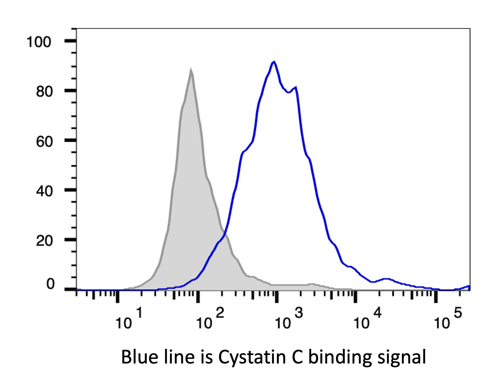
Application: Flow CytometrySample Tested: CHO Chinese hamster ovary cell lineSpecies: MouseVerified Customer | Posted 08/30/2023

There are no reviews that match your criteria.
Protocols
Find general support by application which include: protocols, troubleshooting, illustrated assays, videos and webinars.
- Antigen Retrieval Protocol (PIER)
- Antigen Retrieval for Frozen Sections Protocol
- Appropriate Fixation of IHC/ICC Samples
- Cellular Response to Hypoxia Protocols
- Chromogenic IHC Staining of Formalin-Fixed Paraffin-Embedded (FFPE) Tissue Protocol
- Chromogenic Immunohistochemistry Staining of Frozen Tissue
- ClariTSA™ Fluorophore Kits
- Detection & Visualization of Antibody Binding
- Fluorescent IHC Staining of Frozen Tissue Protocol
- Graphic Protocol for Heat-induced Epitope Retrieval
- Graphic Protocol for the Preparation and Fluorescent IHC Staining of Frozen Tissue Sections
- Graphic Protocol for the Preparation and Fluorescent IHC Staining of Paraffin-embedded Tissue Sections
- Graphic Protocol for the Preparation of Gelatin-coated Slides for Histological Tissue Sections
- IHC Sample Preparation (Frozen sections vs Paraffin)
- Immunofluorescent IHC Staining of Formalin-Fixed Paraffin-Embedded (FFPE) Tissue Protocol
- Immunohistochemistry (IHC) and Immunocytochemistry (ICC) Protocols
- Immunohistochemistry Frozen Troubleshooting
- Immunohistochemistry Paraffin Troubleshooting
- Immunoprecipitation Protocol
- Preparing Samples for IHC/ICC Experiments
- Preventing Non-Specific Staining (Non-Specific Binding)
- Primary Antibody Selection & Optimization
- Protocol for Heat-Induced Epitope Retrieval (HIER)
- Protocol for Making a 4% Formaldehyde Solution in PBS
- Protocol for VisUCyte™ HRP Polymer Detection Reagent
- Protocol for the Preparation & Fixation of Cells on Coverslips
- Protocol for the Preparation and Chromogenic IHC Staining of Frozen Tissue Sections
- Protocol for the Preparation and Chromogenic IHC Staining of Frozen Tissue Sections - Graphic
- Protocol for the Preparation and Chromogenic IHC Staining of Paraffin-embedded Tissue Sections
- Protocol for the Preparation and Chromogenic IHC Staining of Paraffin-embedded Tissue Sections - Graphic
- Protocol for the Preparation and Fluorescent IHC Staining of Frozen Tissue Sections
- Protocol for the Preparation and Fluorescent IHC Staining of Paraffin-embedded Tissue Sections
- Protocol for the Preparation of Gelatin-coated Slides for Histological Tissue Sections
- R&D Systems Quality Control Western Blot Protocol
- TUNEL and Active Caspase-3 Detection by IHC/ICC Protocol
- The Importance of IHC/ICC Controls
- Troubleshooting Guide: Immunohistochemistry
- Troubleshooting Guide: Western Blot Figures
- Western Blot Conditions
- Western Blot Protocol
- Western Blot Protocol for Cell Lysates
- Western Blot Troubleshooting
- Western Blot Troubleshooting Guide
- View all Protocols, Troubleshooting, Illustrated assays and Webinars
Loading...
Associated Pathways
