Galectins are a family of carbohydrate-binding proteins with specificity for N-acetyl-lactosamine-containing glycoproteins. At least 14 mammalian galectins share structural similarities in their carbohydrate recognition domains (CRD), forming three groups often termed prototype (one CRD), tandem-repeat (two CRDs) and chimeric (one CRD, unique N-terminus) (1, 2). All lack classical signal peptides, but are present and active both within and outside of the cell. Galectins are involved in cell adhesion, migration, survival, and apoptosis, and are often up- or down-regulated in cancer (1-3). Galectin-4 is a 36 kDa tandem-repeat galectin found throughout the gastrointestinal tract, but also present in well-differentiated breast and liver carcinomas (3, 4). Each CRD binds a different set of carbohydrate groups, including those found on erythrocyte blood group antigens (3, 5). CRD1 also binds cholesterol 3-sulfate and other sulfatides, which are concentrated within lipid raft membrane microdomains (6, 7). Endocytosed Galectin-4 is thought to play a role in forming the rafts, delivering them to the intestinal apical membrane, and stabilizing highly detergent-resistant "superrafts" (7-9). Human Galectin-4 shares 76%, 77%, 78%, and 80% amino acid (aa) identity with mouse, rat, bovine, and porcine Galectin-4, respectively, with the highest identity occurring within the CRDs. A potential splice variant begins at aa 132 and lacks most of the first CRD (10). Galectin-4 expression is concentrated within microvilli in the gastrointestinal epithelium, where it can interact with CD3 and bind activated T cells in the lamina propria during intestinal inflammation (11, 12). Either pro- or anti-inflammatory activity has been shown, depending on the mouse model used. Galectin-4 can also bind lung, spleen, and kidney macrophages, although its expression is normally low in these tissues (5).

Key Product Details
Species Reactivity
Validated:
Mouse
Cited:
Mouse
Applications
Validated:
Immunohistochemistry, Western Blot
Cited:
Immunohistochemistry, Neutralization
Label
Unconjugated
Antibody Source
Polyclonal Goat IgG
Loading...
Product Specifications
Immunogen
E. coli-derived recombinant mouse Galectin-4
Ala2-Ile326
Accession # Q8K419
Ala2-Ile326
Accession # Q8K419
Specificity
Detects mouse Galectin-4 in direct ELISAs and Western blots.
Clonality
Polyclonal
Host
Goat
Isotype
IgG
Scientific Data Images for Mouse Galectin-4 Antibody
Detection of Galectin‑4 in Mouse Intestine.
Galectin‑4 was detected in immersion fixed paraffin-embedded sections of Mouse Intestine using Goat Anti-Mouse Galectin‑4 Antigen Affinity-purified Polyclonal Antibody (Catalog # AF2128) at 3 µg/mL for 1 hour at room temperature followed by incubation with the Anti-Goat IgG VisUCyte™ HRP Polymer Antibody (Catalog # VC004). Before incubation with the primary antibody, tissue was subjected to heat-induced epitope retrieval using VisUCyte Antigen Retrieval Reagent-Basic (Catalog # VCTS021). Tissue was stained using DAB (brown) and counterstained with hematoxylin (blue). Specific staining was localized to cytoplasm and plasma membrane in intestinal epithelial cells. View our protocol for IHC Staining with VisUCyte HRP Polymer Detection Reagents.Applications for Mouse Galectin-4 Antibody
Application
Recommended Usage
Immunohistochemistry
3-15 µg/mL
Sample: Immersion fixed paraffin-embedded sections of Mouse Intestine
Sample: Immersion fixed paraffin-embedded sections of Mouse Intestine
Western Blot
0.1 µg/mL
Sample: Recombinant Mouse Galectin‑4 (Catalog # 2128-GA)
Sample: Recombinant Mouse Galectin‑4 (Catalog # 2128-GA)
Reviewed Applications
Read 3 reviews rated 4.7 using AF2128 in the following applications:
Formulation, Preparation, and Storage
Purification
Antigen Affinity-purified
Reconstitution
Reconstitute at 0.2 mg/mL in sterile PBS. For liquid material, refer to CoA for concentration.
Loading...
Formulation
Lyophilized from a 0.2 μm filtered solution in PBS with Trehalose. *Small pack size (SP) is supplied either lyophilized or as a 0.2 µm filtered solution in PBS.
Shipping
Lyophilized product is shipped at ambient temperature. Liquid small pack size (-SP) is shipped with polar packs. Upon receipt, store immediately at the temperature recommended below.
Stability & Storage
Use a manual defrost freezer and avoid repeated freeze-thaw cycles.
- 12 months from date of receipt, -20 to -70 °C as supplied.
- 1 month, 2 to 8 °C under sterile conditions after reconstitution.
- 6 months, -20 to -70 °C under sterile conditions after reconstitution.
Calculators
Background: Galectin-4
References
- Yang, R-Y. et al. (2008) Expert Rev. Mol. Med. 10:e17.
- Elola, M.T. et al. (2007) Cell. Mol. Life Sci. 64:1679.
- Huflejt, M.E. and H. Leffler (2004) Glycoconj. J. 20:247.
- Recreche, H. et al. (1997) Eur. J. Biochem. 248:225.
- Markova, V. et al. (2006) Int. J. Mol. Med. 18:65.
- Ideo, H. et al. (2007) J. Biol. Chem. 282:21081.
- Delacour, D. et al. (2005) J. Cell Biol. 169:491.
- Braccia, A. et al. (2003) J. Biol. Chem. 278:15679.
- Stechly, L. et al. (2009) Traffic 10:438.
- Entrez accession # EAW56820.
- Hokama, A. et al. (2004) Immunity 20:681.
- Paclik, D. et al. (2008) PLoS ONE 3:e2629.
Alternate Names
GAL4, Galectin4, LGALS4
Gene Symbol
LGALS4
UniProt
Additional Galectin-4 Products
Product Documents for Mouse Galectin-4 Antibody
Certificate of Analysis
To download a Certificate of Analysis, please enter a lot or batch number in the search box below.
Note: Certificate of Analysis not available for kit components.
Product Specific Notices for Mouse Galectin-4 Antibody
For research use only
Related Research Areas
Citations for Mouse Galectin-4 Antibody
 Powered by Bioz
Customer Reviews for Mouse Galectin-4 Antibody (3)
4.7 out of 5
3 Customer Ratings
Have you used Mouse Galectin-4 Antibody?
Submit a review and receive an Amazon gift card!
$25/€18/£15/$25CAN/¥2500 Yen for a review with an image
$10/€7/£6/$10CAN/¥1110 Yen for a review without an image
Submit a review
Customer Images
Showing
1
-
3 of
3 reviews
Showing All
Filter By:
-
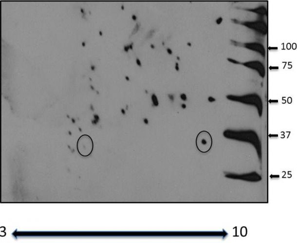
Application: 2D gel, protein modificationSample Tested: Cell LysatesSpecies: HumanVerified Customer | Posted 10/26/2015Although there were mutliple bands, the one circled are galectin-4, the others are interactors isolated on a 2D gel. Using human cell lysates in 2D buffer. <br />Specificity: Specific<br />Sensitivity: Sensitive<br />Buffer: BSA<br />Dilution: 1:1000

-
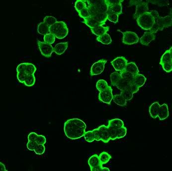
Application: Immunocytochemistry/ImmunofluorescenceSample Tested: Cancer CellsSpecies: HumanVerified Customer | Posted 10/26/2015This antibody is as good as the monoclonal antibody from R&D. <br />Specificity: Specific<br />Sensitivity: Reasonably sensitive<br />Buffer: PBS<br />Dilution: 1:1000
-
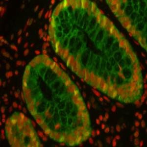
Application: Immunocytochemistry/ImmunofluorescenceSample Tested: Colon tissueSpecies: HumanVerified Customer | Posted 10/26/2015Very specific, Red is propidium iodide the nuclear stain. <br />Specificity: Specific<br />Sensitivity: Reasonably sensitive<br />Buffer: PBS<br />Dilution: 1:500
There are no reviews that match your criteria.
Protocols
Find general support by application which include: protocols, troubleshooting, illustrated assays, videos and webinars.
- Antigen Retrieval Protocol (PIER)
- Antigen Retrieval for Frozen Sections Protocol
- Appropriate Fixation of IHC/ICC Samples
- Cellular Response to Hypoxia Protocols
- Chromogenic IHC Staining of Formalin-Fixed Paraffin-Embedded (FFPE) Tissue Protocol
- Chromogenic Immunohistochemistry Staining of Frozen Tissue
- ClariTSA™ Fluorophore Kits
- Detection & Visualization of Antibody Binding
- Fluorescent IHC Staining of Frozen Tissue Protocol
- Graphic Protocol for Heat-induced Epitope Retrieval
- Graphic Protocol for the Preparation and Fluorescent IHC Staining of Frozen Tissue Sections
- Graphic Protocol for the Preparation and Fluorescent IHC Staining of Paraffin-embedded Tissue Sections
- Graphic Protocol for the Preparation of Gelatin-coated Slides for Histological Tissue Sections
- IHC Sample Preparation (Frozen sections vs Paraffin)
- Immunofluorescent IHC Staining of Formalin-Fixed Paraffin-Embedded (FFPE) Tissue Protocol
- Immunohistochemistry (IHC) and Immunocytochemistry (ICC) Protocols
- Immunohistochemistry Frozen Troubleshooting
- Immunohistochemistry Paraffin Troubleshooting
- Preparing Samples for IHC/ICC Experiments
- Preventing Non-Specific Staining (Non-Specific Binding)
- Primary Antibody Selection & Optimization
- Protocol for Heat-Induced Epitope Retrieval (HIER)
- Protocol for Making a 4% Formaldehyde Solution in PBS
- Protocol for VisUCyte™ HRP Polymer Detection Reagent
- Protocol for the Preparation & Fixation of Cells on Coverslips
- Protocol for the Preparation and Chromogenic IHC Staining of Frozen Tissue Sections
- Protocol for the Preparation and Chromogenic IHC Staining of Frozen Tissue Sections - Graphic
- Protocol for the Preparation and Chromogenic IHC Staining of Paraffin-embedded Tissue Sections
- Protocol for the Preparation and Chromogenic IHC Staining of Paraffin-embedded Tissue Sections - Graphic
- Protocol for the Preparation and Fluorescent IHC Staining of Frozen Tissue Sections
- Protocol for the Preparation and Fluorescent IHC Staining of Paraffin-embedded Tissue Sections
- Protocol for the Preparation of Gelatin-coated Slides for Histological Tissue Sections
- R&D Systems Quality Control Western Blot Protocol
- TUNEL and Active Caspase-3 Detection by IHC/ICC Protocol
- The Importance of IHC/ICC Controls
- Troubleshooting Guide: Immunohistochemistry
- Troubleshooting Guide: Western Blot Figures
- Western Blot Conditions
- Western Blot Protocol
- Western Blot Protocol for Cell Lysates
- Western Blot Troubleshooting
- Western Blot Troubleshooting Guide
- View all Protocols, Troubleshooting, Illustrated assays and Webinars
Loading...