Gas1 (Growth Arrest Specific 1) is one of six structurally unrelated proteins that were identified by their increased expression in growth-arrested cells relative to actively proliferating cells (1, 2). Following mitogenic stimulation, Gas1 expression is transcriptionally suppressed by c-Myc as cells transit from G0 to G1 phases of the cell cycle (3, 4). Overexpression of Gas1 prevents S phase entry and DNA synthesis (5). Gas1-mediated blockade of the cell cycle is p53-dependent but does not require the transactivating domain of p53 (6). The mouse Gas1 cDNA encodes a 343 amino acid (aa) precursor that includes a 38 aa signal sequence, a 277 aa mature protein, and a 28 aa C-terminal propeptide. Gas1 contains Ala-rich and Asp-rich regions as well as an RGD sequence (5). Mature mouse and human Gas1 share 85% aa sequence identity. Mouse Gas1 is a 40 kDa GPI linked glycoprotein that is uniformly distributed on the cell surface (7). In contact inhibited vascular endothelial cells, Gas1 is induced by VE-Cadherin and VEGF expression and mediates the anti-apoptotic effect of VEGF (8). In contrast, Gas1 is induced in hippocampal neurons after NMDA exposure but functions as a pro-apoptotic effector of NMDA-mediated excitotoxicity (9). Gas1 exhibits a range of developmental effects including either promoting or inhibiting growth and differentiation of somite, limb, cerebellar, and eye tissues (10‑14). Gas1 mediates the antagonistic effect of Wnt proteins toward Shh function by binding the N-terminal region of Shh (11). The dependence of Gas1 functions on the cellular context has been addressed by suggesting that Gas1 could function as a co-receptor for GDNF family ligands (15). This speculation is supported by R&D Systems’ data that demonstrate direct binding of Gas1 to Artemin and Neurturin.

Key Product Details
Species Reactivity
Validated:
Mouse
Cited:
Human, Mouse, Rat, Transgenic Mouse
Applications
Validated:
Immunohistochemistry, Western Blot, ELISA Capture (Matched Antibody Pair)
Cited:
Immunohistochemistry, Immunohistochemistry-Paraffin, Western Blot, Immunocytochemistry
Label
Unconjugated
Antibody Source
Polyclonal Goat IgG
Loading...
Product Specifications
Immunogen
Mouse myeloma cell line NS0-derived recombinant mouse Gas1
Leu39-Ser315
Accession # Q01721
Leu39-Ser315
Accession # Q01721
Specificity
Detects mouse Gas1 in ELISAs and Western blots. In sandwich immunoassays, less than 0.1% cross-reactivity with recombinant human Gas1 and recombinant mouse Gas6 is observed.
Clonality
Polyclonal
Host
Goat
Isotype
IgG
Scientific Data Images for Mouse Gas1 Antibody
Gas1 in Mouse Embryo.
Gas1 was detected in immersion fixed frozen sections of mouse embryo (13 d.p.c.) using Goat Anti-Mouse Gas1 Antigen Affinity-purified Polyclonal Antibody (Catalog # AF2644) at 5 µg/mL overnight at 4 °C. Tissue was stained using the Anti-Goat HRP-DAB Cell & Tissue Staining Kit (brown; Catalog # CTS008) and counterstained with hematoxylin (blue). Specific staining was localized to developing brain. View our protocol for Chromogenic IHC Staining of Frozen Tissue Sections.
Detection of Mouse Gas1 by Immunocytochemistry/Immunofluorescence
Immunofluorescent staining of E15.5 Wild type HoxAADD (a–d) and mutant Hoxaadd (Hoxa9,10,11−/−;Hoxd9,10,11−/−) (e–h) forelimbs. Arrowhead: radius, Arrow: ulna, the autopod is oriented to the left of the image. a and e: Six2 immunostaining, showing an increased expression in mutant chondrocytes. b and f: Gas1 staining, showing an increase in mutant limbs that is restricted to cells flanking chondrocytes, consistent with the inclusion of some perichondrial cells in the LCM samples. c and g: Lef1 staining, showing an absence of staining in mutant chondrocytes. d and h: Runx3 staining, showing an absence of staining in mutant chondrocytes Image collected and cropped by CiteAb from the following publication (https://pubmed.ncbi.nlm.nih.gov/26186931), licensed under a CC-BY license. Not internally tested by R&D Systems.
Detection of Mouse Gas1 by Western Blot
Transfection of Hepa 1–6 cells with the growth-arrest specific 1 (Gas1) gene.(A) Anti-HA immunofluorescence of Hepa 1–6 cells transfected with pcDNA3/CAG-HAGas1 in the absence of detergents to preserve the integrity of membranes. Nuclei were counterstained with DAPI. (B) Double immunofluorescent staining anti-HA/anti-BrdU of Hepa 1–6 cells transfected with pcDNA3/CAG-HAGas1. (C) Cell cycle analysis of GFP-positive HEPA 1–6 cells after transfection with pIRES/GFP empty vector. 106 cells (areas indicated in the upper row) were sorted and subjected to cell cycle analysis after propidium iodide staining (lower row). (D) As (C), after transfection with pIRES/HAGas1. (E) Quantitation of the % of cells in the different stages of the cell cycle from the flow cytometry analysis. Experiments were done in triplicate. (F) Analysis of Ccne2 expression in Hepa 1–6 cells overexpressing Gas1. A representative RT-PCR showing Gas1 and Ccne2 expression in cells transfected with either empty pcDNA3/CAG (control) or pcDNA3/CAG-HAGas1 (HAGas1), 15 h after transfection. (G) qRT-PCR to determine CycE2 expression in cells as in (F). qRT-PCR was performed in triplicate from three independent experiments. Values were averaged and normalized to 18S rRNA. **, p<0.01. ***, p<0.001. In A and B the bar represents 11 μm. Image collected and cropped by CiteAb from the following publication (https://pubmed.ncbi.nlm.nih.gov/26161998), licensed under a CC-BY license. Not internally tested by R&D Systems.Applications for Mouse Gas1 Antibody
Application
Recommended Usage
Immunohistochemistry
5-15 µg/mL
Sample: Immersion fixed frozen sections of mouse embryo (13 d.p.c.)
Sample: Immersion fixed frozen sections of mouse embryo (13 d.p.c.)
Western Blot
0.1 µg/mL
Sample: Recombinant Mouse Gas1 (Catalog # 2644-GS)
Sample: Recombinant Mouse Gas1 (Catalog # 2644-GS)
Mouse Gas1 Sandwich Immunoassay
Please Note: Optimal dilutions of this antibody should be experimentally determined.
Reviewed Applications
Read 3 reviews rated 5 using AF2644 in the following applications:
Formulation, Preparation, and Storage
Purification
Antigen Affinity-purified
Reconstitution
Reconstitute at 0.2 mg/mL in sterile PBS. For liquid material, refer to CoA for concentration.
Loading...
Formulation
Lyophilized from a 0.2 μm filtered solution in PBS with Trehalose. *Small pack size (SP) is supplied either lyophilized or as a 0.2 µm filtered solution in PBS.
Shipping
Lyophilized product is shipped at ambient temperature. Liquid small pack size (-SP) is shipped with polar packs. Upon receipt, store immediately at the temperature recommended below.
Stability & Storage
Use a manual defrost freezer and avoid repeated freeze-thaw cycles.
- 12 months from date of receipt, -20 to -70 °C as supplied.
- 1 month, 2 to 8 °C under sterile conditions after reconstitution.
- 6 months, -20 to -70 °C under sterile conditions after reconstitution.
Calculators
Background: Gas1
References
- Schneider, C. et al. (1988) Cell 54:787.
- Mullor, J.L. and A.R. Altaba (2002) BioEssays 24:22.
- Del Sal, G. et al. (1994) Proc. Natl. Acad. Sci. USA 91:1848.
- Lee, T.C. et al. (1997) Proc. Natl. Acad. Sci. USA 94:12886.
- Del Sal, G. et al. (1992) Cell 70:595.
- Del Sal, G. et al. (1995) Mol. Cell. Biol. 15:7152.
- Stebel, M. et al. (2000) FEBS Lett. 481:152.
- Spagnuolo, R. et al. (2004) Blood 103:3005.
- Mellstrom, B. et al. (2002) Mol. Cell Neurosci. 19:417.
- Lee, K.K.H. et al. (2001) Dev. Biol. 234:188.
- Lee, C.S. et al. (2001) Proc. Natl. Acad. Sci. USA 98:11347.
- Liu, Y. et al. (2002) Development 129:5289.
- Liu, Y. et al. (2001) Dev. Biol. 236:30.
- Lee, C.S. et al. (2001) Dev. Biol. 236:17.
- Schueler-Furman, O. et al. (2006) Trends Pharmacol. Sci. 27:72.
Long Name
Growth-arrest-specific Protein 1
Alternate Names
GAS-1, growth arrest-specific 1, Growth arrest-specific gene-1, growth arrest-specific protein 1
Gene Symbol
GAS1
UniProt
Additional Gas1 Products
Product Documents for Mouse Gas1 Antibody
Certificate of Analysis
To download a Certificate of Analysis, please enter a lot or batch number in the search box below.
Note: Certificate of Analysis not available for kit components.
Product Specific Notices for Mouse Gas1 Antibody
For research use only
Related Research Areas
Citations for Mouse Gas1 Antibody
 Powered by Bioz
Customer Reviews for Mouse Gas1 Antibody (3)
5 out of 5
3 Customer Ratings
Have you used Mouse Gas1 Antibody?
Submit a review and receive an Amazon gift card!
$25/€18/£15/$25CAN/¥2500 Yen for a review with an image
$10/€7/£6/$10CAN/¥1110 Yen for a review without an image
Submit a review
Customer Images
Showing
1
-
3 of
3 reviews
Showing All
Filter By:
-
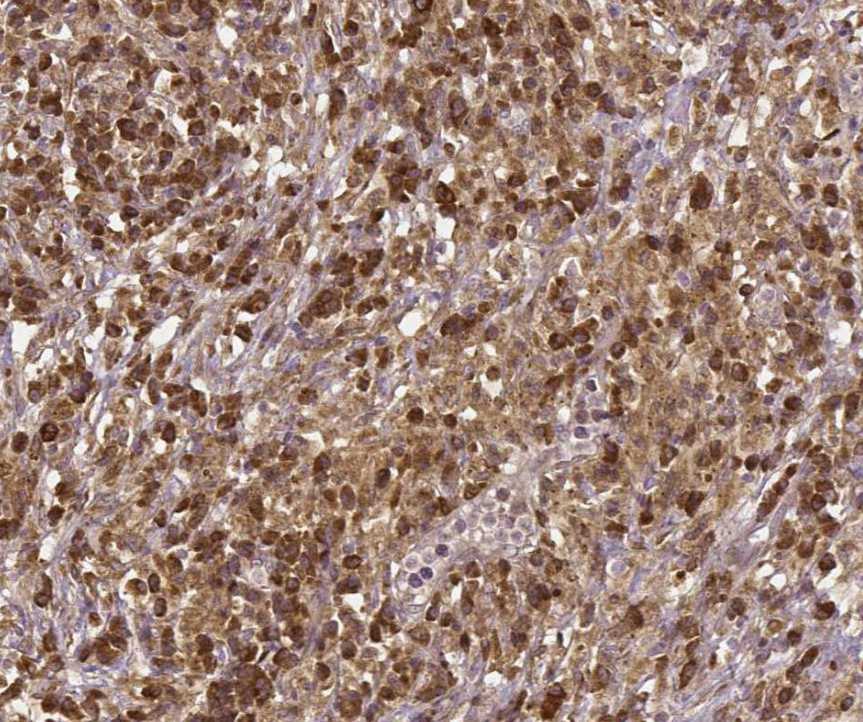
Application: ImmunohistochemistrySample Tested: Melanoma tissueSpecies: HumanVerified Customer | Posted 10/05/2021

-
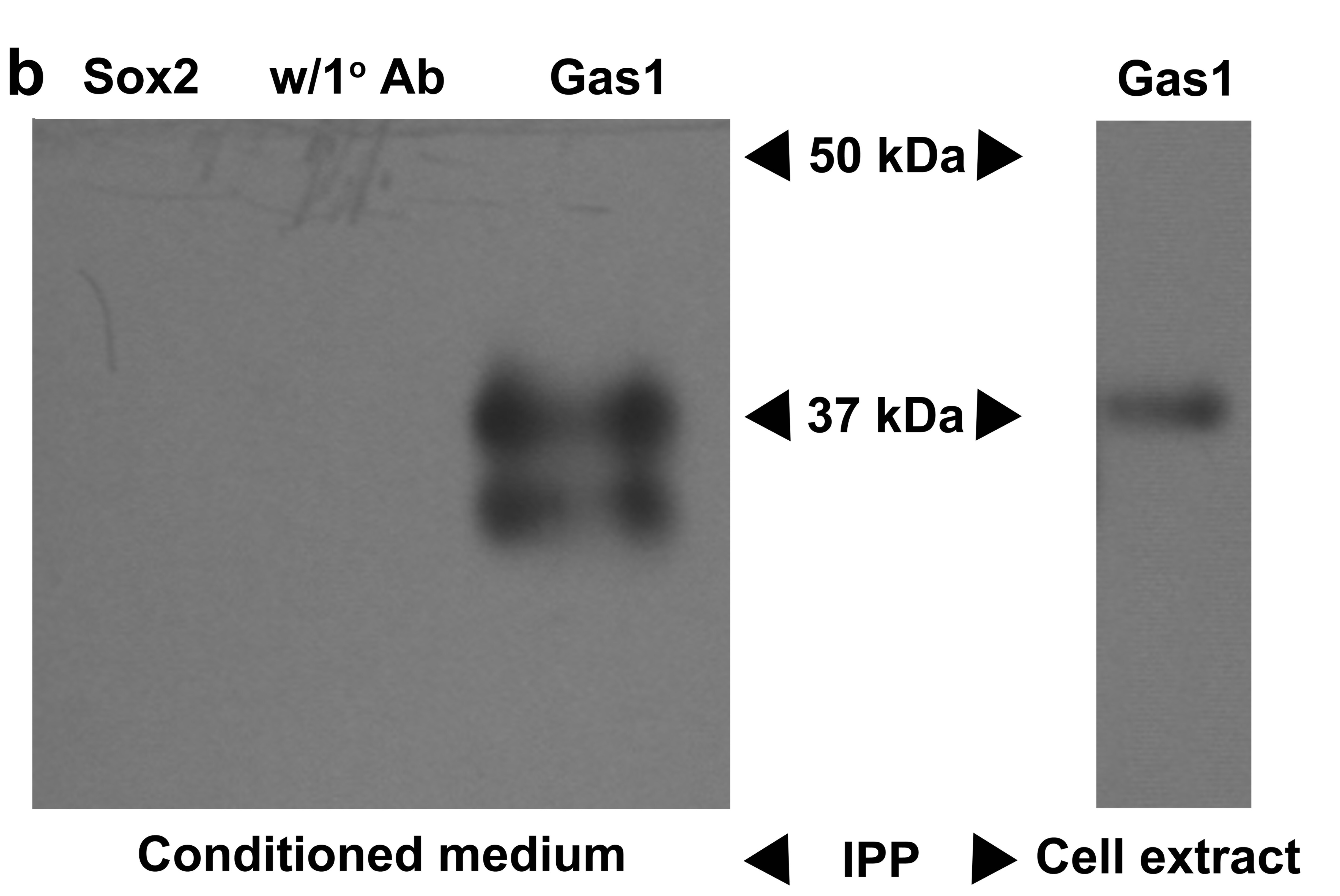
Application: ImmunoprecipitationSample Tested: CELL CONDITIONED MEDIUM and EXTRACT CELLSpecies: Mouse and RatVerified Customer | Posted 01/09/2017This antibody also works perfectly for immunoprecipitation (IPP). In the article of Ayala-Sarmiento et al 2016, DOI:10.1007/s00418-016-1449-0, it is noticeable that this antibody is able to immunoprecipitate both the anchored and the soluble forms of Gas1. A clear and a single band is seen in the immunoprecipitation of the cell extract and two extracellular isoforms of soluble Gas1 are present in the conditioned medium. No signal is detected in the negative control of the IPP, see Figure.
-
Application: Immunocytochemistry/ImmunofluorescenceSample Tested: Embryonic neuronal tissue and Embryonic brainSpecies: MouseVerified Customer | Posted 01/09/2016In the publication of Estudillo et al 2015: Gas1 is present in germinal niches of developing dentate gyrus and cortex. Cell and Tissue Research. It can be appreciated that the antibody gives a congruent and clear signal of GAS1 in embrionic and postnatal brain
There are no reviews that match your criteria.
Protocols
Find general support by application which include: protocols, troubleshooting, illustrated assays, videos and webinars.
- Antigen Retrieval Protocol (PIER)
- Antigen Retrieval for Frozen Sections Protocol
- Appropriate Fixation of IHC/ICC Samples
- Cellular Response to Hypoxia Protocols
- Chromogenic IHC Staining of Formalin-Fixed Paraffin-Embedded (FFPE) Tissue Protocol
- Chromogenic Immunohistochemistry Staining of Frozen Tissue
- ClariTSA™ Fluorophore Kits
- Detection & Visualization of Antibody Binding
- Fluorescent IHC Staining of Frozen Tissue Protocol
- Graphic Protocol for Heat-induced Epitope Retrieval
- Graphic Protocol for the Preparation and Fluorescent IHC Staining of Frozen Tissue Sections
- Graphic Protocol for the Preparation and Fluorescent IHC Staining of Paraffin-embedded Tissue Sections
- Graphic Protocol for the Preparation of Gelatin-coated Slides for Histological Tissue Sections
- IHC Sample Preparation (Frozen sections vs Paraffin)
- Immunofluorescent IHC Staining of Formalin-Fixed Paraffin-Embedded (FFPE) Tissue Protocol
- Immunohistochemistry (IHC) and Immunocytochemistry (ICC) Protocols
- Immunohistochemistry Frozen Troubleshooting
- Immunohistochemistry Paraffin Troubleshooting
- Preparing Samples for IHC/ICC Experiments
- Preventing Non-Specific Staining (Non-Specific Binding)
- Primary Antibody Selection & Optimization
- Protocol for Heat-Induced Epitope Retrieval (HIER)
- Protocol for Making a 4% Formaldehyde Solution in PBS
- Protocol for VisUCyte™ HRP Polymer Detection Reagent
- Protocol for the Preparation & Fixation of Cells on Coverslips
- Protocol for the Preparation and Chromogenic IHC Staining of Frozen Tissue Sections
- Protocol for the Preparation and Chromogenic IHC Staining of Frozen Tissue Sections - Graphic
- Protocol for the Preparation and Chromogenic IHC Staining of Paraffin-embedded Tissue Sections
- Protocol for the Preparation and Chromogenic IHC Staining of Paraffin-embedded Tissue Sections - Graphic
- Protocol for the Preparation and Fluorescent IHC Staining of Frozen Tissue Sections
- Protocol for the Preparation and Fluorescent IHC Staining of Paraffin-embedded Tissue Sections
- Protocol for the Preparation of Gelatin-coated Slides for Histological Tissue Sections
- R&D Systems Quality Control Western Blot Protocol
- TUNEL and Active Caspase-3 Detection by IHC/ICC Protocol
- The Importance of IHC/ICC Controls
- Troubleshooting Guide: Immunohistochemistry
- Troubleshooting Guide: Western Blot Figures
- Western Blot Conditions
- Western Blot Protocol
- Western Blot Protocol for Cell Lysates
- Western Blot Troubleshooting
- Western Blot Troubleshooting Guide
- View all Protocols, Troubleshooting, Illustrated assays and Webinars
Loading...