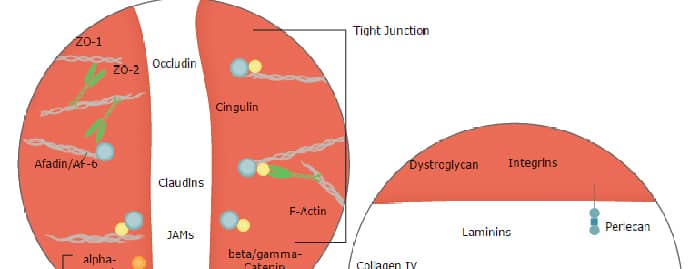
The family of juctional adhesion molecules (JAM), comprising at least three members, are type I transmembrane receptors belonging to the immunoglobulin (Ig) superfamily (1, 2). These proteins are localized in the tight junctions between endothelial cells or epithelial cells. Some family members are also found on blood leukocytes and platelets. JAM-B, alternatively named vascular endothelial JAM (VE-JAM), is expressed prominently on high endothelial venules of lymphoid organs where it is localized to the intercellular boundaries of high endothelial cells. It is also expressed on the endothelium of a variety of non-lymphoid organs, especially the heart and placenta (2, 3, 5). Mouse JAM-B/VE-JAM cDNA predicts a 298 amino acid (aa) precursor protein with a putative 28 aa signal peptide, a 209 aa extracellular region containing two Ig domains, a 23 aa transmembrane domain and a 38 aa cytoplasmic domain containing a PDZ-binding motif and a PKC phosphorylation site (2, 3). Mouse JAM-B shares approximately 79% aa sequence homology with its human homologue. It also shares approximately 35% aa sequence homology with mouse JAM-A or JAM‑C. JAM-B exhibits homotypic interactions, as well as heterotypic interactions with JAM‑C, but not JAM-A (4, 5, 7). It is also a ligand for the Integrin alpha4beta1. However, the JAM-B/alpha4beta1 interaction is facilitated only after prior adhesion of JAM-B to JAM‑C (6). Through its heterotypic interactions with JAM‑C, JAM-B is an adhesive ligand for T, NK, and dendritic cells, and may play a role in regulating leukocyte transmigration (5).
Mouse JAM-B/VE-JAM Antibody (150015)
R&D Systems | Catalog # MAB988


Scientific images may be resized, cropped, or adjusted for brightness or contrast for presentation purposes. These updates are limited to presentation and do not affect the underlying scientific interpretation of the data. Where available, additional source data may be provided upon request.
Key Product Details
Species Reactivity
Validated:
Cited:
Applications
Validated:
Cited:
Label
Antibody Source
Product Specifications
Immunogen
Phe29-Asn236 (predicted)
Accession # Q9JI59
Specificity
Clonality
Host
Isotype
Endotoxin Level
Scientific Data Images for Mouse JAM-B/VE-JAM Antibody (150015)
Cell Adhesion Mediated by JAM‑B/VE‑JAM and Neutral-ization by Mouse JAM‑B/ VE‑JAM Antibody.
Recombinant Mouse JAM-B/VE-JAM Fc Chimera (Catalog # 988-VJ), immobilized onto a microplate previously coated with Goat Anti-Human IgG Fc (Catalog # G-102-C), supports the adhesion of the J45.01 human acute lymphoblastic leukemia T lymphocyte cell line in a dose-dependent manner (orange line), as measured by endogenous cellular lysosomal acid phosphatase activity. Adhesion elicited by Recombinant Mouse JAM-B/VE-JAM Fc Chimera (0.2 µg/mL) is neutralized (green line) by increasing concentrations of Rat Anti-Mouse JAM-B/ VE-JAM Monoclonal Antibody (Catalog # MAB988). The ND50 is typically 0.1-0.5 µg/mL.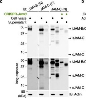
Detection of cm48lzo2cyr2ggmnee5f.jpg by Western Blot
The cleaved JAM-C ectodomain is accumulated in the fat interstitial tissues. (A) Schematic illustration for detecting full-length JAM-C (fJAM-C) and/or soluble JAM-C (sJAM-C) using JAM-C (N) and JAM-C (C) antibodies (Abs) targeting the N- and C-termini, respectively. (B) Knockdown of the Jam3 gene encoding mouse JAM-C in ADSCs using the CRISPR method. (C) Western blot for the indicated proteins in the whole-cell lysates and supernatants of the revealed ADSCs. (D) Western blot for the indicated proteins in the whole-cell lysates of mouse adult spleen tissue (control), adipose tissue and ADSCs. (E) Confocal images of mouse adipose tissue stained with JAM-C (N) and JAM-C (C) Abs. Asterisks indicate lipid droplets in differentiated adipocytes. Squares show the enlarged areas. Scale bar, 50 µm. Image collected and cropped by CiteAb from the following open publication (https://pubmed.ncbi.nlm.nih.gov/33801826), licensed under a CC-BY license. Not internally tested by R&D Systems.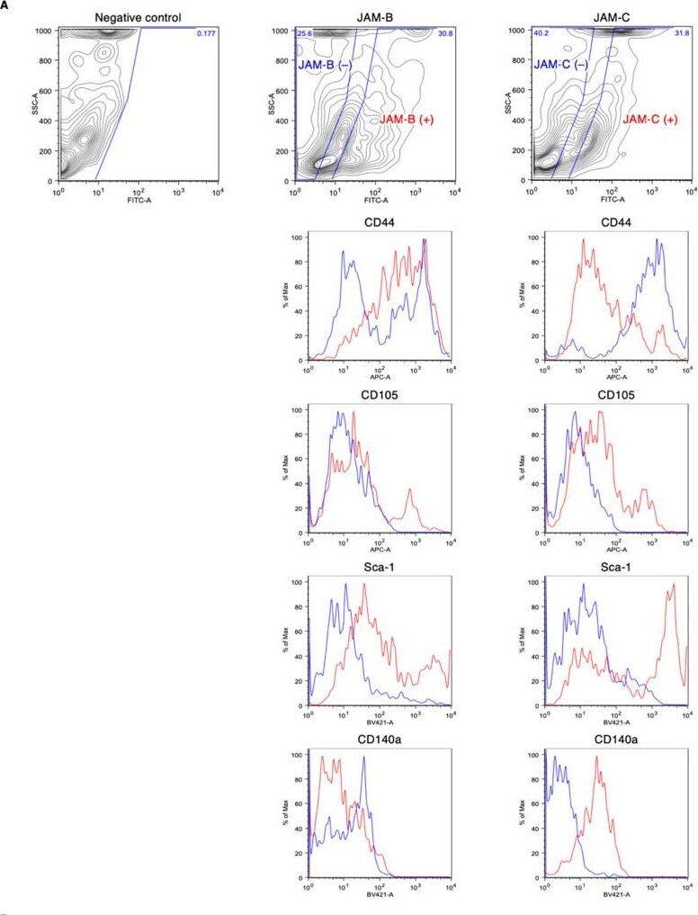
Detection of iz3qwgav7zcxfwh7li0e.jpg by Flow Cytometry
Fluorescence-activated cell sorting (FACS) profiles of the stromal vascular fraction (SVF) of the mouse adipose tissue. (A) Association between the expression of mesenchymal stem cell (MSC) markers CD44, CD105, CD140a and Sca-1 and JAM-B or JAM-C expression. (B) The JAM-C expression in the Sca1 /CD31−/CD45−/Ter119− cell lineage. Image collected and cropped by CiteAb from the following open publication (https://pubmed.ncbi.nlm.nih.gov/33801826), licensed under a CC-BY license. Not internally tested by R&D Systems.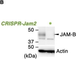
Detection of JAM-B/VE-JAM by Western Blot
A Soluble JAM-C couples with JAM-B to promote the ADSC adhesion and niche function. (A) Knockdown of the Jam2 gene encoding mouse JAM-B in ADSCs using the CRISPR method. (B) Western blot for the indicated proteins in the whole-cell lysates of the revealed ADSCs. (C) Cell adhesion assay for ADSCs grown in the indicated culture conditions. The relative levels are shown in histograms (mean ± SD; n = 8). (D) RT-qPCR for the indicated MSC markers in ADSCs cultivated in the indicated culture conditions. The relative expression levels are shown in the histograms (mean ± SD; n = 4). * p < 0.05; ** p < 0.01; *** p < 0.001. Image collected and cropped by CiteAb from the following open publication (https://pubmed.ncbi.nlm.nih.gov/33801826), licensed under a CC-BY license. Not internally tested by R&D Systems.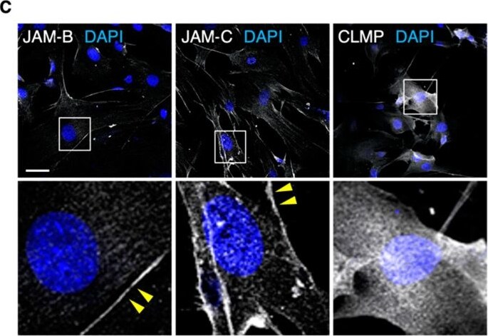
Detection of JAM-B/VE-JAM by Immunocytochemistry/ Immunofluorescence
JAM-B and JAM-C are concentrated on cell membranes of mouse adipose-derived stromal/stem cells (ADSCs). RT-PCR (A) and Western blot (B) for the indicated molecules in ADSCs derived from three mice (#1–3). Mouse kidney (for Jam-4) and spleen tissues (for other junctional adhesion molecules; JAMs) are used as positive controls (Ctrl). P, passage. (C) Confocal images of ADSCs stained for the indicated markers. Arrowheads show JAM-B- and JAM-C-immunoreactive signals on the cell membranes of ADSCs. Squares indicate the enlarged areas. Scale bar, 50 µm. Image collected and cropped by CiteAb from the following open publication (https://pubmed.ncbi.nlm.nih.gov/33801826), licensed under a CC-BY license. Not internally tested by R&D Systems.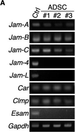
Detection of JAM-B/VE-JAM by Western Blot
JAM-B and JAM-C are concentrated on cell membranes of mouse adipose-derived stromal/stem cells (ADSCs). RT-PCR (A) and Western blot (B) for the indicated molecules in ADSCs derived from three mice (#1–3). Mouse kidney (for Jam-4) and spleen tissues (for other junctional adhesion molecules; JAMs) are used as positive controls (Ctrl). P, passage. (C) Confocal images of ADSCs stained for the indicated markers. Arrowheads show JAM-B- and JAM-C-immunoreactive signals on the cell membranes of ADSCs. Squares indicate the enlarged areas. Scale bar, 50 µm. Image collected and cropped by CiteAb from the following open publication (https://pubmed.ncbi.nlm.nih.gov/33801826), licensed under a CC-BY license. Not internally tested by R&D Systems.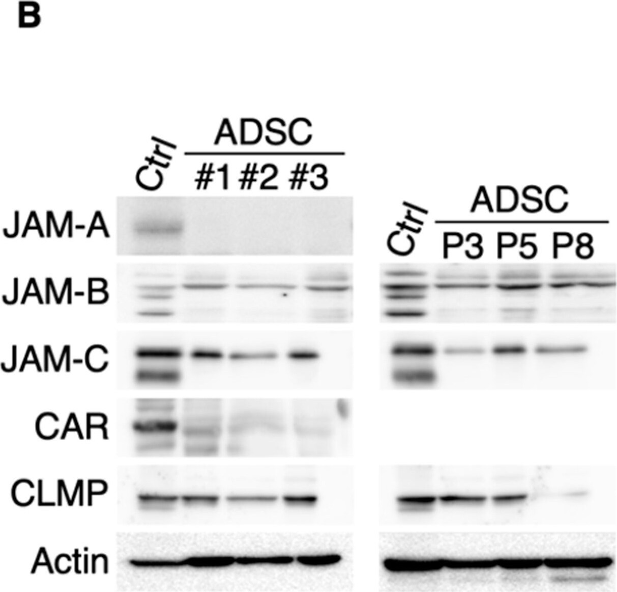
Detection of JAM-B/VE-JAM by Western Blot
JAM-B and JAM-C are concentrated on cell membranes of mouse adipose-derived stromal/stem cells (ADSCs). RT-PCR (A) and Western blot (B) for the indicated molecules in ADSCs derived from three mice (#1–3). Mouse kidney (for Jam-4) and spleen tissues (for other junctional adhesion molecules; JAMs) are used as positive controls (Ctrl). P, passage. (C) Confocal images of ADSCs stained for the indicated markers. Arrowheads show JAM-B- and JAM-C-immunoreactive signals on the cell membranes of ADSCs. Squares indicate the enlarged areas. Scale bar, 50 µm. Image collected and cropped by CiteAb from the following open publication (https://pubmed.ncbi.nlm.nih.gov/33801826), licensed under a CC-BY license. Not internally tested by R&D Systems.Applications for Mouse JAM-B/VE-JAM Antibody (150015)
Western Blot
Sample: Recombinant Mouse JAM-B/VE-JAM Fc Chimera (Catalog # 988-VJ)
Neutralization
Formulation, Preparation, and Storage
Purification
Reconstitution
Reconstitute at 0.5 mg/mL in sterile PBS. For liquid material, refer to CoA for concentration.
Formulation
Shipping
Stability & Storage
- 12 months from date of receipt, -20 to -70 °C as supplied.
- 1 month, 2 to 8 °C under sterile conditions after reconstitution.
- 6 months, -20 to -70 °C under sterile conditions after reconstitution.
Calculators
Background: JAM-B/VE-JAM
References
- Chavakis, T. et al. (2003) Thromb. Haemost. 89:13.
- Aurand-Lions, M. et al. (2001) Blood 98:3699.
- Palmeri, A. et al. (2000) J. Biol. Chem. 275:19139.
- Cunnigham, S. et al. (2000) J. Biol. Chem. 275:34750.
- Liang, T. et al. (2002) J. Immunol. 168:1618.
- Cunningham, A. et al. (2002) J Biol. Chem. 277:27589.
- Arrate, M. et al. (2001) J. Biol. Chem. 276:45826.
Long Name
Alternate Names
Gene Symbol
UniProt
Additional JAM-B/VE-JAM Products
Product Documents for Mouse JAM-B/VE-JAM Antibody (150015)
Certificate of Analysis
To download a Certificate of Analysis, please enter a lot or batch number in the search box below.
Note: Certificate of Analysis not available for kit components.
Product Specific Notices for Mouse JAM-B/VE-JAM Antibody (150015)
For research use only
Citations for Mouse JAM-B/VE-JAM Antibody (150015)
 Powered by Bioz
Customer Reviews for Mouse JAM-B/VE-JAM Antibody (150015)
There are currently no reviews for this product. Be the first to review Mouse JAM-B/VE-JAM Antibody (150015) and earn rewards!
Have you used Mouse JAM-B/VE-JAM Antibody (150015)?
Submit a review and receive an Amazon gift card!
$25/€18/£15/$25CAN/¥2500 for a review with an image
$10/€7/£6/$10CAN/¥1110 for a review without an image
Submit a review
Protocols
Find general support by application which include: protocols, troubleshooting, illustrated assays, videos and webinars.
- Cellular Response to Hypoxia Protocols
- R&D Systems Quality Control Western Blot Protocol
- Troubleshooting Guide: Western Blot Figures
- Western Blot Conditions
- Western Blot Protocol
- Western Blot Protocol for Cell Lysates
- Western Blot Troubleshooting
- Western Blot Troubleshooting Guide
- View all Protocols, Troubleshooting, Illustrated assays and Webinars
Associated Pathways