Osteoactivin (also named GPNMB and DC-HIL) is a 125 kDa, intracellular glycoprotein that is associated with cell endosomal/lysosomal compartments (1, 2). Mouse osteoactivin is synthesized as a type I, transmembrane, 574 amino acid (aa) precursor that contains a 22 aa signal sequence, a 478 aa luminal/extracellular domain, a 23 aa transmembrane segment and a 51 aa cytoplasmic tail. The luminal region contains an N-terminal heparin-binding motif, multiple glycosylation sites, an RGD motif and a 130 aa PKD domain. The intracellular tail also has an RGD motif, plus an ITAM (Y-x-x-I) and lysosomal targeting (L-L) motif. The extracellular/luminal region is 89% and 74% aa identical to the equivalent regions in rat and human, respectively. Cells known to express osteoactivin include osteoblasts, dendritic cells, and melanocytes, plus fetal chondrocytes and stratum basale keratinocytes (2, 3). Osteoactivin is reported to bind to heparan sulfate-proteoglycan, possibly on the surface of fibroblasts and endothelial cells (2). It may also interact with integrins.
Mouse Osteoactivin/GPNMB Antibody
R&D Systems | Catalog # AF2330

Key Product Details
Species Reactivity
Validated:
Mouse
Cited:
Human, Mouse, Transgenic Mouse
Applications
Validated:
Western Blot, ELISA Capture (Matched Antibody Pair), Neutralization
Cited:
Immunohistochemistry, Immunohistochemistry-Paraffin, Western Blot, Neutralization, Immunocytochemistry
Label
Unconjugated
Antibody Source
Polyclonal Goat IgG
Loading...
Product Specifications
Immunogen
Mouse myeloma cell line NS0-derived recombinant mouse Osteoactivin/GPNMB
Lys23-Asn502
Accession # Q8BVA0
Lys23-Asn502
Accession # Q8BVA0
Specificity
Detects mouse Osteoactivin/GPNMB in ELISAs and Western blots. In sandwich immunoassays, less than 0.2% cross-reactivity with recombinant human (rh) Osteoactivin, rhSyndecan-4, and recombinant mouse Syndecan-4 is observed.
Clonality
Polyclonal
Host
Goat
Isotype
IgG
Endotoxin Level
<0.10 EU per 1 μg of the antibody by the LAL method.
Scientific Data Images for Mouse Osteoactivin/GPNMB Antibody
Cell Adhesion Mediated by Osteoactivin/GPNMB and Neutralization by Mouse Osteoactivin/GPNMB Antibody.
Recombinant Mouse Osteoactivin/GPNMB Fc Chimera (Catalog # 2330-AC), immobilized onto a microplate previously coated with Goat Anti-Mouse IgG Fc (Catalog # G-202-C), supports the adhesion of the SVEC4-10 mouse vascular endothelial cell line in a dose-dependent manner (orange line), as measured by endogenous cellular lysosomal acid phosphatase activity. Adhesion elicited by Recombinant Mouse Osteoactivin/GPNMB Fc Chimera (0.25 µg/mL) is neutralized (green line) by increasing concentrations of Goat Anti-Mouse Osteoactivin/ GPNMB Antigen Affinity-purified Polyclonal Antibody (Catalog # AF2330). The ND50 is typically 0.2-0.8 µg/mL.
Detection of Human Osteoactivin/GPNMB by Western Blot
GPNMB and galectin-3 levels are elevated in FTD-GRN brains. a, b GPNMB and galectin-3 levels (ng/mg protein) were measured in frontal lobe tissue lysates generated from cognitively normal controls (CTL; n = 27) and FTD-GRN patients (n = 25). Data analyzed using unpaired t-test. c Representative immunoblots for GPNMB and galectin-3 in frontal lobe lysates from cognitively normal controls (n = 8) and FTD-GRN (n = 8) patients. d GPNMB levels (ng/mL) in CSF samples form cognitively normal controls (n = 14), FTD-GRN (n = 9), FTD-C9orf72 (n = 12) and FTD-MAPT (n = 12) samples quantified by ELISA. Data analyzed using one-way ANOVA. e, f GPNMB immunostaining was performed on frontal lobe tissue sections from cognitively normal controls (n = 5) (e) and FTD-GRN (n = 5) (f) patients. g, h Immunostaining for p-TDP 43 was stained on adjacent sections from identical samples in e, f as marker of FTLD pathology. i GPNMB staining intensity in human brain sections (e, f) were measured and presented as fold change. Representative immunofluorescence staining for cell markers (green) (j, n, r), GPNMB (red) (k, o, s), DAPI (blue) (i, p, t) in paraffin sections of brains from FTD-GRN cases. Iba-1, GFAP, NeuN used for markers of human microglia, astrocytes, and neurons respectively. GPNMB and Iba-1 signals overlap (arrow) (m) whereas, no overlapping signal was observed in co-staining with GFAP or NeuN (q, u). Scale bars were labeled in the images. Data analyzed by unpaired t-test. Scale bars (20 µm) labeled in images and quantitative data are shown as mean ± SEM, *p < 0.05; **p < 0.01; ***p < 0.001; ****p < 0.0001 Image collected and cropped by CiteAb from the following open publication (https://pubmed.ncbi.nlm.nih.gov/33028409), licensed under a CC-BY license. Not internally tested by R&D Systems.
Detection of Mouse Osteoactivin/GPNMB by Immunohistochemistry
GPNMB and galectin-3 co-localize with Iba-1 positive microglia cells in Grn−/− mouse brain. a Representative immunofluorescent co-staining for different cell markers (Iba-1, microglia; GFAP, astrocytes; NeuN, neurons) are shown in green, GPNMB (red), and nuclei (DAPI; blue) in brains of 19-month-old Grn−/− mice. b Representative immunofluorescent co-staining for different cell markers (Iba-1, microglia; GFAP, astrocytes; NeuN, neurons) are shown in green, galectin-3 (red), and nuclei (DAPI; blue) in brains of 19-month-old Grn−/− mice. Scale bars (20 µm) labeled in images Image collected and cropped by CiteAb from the following open publication (https://pubmed.ncbi.nlm.nih.gov/33028409), licensed under a CC-BY license. Not internally tested by R&D Systems.
Mouse Osteoactivin / GPNMB ELISA Standard Curve
Recombinant Mouse Osteoactivin/GPNMB Fc Chimera (Catalog # 2330-AC) was serially diluted and captured by Goat Anti-Mouse Osteoactivin/GPNMB Antigen Affinity-purified Polyclonal Antibody (Catalog # AF2330) coated on a Clear Polystyrene Microplate (Catalog # DY990). Goat Anti-Mouse Osteoactivin/GPNMB Antigen Affinity-purified Polyclonal Antibody (Catalog # AF2330) was biotinylated and incubated with the protein captured on the plate. Detection of the standard curve was achieved by incubating Streptavidin-HRP (Catalog # DY998)
Mouse Osteoactivin / GPNMB ELISA Standard Curve
Recombinant Mouse Osteoactivin/GPNMB Fc Chimera (Catalog # 2330-AC) was serially diluted and captured by Goat Anti-Mouse Osteoactivin/GPNMB Antigen Affinity-purified Polyclonal Antibody (Catalog # AF2330) coated on a Clear Polystyrene Microplate (Catalog # DY990). Goat Anti-Mouse Osteoactivin/GPNMB Antigen Affinity-purified Polyclonal Antibody (Catalog # AF2330) was biotinylated and incubated with the protein captured on the plate. Detection of the standard curve was achieved by incubating Streptavidin-HRP (Catalog # DY998)Applications for Mouse Osteoactivin/GPNMB Antibody
Application
Recommended Usage
Western Blot
0.1 µg/mL
Sample: Recombinant Mouse Osteoactivin/GPNMB Fc Chimera (Catalog # 2330-AC)
Sample: Recombinant Mouse Osteoactivin/GPNMB Fc Chimera (Catalog # 2330-AC)
Neutralization
Measured by its ability to neutralize Osteoactivin/GPNMB-mediated adhesion of the SVEC4‑10 mouse vascular endothelial cell line. The Neutralization Dose (ND50) is typically 0.2-0.8 µg/mL in the presence of 0.25 µg/mL Recombinant Mouse Osteoactivin/GPNMB Fc Chimera.
Mouse Osteoactivin/GPNMB Sandwich Immunoassay
Please Note: Optimal dilutions of this antibody should be experimentally determined.
Reviewed Applications
Read 2 reviews rated 4.5 using AF2330 in the following applications:
Formulation, Preparation, and Storage
Purification
Antigen Affinity-purified
Reconstitution
Reconstitute at 0.2 mg/mL in sterile PBS. For liquid material, refer to CoA for concentration.
Loading...
Formulation
Lyophilized from a 0.2 μm filtered solution in PBS with Trehalose. *Small pack size (SP) is supplied either lyophilized or as a 0.2 µm filtered solution in PBS.
Shipping
Lyophilized product is shipped at ambient temperature. Liquid small pack size (-SP) is shipped with polar packs. Upon receipt, store immediately at the temperature recommended below.
Stability & Storage
Use a manual defrost freezer and avoid repeated freeze-thaw cycles.
- 12 months from date of receipt, -20 to -70 °C as supplied.
- 1 month, 2 to 8 °C under sterile conditions after reconstitution.
- 6 months, -20 to -70 °C under sterile conditions after reconstitution.
Calculators
Background: Osteoactivin/GPNMB
References
- Bachner, D. et al. (2002) Gene Exp. Patterns 1:159.
- Shikano, S. et al. (2001) J. Biol. Chem. 276:8125.
- Owen, T.A. et al. (2003) Crit. Rev. Eukaryot. Gene Expr. 13:205.
Long Name
Glycoprotein Non-Metastatic Melanoma Protein B
Alternate Names
DC-HIL, GPNMB, HGFIN
Gene Symbol
GPNMB
UniProt
Additional Osteoactivin/GPNMB Products
Product Documents for Mouse Osteoactivin/GPNMB Antibody
Certificate of Analysis
To download a Certificate of Analysis, please enter a lot or batch number in the search box below.
Note: Certificate of Analysis not available for kit components.
Product Specific Notices for Mouse Osteoactivin/GPNMB Antibody
For research use only
Related Research Areas
Citations for Mouse Osteoactivin/GPNMB Antibody
 Powered by Bioz
Customer Reviews for Mouse Osteoactivin/GPNMB Antibody (2)
4.5 out of 5
2 Customer Ratings
Have you used Mouse Osteoactivin/GPNMB Antibody?
Submit a review and receive an Amazon gift card!
$25/€18/£15/$25CAN/¥2500 Yen for a review with an image
$10/€7/£6/$10CAN/¥1110 Yen for a review without an image
Submit a review
Customer Images
Showing
1
-
2 of
2 reviews
Showing All
Filter By:
-
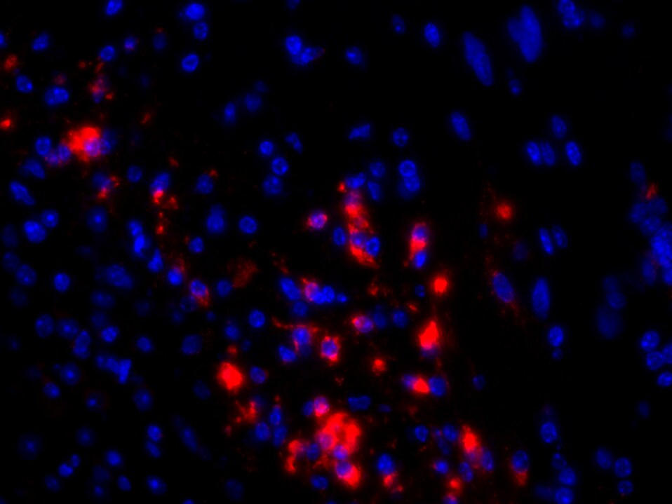
Application: Immunohistochemistry-ParaffinSample Tested: Mouse brainSpecies: MouseVerified Customer | Posted 08/14/2020DAPI bleue and GPNMB red1/100 in PBS
 Bio-Techne ResponseThis review was submitted through the legacy Novus Innovators Program, reflecting a new species or application tested on a primary antibody.
 -
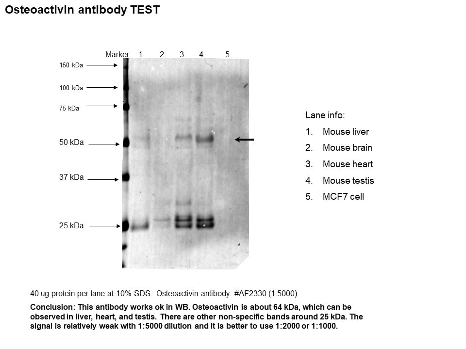
Application: Western BlotSample Tested: Liver, Brain, Mouse heart and TestisSpecies: Mouse and MCF7 cellVerified Customer | Posted 08/30/2017
There are no reviews that match your criteria.
Protocols
Find general support by application which include: protocols, troubleshooting, illustrated assays, videos and webinars.
- Cellular Response to Hypoxia Protocols
- R&D Systems Quality Control Western Blot Protocol
- Troubleshooting Guide: Western Blot Figures
- Western Blot Conditions
- Western Blot Protocol
- Western Blot Protocol for Cell Lysates
- Western Blot Troubleshooting
- Western Blot Troubleshooting Guide
- View all Protocols, Troubleshooting, Illustrated assays and Webinars
Loading...