Advanced glycation endproducts (AGE) are adducts formed by the non-enzymatic glycation or oxidation of macromolecules (1). AGE forms during aging and its formation is accelerated under pathophysiologic states such as diabetes, Alzheimer’s disease, renal failure and immune/inflammatory disorders. Receptor for Advanced Glycation Endoproducts (RAGE), named for its ability to bind AGE, is a multiligand receptor belonging the immunoglobulin (Ig) superfamily. Besides AGE, RAGE binds amyloid beta -peptide, S100/calgranulin family proteins, high mobility group B1 (HMGB1, also know as amphoterin) and leukocyte integrins (1, 2). The mouse RAGE gene encodes a 403 amino acid (aa) type I transmembrane glycoprotein with a 22 aa signal peptide, a 319 aa extracellular domain containing an Ig-like V-type domain and two Ig-like C-type domains, a 21 aa transmembrane domain and a 41 aa cytoplasmic domain (3). The V-type domain and the cytoplasmic domain are important for ligand binding and for intracellular signaling, respectively. Two alternative splice variants, lacking the V-type domain or the cytoplasmic tail, are known (1, 4). RAGE is highly expressed in the embryonic central nervous system (5). In adult tissues, RAGE is expressed at low levels in multiple tissues including endothelial and smooth muscle cells, mononuclear phagocytes, pericytes, microglia, neurons, cardiac myocytes and hepatocytes (6). The expression of RAGE is upregulated upon ligand interaction. Depending on the cellular context and interacting ligand, RAGE activation can trigger differential signaling pathways that affect divergent pathways of gene expression (1, 7). RAGE activation modulates varied essential cellular responses (including inflammation, immunity, proliferation, cellular adhesion and migration) that contribute to cellular dysfunction associated with chronic diseases such as diabetes, cancer, amyloidoses and immune or inflammatory disorders (1).
Mouse RAGE/AGER Antibody (697005)
R&D Systems | Catalog # MAB11794

Key Product Details
Species Reactivity
Validated:
Mouse
Cited:
Transgenic Mouse
Applications
Validated:
Immunohistochemistry
Cited:
Immunohistochemistry
Label
Unconjugated
Antibody Source
Monoclonal Rat IgG2B Clone # 697005
Loading...
Product Specifications
Immunogen
Mouse myeloma cell line NS0-derived recombinant mouse RAGE
Gly23-Leu342
Accession # Q62151
Gly23-Leu342
Accession # Q62151
Specificity
Detects mouse RAGE in direct ELISAs.
In direct ELISAs, 100% cross-reactivity
with recombinant rat RAGE, 20% - 50% cross-reactivity with recombinant human
RAGE, and no cross-reactivity with recombinant canine RAGE is observed.
Clonality
Monoclonal
Host
Rat
Isotype
IgG2B
Scientific Data Images for Mouse RAGE/AGER Antibody (697005)
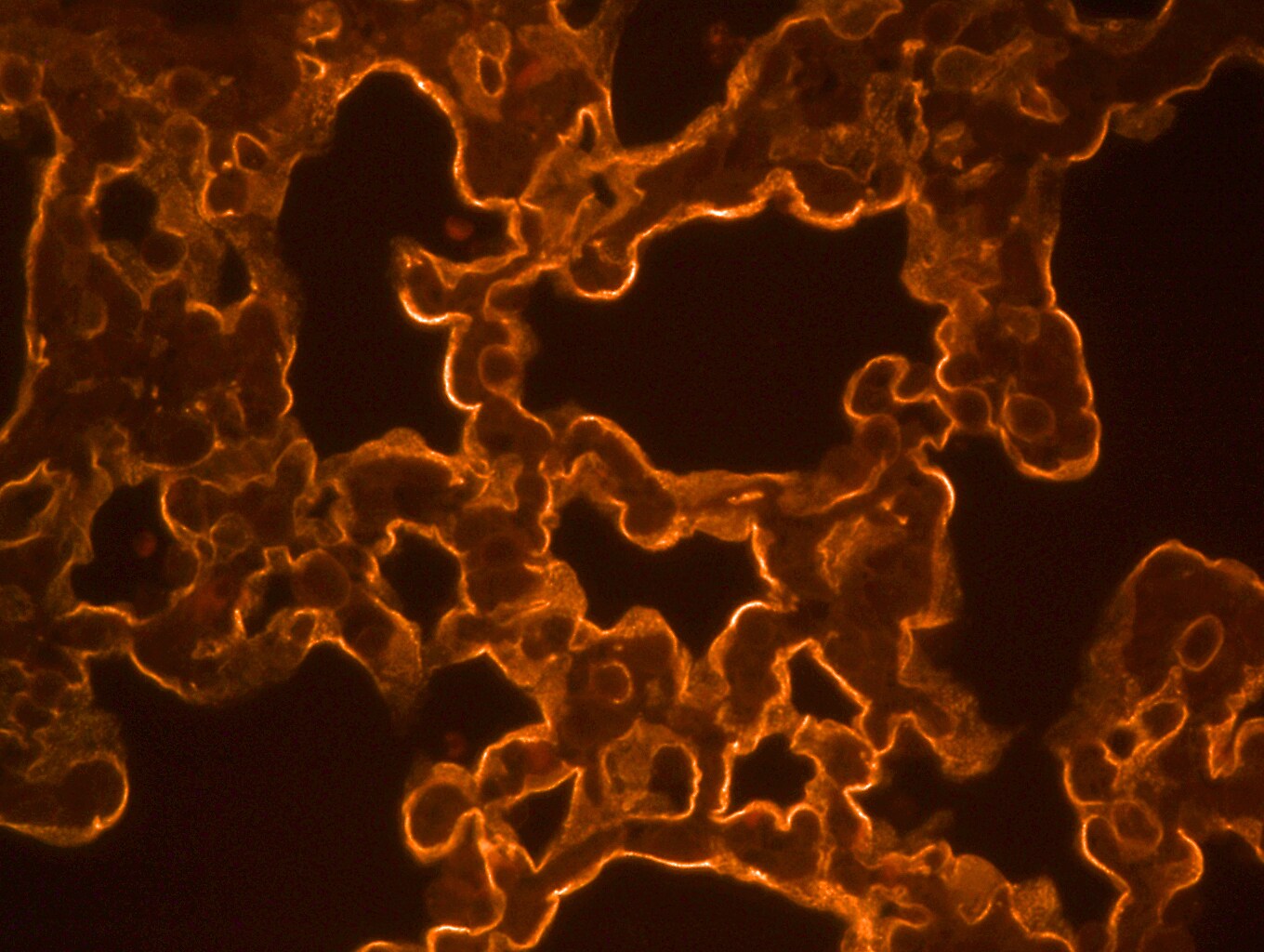
RAGE in Mouse Spinal Cord.
RAGE was detected in perfusion fixed frozen sections of mouse spinal cord using Rat Anti-Mouse RAGE Monoclonal Antibody (Catalog # MAB11794) at 25 µg/mL overnight at 4 °C. Tissue was stained using the NorthernLights™ 557-conjugated Anti-Rat IgG Secondary Antibody (red; Catalog # NL013) and counterstained with DAPI (blue). Specific staining was localized to the plasma membranes of motor neurons. View our protocol for Fluorescent IHC Staining of Frozen Tissue Sections.Applications for Mouse RAGE/AGER Antibody (697005)
Application
Recommended Usage
Immunohistochemistry
8-25 µg/mL
Sample: Perfusion fixed frozen sections of mouse spinal cord
Sample: Perfusion fixed frozen sections of mouse spinal cord
Reviewed Applications
Read 1 review rated 4 using MAB11794 in the following applications:
Formulation, Preparation, and Storage
Purification
Protein A or G purified from hybridoma culture supernatant
Reconstitution
Sterile PBS to a final concentration of 0.5 mg/mL. For liquid material, refer to CoA for concentration.
Loading...
Formulation
Lyophilized from a 0.2 μm filtered solution in PBS with Trehalose. *Small pack size (SP) is supplied either lyophilized or as a 0.2 µm filtered solution in PBS.
Shipping
Lyophilized product is shipped at ambient temperature. Liquid small pack size (-SP) is shipped with polar packs. Upon receipt, store immediately at the temperature recommended below.
Stability & Storage
Use a manual defrost freezer and avoid repeated freeze-thaw cycles.
- 12 months from date of receipt, -20 to -70 °C as supplied.
- 1 month, 2 to 8 °C under sterile conditions after reconstitution.
- 6 months, -20 to -70 °C under sterile conditions after reconstitution.
Calculators
Background: RAGE/AGER
References
- Schmidt, A. et al. (2001) J. Clin. Invest. 108:949.
- Chavakis, T. et al. (2003) J. Exp. Med. 198:507.
- Renard, C. et al. (1997) Mol. Pharmacol. 52:54.
- Yonekura, H. et al. (2003) Biochem. J. 370:1097.
- Hori, O. et al. (1995) J. Biol. Chem. 270:25752.
- Brett, J. et al. (1993) Am. J. Pathol. 143:1699.
- Valencia, J.V. et al. (2004) Diabetes 53:743.
Long Name
Receptor for Advanced Glycation End Products
Alternate Names
AGER, SCARJ1
Gene Symbol
AGER
UniProt
Additional RAGE/AGER Products
Product Documents for Mouse RAGE/AGER Antibody (697005)
Certificate of Analysis
To download a Certificate of Analysis, please enter a lot or batch number in the search box below.
Note: Certificate of Analysis not available for kit components.
Product Specific Notices for Mouse RAGE/AGER Antibody (697005)
For research use only
Citations for Mouse RAGE/AGER Antibody (697005)
 Powered by Bioz
Customer Reviews for Mouse RAGE/AGER Antibody (697005) (1)
4 out of 5
1 Customer Rating
Have you used Mouse RAGE/AGER Antibody (697005)?
Submit a review and receive an Amazon gift card!
$25/€18/£15/$25CAN/¥2500 Yen for a review with an image
$10/€7/£6/$10CAN/¥1110 Yen for a review without an image
Submit a review
Customer Images
Showing
1
-
1 of
1 review
Showing All
Filter By:
-
Application: ImmunohistochemistrySample Tested: Lung tissueSpecies: MouseVerified Customer | Posted 07/14/2016Used on paraffin sections of mouse lung at a dilution of 1:100 Antigen retrieval performed Secondary Ab (Rhodamine) used at a dilution of 1:100

There are no reviews that match your criteria.
Protocols
Find general support by application which include: protocols, troubleshooting, illustrated assays, videos and webinars.
- Antigen Retrieval Protocol (PIER)
- Antigen Retrieval for Frozen Sections Protocol
- Appropriate Fixation of IHC/ICC Samples
- Cellular Response to Hypoxia Protocols
- Chromogenic IHC Staining of Formalin-Fixed Paraffin-Embedded (FFPE) Tissue Protocol
- Chromogenic Immunohistochemistry Staining of Frozen Tissue
- ClariTSA™ Fluorophore Kits
- Detection & Visualization of Antibody Binding
- Fluorescent IHC Staining of Frozen Tissue Protocol
- Graphic Protocol for Heat-induced Epitope Retrieval
- Graphic Protocol for the Preparation and Fluorescent IHC Staining of Frozen Tissue Sections
- Graphic Protocol for the Preparation and Fluorescent IHC Staining of Paraffin-embedded Tissue Sections
- Graphic Protocol for the Preparation of Gelatin-coated Slides for Histological Tissue Sections
- IHC Sample Preparation (Frozen sections vs Paraffin)
- Immunofluorescent IHC Staining of Formalin-Fixed Paraffin-Embedded (FFPE) Tissue Protocol
- Immunohistochemistry (IHC) and Immunocytochemistry (ICC) Protocols
- Immunohistochemistry Frozen Troubleshooting
- Immunohistochemistry Paraffin Troubleshooting
- Preparing Samples for IHC/ICC Experiments
- Preventing Non-Specific Staining (Non-Specific Binding)
- Primary Antibody Selection & Optimization
- Protocol for Heat-Induced Epitope Retrieval (HIER)
- Protocol for Making a 4% Formaldehyde Solution in PBS
- Protocol for VisUCyte™ HRP Polymer Detection Reagent
- Protocol for the Preparation & Fixation of Cells on Coverslips
- Protocol for the Preparation and Chromogenic IHC Staining of Frozen Tissue Sections
- Protocol for the Preparation and Chromogenic IHC Staining of Frozen Tissue Sections - Graphic
- Protocol for the Preparation and Chromogenic IHC Staining of Paraffin-embedded Tissue Sections
- Protocol for the Preparation and Chromogenic IHC Staining of Paraffin-embedded Tissue Sections - Graphic
- Protocol for the Preparation and Fluorescent IHC Staining of Frozen Tissue Sections
- Protocol for the Preparation and Fluorescent IHC Staining of Paraffin-embedded Tissue Sections
- Protocol for the Preparation of Gelatin-coated Slides for Histological Tissue Sections
- TUNEL and Active Caspase-3 Detection by IHC/ICC Protocol
- The Importance of IHC/ICC Controls
- Troubleshooting Guide: Immunohistochemistry
- View all Protocols, Troubleshooting, Illustrated assays and Webinars


