Resistin, also known as adipocyte-specific secretory factor (ADSF) and found in inflammatory zone 3 (FIZZ3), is a member of a family of secreted cysteine-rich peptide hormones that also includes Resistin-like molecules RELM alpha, beta, and gamma. These molecules play important roles in inflammation, glucose metabolism, and insulin resistance. Mature mouse Resistin is a 12 kDa protein with an N-terminal alpha -helical domain and a C-terminal beta -sandwich domain that is stabilized by multiple intrachain disulfide bonds. Resistin circulates as noncovalent trimers and disulfide-linked hexamers, with the trimeric form showing greater bioactivity. Resistin can also form multimers with RELM beta. Mature mouse Resistin shares 56% and 72% amino acid (aa) sequence identity with human and rat Resistin, respectively. It shares 34% - 42% aa sequence identity with mouse RELM alpha, beta, and gamma. In rodents, Resistin is expressed by adipocytes and in the pituitary and arcuate nucleus of the hypothalamus. It is upregulated during adipogenesis, in obesity, and by insulin or a high carbohydrate diet. This is in contrast to human Resistin which is produced by macrophages and monocytes but not by adipocytes. Mouse Resistin induces proinflammatory molecule production in adipocytes and promotes hepatic gluconeogenesis and insulin resistance. Human Resistin promotes lipolysis by human and mouse adipocytes, but mouse Resistin does not promote lipolysis by adipocytes of either species. Both mouse and human Resistin promote vascular endothelial cell sprouting in vitro and inflammatory reactions in vivo.

Key Product Details
Species Reactivity
Validated:
Mouse
Cited:
Mouse
Applications
Validated:
Immunohistochemistry, Western Blot
Cited:
Western Blot
Label
Unconjugated
Antibody Source
Polyclonal Goat IgG
Loading...
Product Specifications
Immunogen
E. coli-derived recombinant mouse Resistin (R&D Systems, Catalog # 1069-RN)
Ser21-Ser114
Accession # Q99P87
Ser21-Ser114
Accession # Q99P87
Specificity
Detects mouse Resistin in direct ELISAs and Western blots. In this format, less than 2% cross-reactivity with recombinant human Resistin and recombinant mouse RELM beta is observed.
Clonality
Polyclonal
Host
Goat
Isotype
IgG
Scientific Data Images for Mouse Resistin Antibody
Resistin in Mouse Brain.
Resistin was detected in perfusion fixed frozen sections of mouse brain (caudate putamen) using 15 µg/mL Mouse Resistin Antigen Affinity-purified Polyclonal Antibody (Catalog # AF1069) overnight at 4 °C. Tissue was stained with the Anti-Goat HRP-DAB Cell & Tissue Staining Kit (brown; Catalog # CTS008) and counterstained with hematoxylin (blue). View our protocol for Chromogenic IHC Staining of Frozen Tissue Sections.Applications for Mouse Resistin Antibody
Application
Recommended Usage
Immunohistochemistry
5-15 µg/mL
Sample: Perfusion fixed frozen sections of mouse brain (caudate putamen)
Sample: Perfusion fixed frozen sections of mouse brain (caudate putamen)
Western Blot
0.1 µg/mL
Sample: Recombinant Mouse Resistin (Catalog # 1069-RN)
Sample: Recombinant Mouse Resistin (Catalog # 1069-RN)
Reviewed Applications
Read 1 review rated 4 using AF1069 in the following applications:
Formulation, Preparation, and Storage
Purification
Antigen Affinity-purified
Reconstitution
Reconstitute at 0.2 mg/mL in sterile PBS. For liquid material, refer to CoA for concentration.
Loading...
Formulation
Lyophilized from a 0.2 μm filtered solution in PBS with Trehalose. *Small pack size (SP) is supplied either lyophilized or as a 0.2 µm filtered solution in PBS.
Shipping
Lyophilized product is shipped at ambient temperature. Liquid small pack size (-SP) is shipped with polar packs. Upon receipt, store immediately at the temperature recommended below.
Stability & Storage
Use a manual defrost freezer and avoid repeated freeze-thaw cycles.
- 12 months from date of receipt, -20 to -70 °C as supplied.
- 1 month, 2 to 8 °C under sterile conditions after reconstitution.
- 6 months, -20 to -70 °C under sterile conditions after reconstitution.
Calculators
Background: Resistin
Alternate Names
ADSF, FIZZ3, HXCP1, RETN
Gene Symbol
RETN
UniProt
Additional Resistin Products
Product Documents for Mouse Resistin Antibody
Certificate of Analysis
To download a Certificate of Analysis, please enter a lot or batch number in the search box below.
Note: Certificate of Analysis not available for kit components.
Product Specific Notices for Mouse Resistin Antibody
For research use only
Related Research Areas
Citations for Mouse Resistin Antibody
 Powered by Bioz
Customer Reviews for Mouse Resistin Antibody (1)
4 out of 5
1 Customer Rating
Have you used Mouse Resistin Antibody?
Submit a review and receive an Amazon gift card!
$25/€18/£15/$25CAN/¥2500 Yen for a review with an image
$10/€7/£6/$10CAN/¥1110 Yen for a review without an image
Submit a review
Customer Images
Showing
1
-
1 of
1 review
Showing All
Filter By:
-
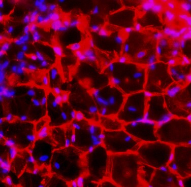
Application: Immunocytochemistry/ImmunofluorescenceSample Tested: Adipose tissueSpecies: MouseVerified Customer | Posted 05/26/2018

There are no reviews that match your criteria.
Protocols
Find general support by application which include: protocols, troubleshooting, illustrated assays, videos and webinars.
- Antigen Retrieval Protocol (PIER)
- Antigen Retrieval for Frozen Sections Protocol
- Appropriate Fixation of IHC/ICC Samples
- Cellular Response to Hypoxia Protocols
- Chromogenic IHC Staining of Formalin-Fixed Paraffin-Embedded (FFPE) Tissue Protocol
- Chromogenic Immunohistochemistry Staining of Frozen Tissue
- ClariTSA™ Fluorophore Kits
- Detection & Visualization of Antibody Binding
- Fluorescent IHC Staining of Frozen Tissue Protocol
- Graphic Protocol for Heat-induced Epitope Retrieval
- Graphic Protocol for the Preparation and Fluorescent IHC Staining of Frozen Tissue Sections
- Graphic Protocol for the Preparation and Fluorescent IHC Staining of Paraffin-embedded Tissue Sections
- Graphic Protocol for the Preparation of Gelatin-coated Slides for Histological Tissue Sections
- IHC Sample Preparation (Frozen sections vs Paraffin)
- Immunofluorescent IHC Staining of Formalin-Fixed Paraffin-Embedded (FFPE) Tissue Protocol
- Immunohistochemistry (IHC) and Immunocytochemistry (ICC) Protocols
- Immunohistochemistry Frozen Troubleshooting
- Immunohistochemistry Paraffin Troubleshooting
- Preparing Samples for IHC/ICC Experiments
- Preventing Non-Specific Staining (Non-Specific Binding)
- Primary Antibody Selection & Optimization
- Protocol for Heat-Induced Epitope Retrieval (HIER)
- Protocol for Making a 4% Formaldehyde Solution in PBS
- Protocol for VisUCyte™ HRP Polymer Detection Reagent
- Protocol for the Preparation & Fixation of Cells on Coverslips
- Protocol for the Preparation and Chromogenic IHC Staining of Frozen Tissue Sections
- Protocol for the Preparation and Chromogenic IHC Staining of Frozen Tissue Sections - Graphic
- Protocol for the Preparation and Chromogenic IHC Staining of Paraffin-embedded Tissue Sections
- Protocol for the Preparation and Chromogenic IHC Staining of Paraffin-embedded Tissue Sections - Graphic
- Protocol for the Preparation and Fluorescent IHC Staining of Frozen Tissue Sections
- Protocol for the Preparation and Fluorescent IHC Staining of Paraffin-embedded Tissue Sections
- Protocol for the Preparation of Gelatin-coated Slides for Histological Tissue Sections
- R&D Systems Quality Control Western Blot Protocol
- TUNEL and Active Caspase-3 Detection by IHC/ICC Protocol
- The Importance of IHC/ICC Controls
- Troubleshooting Guide: Immunohistochemistry
- Troubleshooting Guide: Western Blot Figures
- Western Blot Conditions
- Western Blot Protocol
- Western Blot Protocol for Cell Lysates
- Western Blot Troubleshooting
- Western Blot Troubleshooting Guide
- View all Protocols, Troubleshooting, Illustrated assays and Webinars
Loading...
Associated Pathways
