Key Product Details
Species Reactivity
Validated:
Human
Cited:
Human
Applications
Validated:
Immunohistochemistry, Immunohistochemistry-Paraffin, Western Blot, Flow Cytometry, Immunofluorescence, Immunocytochemistry/ Immunofluorescence
Cited:
Western Blot, IF/IHC
Label
Unconjugated
Antibody Source
Monoclonal Mouse IgG1 kappa Clone # CLH2
Loading...
Product Specifications
Immunogen
A synthetic peptide of human MUC5AC tandem repeat (Uniprot: P98088)
Localization
Cytoplasmic
Specificity
Mucin 5AC glycoprotein (MUC5AC) is a HMW glycoprotein belonging to the superfamily of mucins. Mucins are produced by epithelial cells and can be divided into two families; secretory mucins and membrane bound mucins. MUC5AC is a mucus-forming, secreted mucin that is found in normal gastric and tracheo-bronchial mucosa, but absent from normal colon. MUC5AC expression is present in primary ovarian mucinous cancer but usually absent in colorectal adenocarcinoma, thus showing an expression pattern opposite to MUC2. Together with a panel of antibodies, Anti-MUC5AC may be useful for differential identification of primary mucinous ovarian tumors from colon adenocarcinoma metastatic to the ovary. MUC5AC antibodies may also be useful for identification of intestinal metaplasia as well as in the identification of pancreatic carcinoma and pre-cancerous changes vs. normal pancreas. The antibody recognizes the sequence TTSTTSAP within the tandem repeat of MUC5AC. It further recognizes glycosylated as well as unglycosylated MUC5AC.
Clonality
Monoclonal
Host
Mouse
Isotype
IgG1 kappa
Description
200ug/ml of antibody purified from Bioreactor Concentrate by Protein A or G. Prepared in 10 mM PBS with 0.05% BSA & 0.05% azide. Also available WITHOUT BSA & azide at 1.0 mg/ml. (NBP3-11458)
Antibody with azide - store at 2 to 8C. Antibody without azide - store at -20 to -80C.
Antibody with azide - store at 2 to 8C. Antibody without azide - store at -20 to -80C.
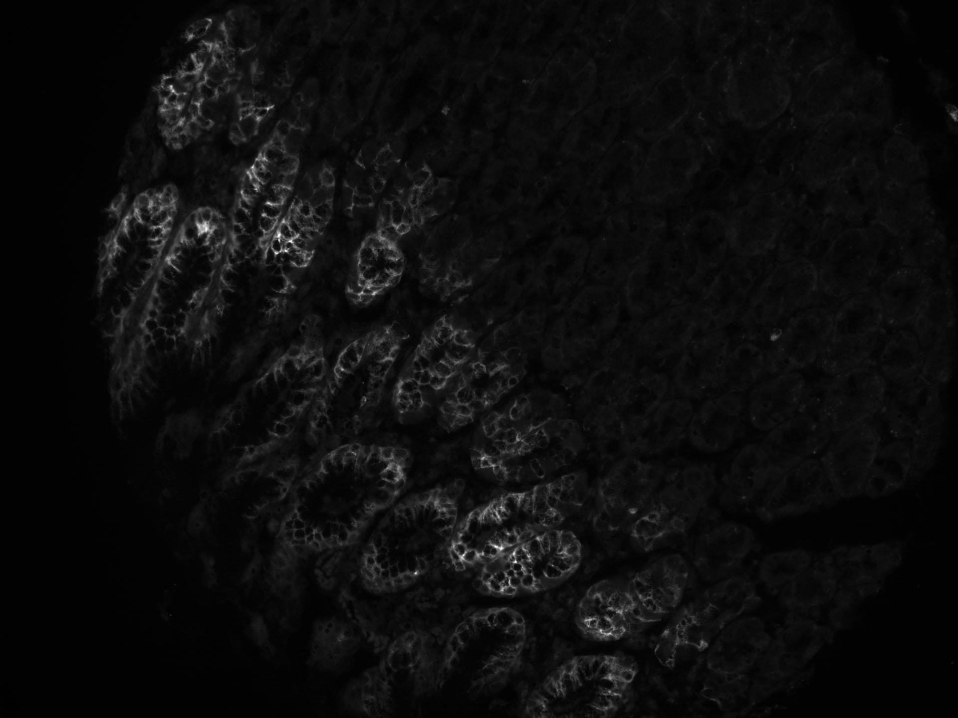
Scientific Data Images for MUC5AC Antibody (CLH2)
![Immunocytochemistry/ Immunofluorescence: MUC5AC Antibody (CLH2) [NBP2-44455]](https://resources.rndsystems.com/images/products/MUC5AC-Antibody-CLH2-Immunocytochemistry-Immunofluorescence-NBP2-44455-img0003.jpg "Immunocytochemistry/ Immunofluorescence: MUC5AC Antibody (CLH2) [NBP2-44455]")
Immunocytochemistry/ Immunofluorescence: MUC5AC Antibody (CLH2) [NBP2-44455]
Immunocytochemistry/Immunofluorescence: MUC5AC Antibody (CLH2) [NBP2-44455] - Human stomach tissue (FFPE). Antibody at 1:100. ICC/IF image submitted by a verified customer review.![Immunohistochemistry-Paraffin: MUC5AC Antibody (CLH2) [NBP2-44455]](https://resources.rndsystems.com/images/products/MUC5AC-Antibody-CLH2-Immunohistochemistry-Paraffin-NBP2-44455-img0002.jpg "Immunohistochemistry-Paraffin: MUC5AC Antibody (CLH2) [NBP2-44455]")
Immunohistochemistry-Paraffin: MUC5AC Antibody (CLH2) [NBP2-44455]
Immunohistochemistry-Paraffin: MUC5AC Antibody (CLH2) [NBP2-44455] - Formalin-fixed, paraffin-embedded human Gastric Carcinoma stained with MUC5AC Antibody (CLH2). Courtesy of Dr. Leonor David, IPATIMUP and Medical Faculty, University of Porto, Portugal.Applications for MUC5AC Antibody (CLH2)
Application
Recommended Usage
Flow Cytometry
1-2 ug/million cells
Immunocytochemistry/ Immunofluorescence
1-2 ug/ml
Immunofluorescence
1 - 2 ug/ml
Immunohistochemistry-Paraffin
1-2 ug/ml
Application Notes
Immunohistochemistry (Formalin-fixed): 1-2ug/ml for 30 minutes at RT. Staining of formalin-fixed tissues requires heating tissue sections in 10mM Tris with 1mM EDTA, pH 9.0, for 45 min at 95C followed by cooling at RT for 20 minutes.
Optimal dilution for a specific application should be determined.
Use in WB reported in scientific literature PMID: 32709695
Optimal dilution for a specific application should be determined.
Use in WB reported in scientific literature PMID: 32709695
Reviewed Applications
Read 1 review rated 5 using NBP2-44455 in the following applications:
Flow Cytometry Panel Builder
Bio-Techne Knows Flow Cytometry
Save time and reduce costly mistakes by quickly finding compatible reagents using the Panel Builder Tool.
Advanced Features
- Spectra Viewer - Custom analysis of spectra from multiple fluorochromes
- Spillover Popups - Visualize the spectra of individual fluorochromes
- Antigen Density Selector - Match fluorochrome brightness with antigen density

Formulation, Preparation, and Storage
Purification
Protein A or G purified
Formulation
10 mM PBS with 0.05% BSA
Preservative
0.05% Sodium Azide
Concentration
0.2 mg/ml
Shipping
The product is shipped with polar packs. Upon receipt, store it immediately at the temperature recommended below.
Stability & Storage
Store at 4C.
Background: MUC5AC
Given that MUC5AC is the primary mucin produced and secreted by cells lining airway, it is understandable that its expression is dysregulated in a number of respiratory diseases (1,3,5,6). For instance, MUC5AC is overexpressed in asthma and the protein is also more highly glycosylated, specifically fucosylated, in the disease state (1,3,7). Additionally, its overproduction is a key feature in chronic obstructive pulmonary disease (COPD) contributing to mucosal blockage (6). Multiple pathways are associated with overproduction of MUC5AC including NFkappaB, the primary pathway for production and secretion, and also the MAPK and STAT6 pathways (3). In addition to conventional, synthetic therapeutics agents, researchers are exploring natural MUC5AC inhibitors such as flavonoids, glycosides, and steroids to treat associated disorders (3).
References
1. Krishn, S. R., Ganguly, K., Kaur, S., & Batra, S. K. (2018). Ramifications of secreted mucin MUC5AC in malignant journey: a holistic view. Carcinogenesis. https://doi.org/10.1093/carcin/bgy019
2. Ballester, B., Milara, J., & Cortijo, J. (2019). Mucins as a New Frontier in Pulmonary Fibrosis. Journal of clinical medicine. https://doi.org/10.3390/jcm8091447
3. Samsuzzaman, M., Uddin, M. S., Shah, M. A., & Mathew, B. (2019). Natural inhibitors on airway mucin: Molecular insight into the therapeutic potential targeting MUC5AC expression and production. Life sciences. https://doi.org/10.1016/j.lfs.2019.05.041
4. Uniprot (P98088)
5. Li, J., & Ye, Z. (2020). The Potential Role and Regulatory Mechanisms of MUC5AC in Chronic Obstructive Pulmonary Disease. Molecules (Basel, Switzerland). https://doi.org/10.3390/molecules25194437
6. Bonser, L. R., & Erle, D. J. (2017). Airway Mucus and Asthma: The Role of MUC5AC and MUC5B. Journal of clinical medicine. https://doi.org/10.3390/jcm6120112
Alternate Names
gastric mucin, leB, lewis B blood group antigen, major airway glycoprotein, MUC5, mucin 5, subtypes A and C, tracheobronchial/gastric, mucin 5AC, oligomeric mucus/gel-forming, mucin 5AC, oligomeric mucus/gel-forming pseudogene, mucin-5 subtype AC, tracheobronchial, mucin-5AC, TBM, tracheobronchial mucin
Gene Symbol
MUC5AC
UniProt
Additional MUC5AC Products
Product Documents for MUC5AC Antibody (CLH2)
Certificate of Analysis
To download a Certificate of Analysis, please enter a lot or batch number in the search box below.
Product Specific Notices for MUC5AC Antibody (CLH2)
This product is for research use only and is not approved for use in humans or in clinical diagnosis. Primary Antibodies are guaranteed for 1 year from date of receipt.
Citations for MUC5AC Antibody (CLH2)
 Powered by Bioz
Customer Reviews for MUC5AC Antibody (CLH2) (1)
5 out of 5
1 Customer Rating
Have you used MUC5AC Antibody (CLH2)?
Submit a review and receive an Amazon gift card!
$25/€18/£15/$25CAN/¥2500 Yen for a review with an image
$10/€7/£6/$10CAN/¥1110 Yen for a review without an image
Submit a review
Customer Images
Showing
1
-
1 of
1 review
Showing All
Filter By:
-
Application: Immunofluorescence - paraffinSample Tested: Stomach tissueSpecies: HumanVerified Customer | Posted 03/07/2020MUC5AC, Human stomach FFPE tissue, 1:100pH9 antigen retrieval

There are no reviews that match your criteria.
Protocols
Find general support by application which include: protocols, troubleshooting, illustrated assays, videos and webinars.
- 7-Amino Actinomycin D (7-AAD) Cell Viability Flow Cytometry Protocol
- Antigen Retrieval Protocol (PIER)
- Antigen Retrieval for Frozen Sections Protocol
- Appropriate Fixation of IHC/ICC Samples
- Cellular Response to Hypoxia Protocols
- Chromogenic IHC Staining of Formalin-Fixed Paraffin-Embedded (FFPE) Tissue Protocol
- Chromogenic Immunohistochemistry Staining of Frozen Tissue
- ClariTSA™ Fluorophore Kits
- Detection & Visualization of Antibody Binding
- Extracellular Membrane Flow Cytometry Protocol
- Flow Cytometry Protocol for Cell Surface Markers
- Flow Cytometry Protocol for Staining Membrane Associated Proteins
- Flow Cytometry Staining Protocols
- Flow Cytometry Troubleshooting Guide
- Fluorescent IHC Staining of Frozen Tissue Protocol
- Graphic Protocol for Heat-induced Epitope Retrieval
- Graphic Protocol for the Preparation and Fluorescent IHC Staining of Frozen Tissue Sections
- Graphic Protocol for the Preparation and Fluorescent IHC Staining of Paraffin-embedded Tissue Sections
- Graphic Protocol for the Preparation of Gelatin-coated Slides for Histological Tissue Sections
- ICC Cell Smear Protocol for Suspension Cells
- ICC Immunocytochemistry Protocol Videos
- ICC for Adherent Cells
- IHC Sample Preparation (Frozen sections vs Paraffin)
- Immunocytochemistry (ICC) Protocol
- Immunocytochemistry Troubleshooting
- Immunofluorescence of Organoids Embedded in Cultrex Basement Membrane Extract
- Immunofluorescent IHC Staining of Formalin-Fixed Paraffin-Embedded (FFPE) Tissue Protocol
- Immunohistochemistry (IHC) and Immunocytochemistry (ICC) Protocols
- Immunohistochemistry Frozen Troubleshooting
- Immunohistochemistry Paraffin Troubleshooting
- Intracellular Flow Cytometry Protocol Using Alcohol (Methanol)
- Intracellular Flow Cytometry Protocol Using Detergents
- Intracellular Nuclear Staining Flow Cytometry Protocol Using Detergents
- Intracellular Staining Flow Cytometry Protocol Using Alcohol Permeabilization
- Intracellular Staining Flow Cytometry Protocol Using Detergents to Permeabilize Cells
- Preparing Samples for IHC/ICC Experiments
- Preventing Non-Specific Staining (Non-Specific Binding)
- Primary Antibody Selection & Optimization
- Propidium Iodide Cell Viability Flow Cytometry Protocol
- Protocol for Heat-Induced Epitope Retrieval (HIER)
- Protocol for Liperfluo
- Protocol for Making a 4% Formaldehyde Solution in PBS
- Protocol for VisUCyte™ HRP Polymer Detection Reagent
- Protocol for the Characterization of Human Th22 Cells
- Protocol for the Characterization of Human Th9 Cells
- Protocol for the Fluorescent ICC Staining of Cell Smears - Graphic
- Protocol for the Fluorescent ICC Staining of Cultured Cells on Coverslips - Graphic
- Protocol for the Preparation & Fixation of Cells on Coverslips
- Protocol for the Preparation and Chromogenic IHC Staining of Frozen Tissue Sections
- Protocol for the Preparation and Chromogenic IHC Staining of Frozen Tissue Sections - Graphic
- Protocol for the Preparation and Chromogenic IHC Staining of Paraffin-embedded Tissue Sections
- Protocol for the Preparation and Chromogenic IHC Staining of Paraffin-embedded Tissue Sections - Graphic
- Protocol for the Preparation and Fluorescent ICC Staining of Cells on Coverslips
- Protocol for the Preparation and Fluorescent ICC Staining of Non-adherent Cells
- Protocol for the Preparation and Fluorescent ICC Staining of Stem Cells on Coverslips
- Protocol for the Preparation and Fluorescent IHC Staining of Frozen Tissue Sections
- Protocol for the Preparation and Fluorescent IHC Staining of Paraffin-embedded Tissue Sections
- Protocol for the Preparation of Gelatin-coated Slides for Histological Tissue Sections
- Protocol for the Preparation of a Cell Smear for Non-adherent Cell ICC - Graphic
- Protocol: Annexin V and PI Staining by Flow Cytometry
- Protocol: Annexin V and PI Staining for Apoptosis by Flow Cytometry
- R&D Systems Quality Control Western Blot Protocol
- TUNEL and Active Caspase-3 Detection by IHC/ICC Protocol
- The Importance of IHC/ICC Controls
- Troubleshooting Guide: Fluorokine Flow Cytometry Kits
- Troubleshooting Guide: Immunohistochemistry
- Troubleshooting Guide: Western Blot Figures
- Western Blot Conditions
- Western Blot Protocol
- Western Blot Protocol for Cell Lysates
- Western Blot Troubleshooting
- Western Blot Troubleshooting Guide
- View all Protocols, Troubleshooting, Illustrated assays and Webinars
Loading...