Nanog Antibody - BSA Free
Novus Biologicals | Catalog # NB100-588
![Western Blot: Nanog Antibody [NB100-588]](https://resources.rndsystems.com/images/products/Nanog-Antibody-Western-Blot-NB100-588-img0010.jpg "Western Blot: Nanog Antibody [NB100-588]")
Key Product Details
Validated by
Independent Antibodies, Biological Validation
Species Reactivity
Validated:
Human, Mouse
Cited:
Human, Mouse
Applications
Validated:
Western Blot, Flow Cytometry, Immunocytochemistry/ Immunofluorescence, Immunoprecipitation
Cited:
Western Blot, Immunocytochemistry/ Immunofluorescence
Label
Unconjugated
Antibody Source
Polyclonal Rabbit IgG
Format
BSA Free
Loading...
Product Specifications
Immunogen
A synthetic peptide which maps to a region between residues 250 and the C-terminus (residue 305) of mouse Nanog homeobox using the numbering given in TrEMBL entry Q80Z64 (GeneID 71950).
Reactivity Notes
Human reactivity reported in (PMID: 28228260), Customer review.
Marker
Embryonic Stem Cell Marker
Clonality
Polyclonal
Host
Rabbit
Isotype
IgG
Scientific Data Images for Nanog Antibody - BSA Free
![Flow Cytometry: Nanog Antibody [NB100-588]](https://resources.rndsystems.com/images/products/Nanog-Antibody-Flow-Cytometry-NB100-588-img0005.jpg "Flow Cytometry: Nanog Antibody [NB100-588]")
Flow Cytometry: Nanog Antibody [NB100-588]
Flow Cytometry: Nanog Antibody [NB100-588] - FACS analysis of Nanog expression in primary mouse hepatic stellate cell cultured for 7 days. Image from verified customer review.![Western Blot: Nanog Antibody [NB100-588]](https://resources.rndsystems.com/images/products/Nanog-Antibody-Western-Blot-NB100-588-img0004.jpg "Western Blot: Nanog Antibody [NB100-588]")
![Western Blot: Nanog Antibody [NB100-588]](https://resources.rndsystems.com/images/products/Nanog-Antibody-Western-Blot-NB100-588-img0007.jpg "Western Blot: Nanog Antibody [NB100-588]")
Western Blot: Nanog Antibody [NB100-588]
Western Blot: Nanog Antibody [NB100-588] - The expression of pluripotency factor Nanog in human head and neck cancer cell lines cell lines FaDu and HN5. WB from a verified customer review.![Western Blot: Nanog Antibody [NB100-588]](https://resources.rndsystems.com/images/products/Nanog-Antibody-Western-Blot-NB100-588-img0008.jpg "Western Blot: Nanog Antibody [NB100-588]")
Western Blot: Nanog Antibody [NB100-588]
Western Blot: Nanog Antibody [NB100-588] - Detection of mouse Nanog by western blot. Samples: Whole cell lysate (50 ug) from F9 cells prepared using NETN lysis buffer. Antibody: Affinity purified rabbit anti-Nanog antibody NB100-588 used for WB at 0.1 ug/ml. Detection: Chemiluminescence with an exposure time of 30 seconds.![Western Blot: Nanog Antibody [NB100-588]](https://resources.rndsystems.com/images/products/Nanog-Antibody-Western-Blot-NB100-588-img0009.jpg "Western Blot: Nanog Antibody [NB100-588]")
![Flow Cytometry: Nanog Antibody [NB100-588]](https://resources.rndsystems.com/images/products/Nanog-Antibody-Flow-Cytometry-NB100-588-img0003.jpg "Flow Cytometry: Nanog Antibody [NB100-588]")
Flow Cytometry: Nanog Antibody [NB100-588]
Flow Cytometry: Nanog Antibody [NB100-588] - staining of NTERA-2 cells using NB100-588 at a 1:50 dilution detected using Dylight-488 conjugated goat anti-rabbit secondary antibody.
Western Blot: Nanog Antibody [NB100-588] -
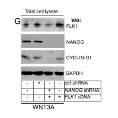
Western Blot: Nanog Antibody [NB100-588] - HIFs are required for hypoxia-induced expression of pluripotency factorsA-C. Breast cancer cell lines were exposed to 20% or 1% O2 for 24 h & NANOG (A), KLF4 (B), & SOX2 (C) mRNA levels were determined by RT-qPCR, relative to 18S rRNA, & normalized to the mean value for MDA-MB-231 cells (MDA231) at 20% O2 (mean ± SEM; n = 3). *P < 0.05, **P < 0.01, ***P < 0.001 vs. same cell line at 20% O2 by Student's t test. D & E. HCC-1954 (D) & MCF-7 (E) subclones, which were stably transfected with an expression vector encoding a non-targeting control (NTC) shRNA, or vector encoding shRNA targeting HIF-1 alpha (sh1 alpha ) or HIF-2 alpha (sh2 alpha ), or vectors encoding shRNAs targeting both HIF-1 alpha & HIF-2 alpha (DKD), were exposed to 20% or 1% O2 for 24 h & RT-qPCR was performed to determine NANOG (D) or KLF4 (E) mRNA levels relative to 18S rRNA. The results were normalized to NTC at 20% O2 (mean ± SEM; n = 3). *P < 0.05, **P < 0.01, ***P < 0.001 vs. NTC at 20% O2; #P < 0.05, ##P < 0.01, ###P < 0.001 vs. NTC at 1% O2 by ANOVA. F. ZR75.1 cells treated with vehicle or digoxin (200 nM) were exposed to 20% or 1% O2 for 24 h & SOX2 mRNA was measured (mean ± SEM; n = 3). *P < 0.05, **P < 0.01 vs. NTC at 20% O2; ###P < 0.001 vs. NTC at 1% O2 by ANOVA. G & H. NTC & DKD subclones of HCC-1954 (G) & MCF-7 (H) were exposed to 20% or 1% O2 for 48 h, whole cell lysates were prepared, & immunoblot assays were performed to analyze HIF-1 alpha, HIF-2 alpha, NANOG & KLF4 protein expression. Actin was also analyzed as a loading control. I. ZR75.1 cells were treated with vehicle or digoxin (200 nM), exposed to 20% or 1% O2 for 48 h, & HIF-1 alpha, NANOG & SOX2 immunoblot assays were performed. Image collected & cropped by CiteAb from the following publication (https://www.oncotarget.com/lookup/doi/10.18632/oncotarget.11743), licensed under a CC-BY license. Not internally tested by Novus Biologicals.Applications for Nanog Antibody - BSA Free
Application
Recommended Usage
Flow Cytometry
1:150
Immunocytochemistry/ Immunofluorescence
1:10-1:2000
Immunoprecipitation
2-5 ug/mg lysate
Western Blot
1:2000-1:10000
Application Notes
WB from a verified customer review, PMID ( 28228260). ICC/IF reported in (PMID: 22505032). Nanog antibody validated for FLOW from a verified customer review.
Reviewed Applications
Read 4 reviews rated 3.8 using NB100-588 in the following applications:
Flow Cytometry Panel Builder
Bio-Techne Knows Flow Cytometry
Save time and reduce costly mistakes by quickly finding compatible reagents using the Panel Builder Tool.
Advanced Features
- Spectra Viewer - Custom analysis of spectra from multiple fluorochromes
- Spillover Popups - Visualize the spectra of individual fluorochromes
- Antigen Density Selector - Match fluorochrome brightness with antigen density

Formulation, Preparation, and Storage
Purification
Immunogen affinity purified
Formulation
Tris-Citrate/Phosphate (pH 7.0 - 8.0)
Format
BSA Free
Preservative
0.09% Sodium Azide
Concentration
1.0 mg/ml
Shipping
The product is shipped with polar packs. Upon receipt, store it immediately at the temperature recommended below.
Stability & Storage
Store at 4C. Do not freeze.
Background: Nanog
Long Name
Nanog Homeobox
Alternate Names
FLJ12581, FLJ40451, hNanog, homeobox protein NANOG, Homeobox transcription factor Nanog, homeobox transcription factor Nanog-delta 48, Nanog homeobox
Entrez Gene IDs
71950 (Mouse)
Gene Symbol
NANOG
UniProt
Additional Nanog Products
Product Documents for Nanog Antibody - BSA Free
Certificate of Analysis
To download a Certificate of Analysis, please enter a lot or batch number in the search box below.
Product Specific Notices for Nanog Antibody - BSA Free
This product is for research use only and is not approved for use in humans or in clinical diagnosis. Primary Antibodies are guaranteed for 1 year from date of receipt.
Citations for Nanog Antibody - BSA Free
 Powered by Bioz
Customer Reviews for Nanog Antibody - BSA Free (4)
3.8 out of 5
4 Customer Ratings
Have you used Nanog Antibody - BSA Free?
Submit a review and receive an Amazon gift card!
$25/€18/£15/$25CAN/¥2500 Yen for a review with an image
$10/€7/£6/$10CAN/¥1110 Yen for a review without an image
Submit a review
Customer Images
-(01-ml)_NB100-588_7181.png)
Showing
1
-
4 of
4 reviews
Showing All
Filter By:
-
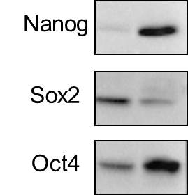
Application: Western BlotSample Tested: Human head and neck cancer cell linesSpecies: HumanVerified Customer | Posted 10/11/2018The expression of pluripotency factors Nanog, Sox2, and Oct4 in human HNSCC cell lines FaDu and HN5.

-
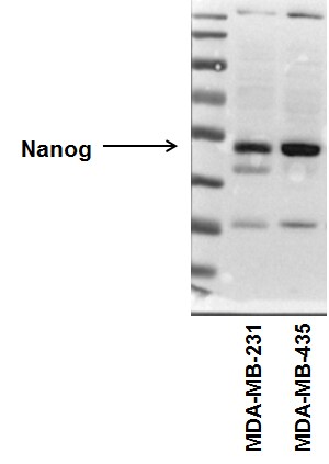
Application: Western BlotSample Tested: Human cancer cell whole cell lysateSpecies: HumanVerified Customer | Posted 08/22/2015Nanog expression in human breast cancer cell lines. NB100-588
-
Application: Flow CytometrySample Tested:Species: MouseVerified Customer | Posted 04/29/2014FACS analysis of Nanog expression in primary mouse hepatic stellate cell cultured for 7 days.
-
Application: Western BlotSample Tested: HUVEC, MCF7, JURKAT, Sample Amount: 2-30 ugSpecies: HumanVerified Customer | Posted 06/08/2011
There are no reviews that match your criteria.
Protocols
Find general support by application which include: protocols, troubleshooting, illustrated assays, videos and webinars.
- 7-Amino Actinomycin D (7-AAD) Cell Viability Flow Cytometry Protocol
- Appropriate Fixation of IHC/ICC Samples
- Cellular Response to Hypoxia Protocols
- ClariTSA™ Fluorophore Kits
- Detection & Visualization of Antibody Binding
- Extracellular Membrane Flow Cytometry Protocol
- Flow Cytometry Protocol for Cell Surface Markers
- Flow Cytometry Protocol for Staining Membrane Associated Proteins
- Flow Cytometry Staining Protocols
- Flow Cytometry Troubleshooting Guide
- ICC Cell Smear Protocol for Suspension Cells
- ICC Immunocytochemistry Protocol Videos
- ICC for Adherent Cells
- Immunocytochemistry (ICC) Protocol
- Immunocytochemistry Troubleshooting
- Immunofluorescence of Organoids Embedded in Cultrex Basement Membrane Extract
- Immunohistochemistry (IHC) and Immunocytochemistry (ICC) Protocols
- Immunoprecipitation Protocol
- Intracellular Flow Cytometry Protocol Using Alcohol (Methanol)
- Intracellular Flow Cytometry Protocol Using Detergents
- Intracellular Nuclear Staining Flow Cytometry Protocol Using Detergents
- Intracellular Staining Flow Cytometry Protocol Using Alcohol Permeabilization
- Intracellular Staining Flow Cytometry Protocol Using Detergents to Permeabilize Cells
- Preparing Samples for IHC/ICC Experiments
- Preventing Non-Specific Staining (Non-Specific Binding)
- Primary Antibody Selection & Optimization
- Propidium Iodide Cell Viability Flow Cytometry Protocol
- Protocol for Liperfluo
- Protocol for VisUCyte™ HRP Polymer Detection Reagent
- Protocol for the Characterization of Human Th22 Cells
- Protocol for the Characterization of Human Th9 Cells
- Protocol for the Fluorescent ICC Staining of Cell Smears - Graphic
- Protocol for the Fluorescent ICC Staining of Cultured Cells on Coverslips - Graphic
- Protocol for the Preparation and Fluorescent ICC Staining of Cells on Coverslips
- Protocol for the Preparation and Fluorescent ICC Staining of Non-adherent Cells
- Protocol for the Preparation and Fluorescent ICC Staining of Stem Cells on Coverslips
- Protocol for the Preparation of a Cell Smear for Non-adherent Cell ICC - Graphic
- Protocol: Annexin V and PI Staining by Flow Cytometry
- Protocol: Annexin V and PI Staining for Apoptosis by Flow Cytometry
- R&D Systems Quality Control Western Blot Protocol
- TUNEL and Active Caspase-3 Detection by IHC/ICC Protocol
- The Importance of IHC/ICC Controls
- Troubleshooting Guide: Fluorokine Flow Cytometry Kits
- Troubleshooting Guide: Western Blot Figures
- Western Blot Conditions
- Western Blot Protocol
- Western Blot Protocol for Cell Lysates
- Western Blot Troubleshooting
- Western Blot Troubleshooting Guide
- View all Protocols, Troubleshooting, Illustrated assays and Webinars
Loading...
Associated Pathways
