NDC80 Antibody (9G3.23) - Azide and BSA Free
Novus Biologicals | Catalog # NB100-338
![Western Blot: NDC80 Antibody (9G3.23) [NB100-338]](https://resources.rndsystems.com/images/products/NDC80-Antibody-9G3-23-Western-Blot-NB100-338-img0016.jpg "Western Blot: NDC80 Antibody (9G3.23) [NB100-338]")
Loading...
Key Product Details
Species Reactivity
Validated:
Human, Mouse, Hamster, Marsupial
Cited:
Human
Applications
Validated:
Immunohistochemistry, Immunohistochemistry-Frozen, Western Blot, Flow Cytometry, Immunocytochemistry/ Immunofluorescence, Immunoprecipitation, Proximity Ligation Assay
Cited:
Western Blot, Immunocytochemistry/ Immunofluorescence
Label
Unconjugated
Antibody Source
Monoclonal Mouse IgG2A Clone # 9G3.23
Format
Azide and BSA Free
Loading...
Product Specifications
Immunogen
Human NDC80 protein consisting of amino acids 56-642.
Reactivity Notes
Kangaroo rat (100%).
Localization
Nuclear
Clonality
Monoclonal
Host
Mouse
Isotype
IgG2A
Theoretical MW
80 kDa.
Disclaimer note: The observed molecular weight of the protein may vary from the listed predicted molecular weight due to post translational modifications, post translation cleavages, relative charges, and other experimental factors.
Disclaimer note: The observed molecular weight of the protein may vary from the listed predicted molecular weight due to post translational modifications, post translation cleavages, relative charges, and other experimental factors.
Scientific Data Images for NDC80 Antibody (9G3.23) - Azide and BSA Free
Western Blot: NDC80 Antibody (9G3.23) [NB100-338]
Western Blot: NDC80 Antibody (9G3.23) [NB100-338] - HEC1 antibody [9G3.23] detects HEC1 protein by western blot analysis. Various whole cell extracts and HeLa nuclear extracts (30 ug) were separated by 7.5% SDS-PAGE, and the membrane was blotted with HEC1 antibody [9G3.23] diluted at 1:1000. The HRP-conjugated anti-mouse IgG antibody (NBP2-193821) was used to detect the primary antibody, and the signal was developed with Trident femto Western HRP Substrate.![Western Blot: NDC80 Antibody (9G3.23) [NB100-338]](https://resources.rndsystems.com/images/products/NDC80-Antibody-9G3-23-Western-Blot-NB100-338-img0007.jpg "Western Blot: NDC80 Antibody (9G3.23) [NB100-338]")
Western Blot: NDC80 Antibody (9G3.23) [NB100-338]
Western Blot: NDC80 Antibody (9G3.23) [NB100-338] - WB analysis of NDC80 in U2OS Whole Cell Lysate. Verified customer review from 1DegreeBio.![Immunocytochemistry/ Immunofluorescence: NDC80 Antibody (9G3.23) [NB100-338]](https://resources.rndsystems.com/images/products/NDC80-Antibody-9G3-23-Immunocytochemistry-Immunofluorescence-NB100-338-img0003.jpg "Immunocytochemistry/ Immunofluorescence: NDC80 Antibody (9G3.23) [NB100-338]")
Immunocytochemistry/ Immunofluorescence: NDC80 Antibody (9G3.23) [NB100-338]
Immunocytochemistry/Immunofluorescence: NDC80 Antibody (9G3.23) [NB100-338] - Mouse anti-Hec1 and rabbit anti-Hec1 (phospho Ser165) antibodies were used to co-stain MCF10A cells. Insets show kinetochores of misaligned chromosomes. DAPI staining shows chromatin.![Immunocytochemistry/ Immunofluorescence: NDC80 Antibody (9G3.23) [NB100-338]](https://resources.rndsystems.com/images/products/NDC80-Antibody-9G3-23-Immunocytochemistry-Immunofluorescence-NB100-338-img0004.jpg "Immunocytochemistry/ Immunofluorescence: NDC80 Antibody (9G3.23) [NB100-338]")
Immunocytochemistry/ Immunofluorescence: NDC80 Antibody (9G3.23) [NB100-338]
Immunocytochemistry/Immunofluorescence: NDC80 Antibody (9G3.23) [NB100-338] - IF analysis of NDC80 in fixed human cell lines of RPE1, H4, and 42MGBA. Image courtesy of anonymous customer product review.![Immunocytochemistry/ Immunofluorescence: NDC80 Antibody (9G3.23) [NB100-338]](https://resources.rndsystems.com/images/products/NDC80-Antibody-9G3-23-Immunocytochemistry-Immunofluorescence-NB100-338-img0008.jpg "Immunocytochemistry/ Immunofluorescence: NDC80 Antibody (9G3.23) [NB100-338]")
Immunocytochemistry/ Immunofluorescence: NDC80 Antibody (9G3.23) [NB100-338]
Immunocytochemistry/Immunofluorescence: NDC80 Antibody (9G3.23) [NB100-338] - IF analysis of NDC80 in PTK1 cells.Verified customer review from 1DegreeBio. [NB100-338] -")
Immunocytochemistry/ Immunofluorescence: NDC80 Antibody (9G3.23) [NB100-338] -
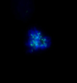
Immunocytochemistry/ Immunofluorescence: NDC80 Antibody (9G3.23) [NB100-338] - NDC80 antibody [9G3.23] detects NDC80 protein at kinetochore by immunofluorescent analysis.Sample: HeLa cells were fixed in 4% paraformaldehyde at RT for 15 min.
Green: NDC80 stained by NDC80 antibody [9G3.23] (NB100-338) diluted at 1:500.
Blue: Fluoroshield with DAPI.
 [NB100-338] -")
Immunocytochemistry/ Immunofluorescence: NDC80 Antibody (9G3.23) [NB100-338] -
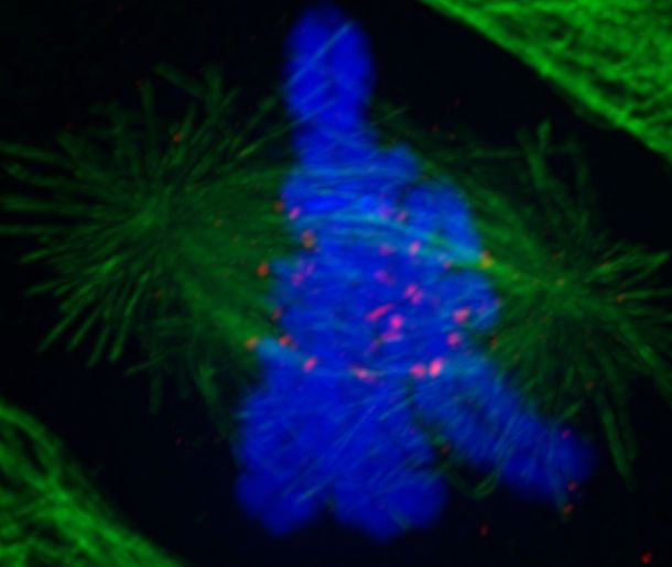
Immunocytochemistry/ Immunofluorescence: NDC80 Antibody (9G3.23) [NB100-338] - NDC80 antibody [9G3.23] detects NDC80 protein at kinetochore by immunofluorescent analysis.Sample: HeLa cells were fixed in 4% paraformaldehyde at RT for 15 min.
Green: NDC80 stained by NDC80 antibody [9G3.23] (NB100-338) diluted at 1:1000.
Red: alpha Tubulin, a cytoskeleton marker, stained by alpha Tubulin antibody diluted at 1:1000.
Blue: Fluoroshield with DAPI.
 [NB100-338] -")
Immunocytochemistry/ Immunofluorescence: NDC80 Antibody (9G3.23) [NB100-338] -
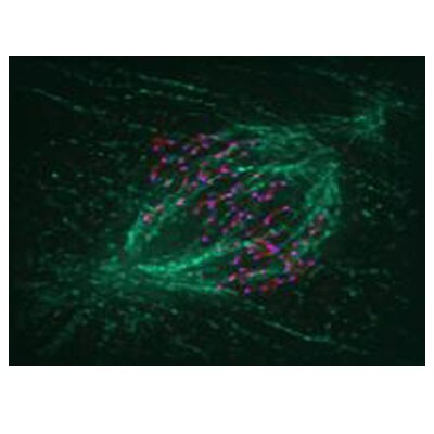
Immunocytochemistry/ Immunofluorescence: NDC80 Antibody (9G3.23) [NB100-338] - NDC80 antibody [9G3.23] detects NDC80 protein at kinetochore by immunofluorescent analysis.Sample: HeLa cells were fixed in 4% paraformaldehyde at RT for 15 min.
Green: NDC80 stained by NDC80 antibody [9G3.23] (NB100-338) diluted at 1:500.
Red: alpha Tubulin 4a, a cytoskeleton marker, stained by alpha Tubulin 4a antibody diluted at 1:500.
Blue: Hoechst 33342 staining.
 [NB100-338] -")
Immunocytochemistry/ Immunofluorescence: NDC80 Antibody (9G3.23) [NB100-338] -
Immunocytochemistry/ Immunofluorescence: NDC80 Antibody (9G3.23) [NB100-338] - NDC80 antibody [9G3.23] detects NDC80 protein at kinetochore by immunofluorescent analysis.Sample: HeLa cells were fixed in 4% paraformaldehyde at RT for 15 min.
Green: NDC80 stained by NDC80 antibody [9G3.23] (NB100-338) diluted at 1:500.
Red: beta Tubulin, a cytoskeleton marker, stained by beta Tubulin antibody diluted at 1:1000.
Blue: Fluoroshield with DAPI.
 [NB100-338] -")
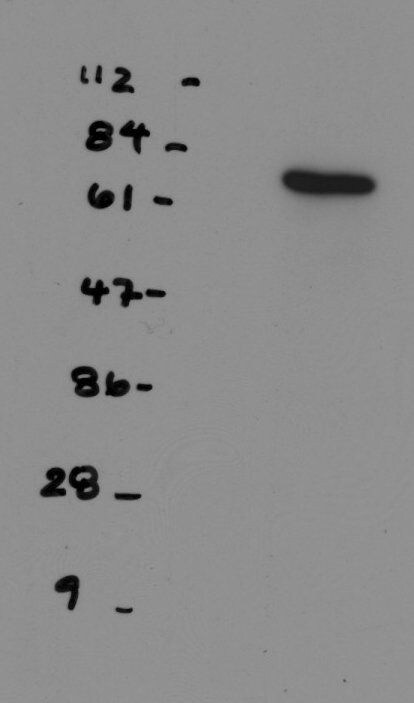
Western Blot: NDC80 Antibody (9G3.23) [NB100-338] -
Various whole cell extracts (30 ug) were separated by 7.5% SDS-PAGE, and the membrane was blotted with NDC80 antibody [9G3.23] (NB100-338) diluted at 1:1000. The HRP-conjugated anti-mouse IgG antibody was used to detect the primary antibody, and the signal was developed with Trident ECL plus-Enhanced.Applications for NDC80 Antibody (9G3.23) - Azide and BSA Free
Application
Recommended Usage
Immunocytochemistry/ Immunofluorescence
1:100-1:1000
Immunohistochemistry
1:10 - 1:500
Immunohistochemistry-Frozen
1:10 - 1:500
Immunoprecipitation
1-5 ug/ml
Western Blot
1:500-1:3000
Reviewed Applications
Read 5 reviews rated 4.6 using NB100-338 in the following applications:
Flow Cytometry Panel Builder
Bio-Techne Knows Flow Cytometry
Save time and reduce costly mistakes by quickly finding compatible reagents using the Panel Builder Tool.
Advanced Features
- Spectra Viewer - Custom analysis of spectra from multiple fluorochromes
- Spillover Popups - Visualize the spectra of individual fluorochromes
- Antigen Density Selector - Match fluorochrome brightness with antigen density

Formulation, Preparation, and Storage
Purification
Protein A purified
Formulation
PBS
Format
Azide and BSA Free
Preservative
No Preservative
Concentration
Concentrations vary lot to lot. See vial label for concentration. If unlisted please contact technical services.
Shipping
The product is shipped with polar packs. Upon receipt, store it immediately at the temperature recommended below.
Stability & Storage
Store at 4C short term. Aliquot and store at -20C long term. Avoid freeze-thaw cycles.
Background: NDC80
Alternate Names
HEC1KNTC2, HECRetinoblastoma-associated protein HEC, Highly expressed in cancer protein, highly expressed in cancer, rich in leucine heptad repeats, highly expressed in cancer, rich in leucine heptad repeats (yeast), HsHec1, hsNDC80, kinetochore associated 2, Kinetochore protein Hec1, kinetochore protein NDC80 homolog, Kinetochore-associated protein 2, NDC80 homolog, kinetochore complex component (S. cerevisiae), TID3
Entrez Gene IDs
10403 (Human)
Gene Symbol
NDC80
UniProt
Additional NDC80 Products
Product Documents for NDC80 Antibody (9G3.23) - Azide and BSA Free
Certificate of Analysis
To download a Certificate of Analysis, please enter a lot or batch number in the search box below.
Product Specific Notices for NDC80 Antibody (9G3.23) - Azide and BSA Free
This product is for research use only and is not approved for use in humans or in clinical diagnosis. Primary Antibodies are guaranteed for 1 year from date of receipt.
Citations for NDC80 Antibody (9G3.23) - Azide and BSA Free
 Powered by Bioz
Customer Reviews for NDC80 Antibody (9G3.23) - Azide and BSA Free (5)
4.6 out of 5
5 Customer Ratings
Have you used NDC80 Antibody (9G3.23) - Azide and BSA Free?
Submit a review and receive an Amazon gift card!
$25/€18/£15/$25CAN/¥2500 Yen for a review with an image
$10/€7/£6/$10CAN/¥1110 Yen for a review without an image
Submit a review
Customer Images
Showing
1
-
5 of
5 reviews
Showing All
Filter By:
-
Application: ImmunofluorescenceSample Tested: RPE1 cellsSpecies: HumanVerified Customer | Posted 04/21/2016RPE1 cell stained with Ndc80

-
Application: Western BlotSample Tested: U2OS whole cell lysateSpecies: HumanVerified Customer | Posted 11/06/2013
-
Application: ImmunofluorescenceSample Tested: PTK1 cellsSpecies: OtherVerified Customer | Posted 11/06/2013
-
Application: ImmunofluorescenceSample Tested:Species: HumanVerified Customer | Posted 10/04/2013
-
Application: ImmunofluorescenceSample Tested: Fixed cell lines (RPE1, H4, 42MGBA)Species: HumanVerified Customer | Posted 06/06/2012Metaphase Spindle (alpha tubulin = green, anti-centromere = red, Hec1 = purple
There are no reviews that match your criteria.
Protocols
Find general support by application which include: protocols, troubleshooting, illustrated assays, videos and webinars.
- 7-Amino Actinomycin D (7-AAD) Cell Viability Flow Cytometry Protocol
- Antigen Retrieval Protocol (PIER)
- Antigen Retrieval for Frozen Sections Protocol
- Appropriate Fixation of IHC/ICC Samples
- Cellular Response to Hypoxia Protocols
- Chromogenic IHC Staining of Formalin-Fixed Paraffin-Embedded (FFPE) Tissue Protocol
- Chromogenic Immunohistochemistry Staining of Frozen Tissue
- ClariTSA™ Fluorophore Kits
- Detection & Visualization of Antibody Binding
- Extracellular Membrane Flow Cytometry Protocol
- Flow Cytometry Protocol for Cell Surface Markers
- Flow Cytometry Protocol for Staining Membrane Associated Proteins
- Flow Cytometry Staining Protocols
- Flow Cytometry Troubleshooting Guide
- Fluorescent IHC Staining of Frozen Tissue Protocol
- Graphic Protocol for Heat-induced Epitope Retrieval
- Graphic Protocol for the Preparation and Fluorescent IHC Staining of Frozen Tissue Sections
- Graphic Protocol for the Preparation and Fluorescent IHC Staining of Paraffin-embedded Tissue Sections
- Graphic Protocol for the Preparation of Gelatin-coated Slides for Histological Tissue Sections
- ICC Cell Smear Protocol for Suspension Cells
- ICC Immunocytochemistry Protocol Videos
- ICC for Adherent Cells
- IHC Sample Preparation (Frozen sections vs Paraffin)
- Immunocytochemistry (ICC) Protocol
- Immunocytochemistry Troubleshooting
- Immunofluorescence of Organoids Embedded in Cultrex Basement Membrane Extract
- Immunofluorescent IHC Staining of Formalin-Fixed Paraffin-Embedded (FFPE) Tissue Protocol
- Immunohistochemistry (IHC) and Immunocytochemistry (ICC) Protocols
- Immunohistochemistry Frozen Troubleshooting
- Immunohistochemistry Paraffin Troubleshooting
- Immunoprecipitation Protocol
- Intracellular Flow Cytometry Protocol Using Alcohol (Methanol)
- Intracellular Flow Cytometry Protocol Using Detergents
- Intracellular Nuclear Staining Flow Cytometry Protocol Using Detergents
- Intracellular Staining Flow Cytometry Protocol Using Alcohol Permeabilization
- Intracellular Staining Flow Cytometry Protocol Using Detergents to Permeabilize Cells
- Preparing Samples for IHC/ICC Experiments
- Preventing Non-Specific Staining (Non-Specific Binding)
- Primary Antibody Selection & Optimization
- Propidium Iodide Cell Viability Flow Cytometry Protocol
- Protocol for Heat-Induced Epitope Retrieval (HIER)
- Protocol for Liperfluo
- Protocol for Making a 4% Formaldehyde Solution in PBS
- Protocol for VisUCyte™ HRP Polymer Detection Reagent
- Protocol for the Characterization of Human Th22 Cells
- Protocol for the Characterization of Human Th9 Cells
- Protocol for the Fluorescent ICC Staining of Cell Smears - Graphic
- Protocol for the Fluorescent ICC Staining of Cultured Cells on Coverslips - Graphic
- Protocol for the Preparation & Fixation of Cells on Coverslips
- Protocol for the Preparation and Chromogenic IHC Staining of Frozen Tissue Sections
- Protocol for the Preparation and Chromogenic IHC Staining of Frozen Tissue Sections - Graphic
- Protocol for the Preparation and Chromogenic IHC Staining of Paraffin-embedded Tissue Sections
- Protocol for the Preparation and Chromogenic IHC Staining of Paraffin-embedded Tissue Sections - Graphic
- Protocol for the Preparation and Fluorescent ICC Staining of Cells on Coverslips
- Protocol for the Preparation and Fluorescent ICC Staining of Non-adherent Cells
- Protocol for the Preparation and Fluorescent ICC Staining of Stem Cells on Coverslips
- Protocol for the Preparation and Fluorescent IHC Staining of Frozen Tissue Sections
- Protocol for the Preparation and Fluorescent IHC Staining of Paraffin-embedded Tissue Sections
- Protocol for the Preparation of Gelatin-coated Slides for Histological Tissue Sections
- Protocol for the Preparation of a Cell Smear for Non-adherent Cell ICC - Graphic
- Protocol: Annexin V and PI Staining by Flow Cytometry
- Protocol: Annexin V and PI Staining for Apoptosis by Flow Cytometry
- R&D Systems Quality Control Western Blot Protocol
- TUNEL and Active Caspase-3 Detection by IHC/ICC Protocol
- The Importance of IHC/ICC Controls
- Troubleshooting Guide: Fluorokine Flow Cytometry Kits
- Troubleshooting Guide: Immunohistochemistry
- Troubleshooting Guide: Western Blot Figures
- Western Blot Conditions
- Western Blot Protocol
- Western Blot Protocol for Cell Lysates
- Western Blot Troubleshooting
- Western Blot Troubleshooting Guide
- View all Protocols, Troubleshooting, Illustrated assays and Webinars
Loading...