Neutrophils Antibody (RM0028-3G23) - Azide and BSA Free
Novus Biologicals | Catalog # NBP2-12439
![Immunocytochemistry/ Immunofluorescence: Neutrophils Antibody (RM0028-3G23) - Azide and BSA Free [NBP2-12439]](https://resources.rndsystems.com/images/products/Neutrophils-Antibody-RM0028-3G23-Immunofluorescence-NBP2-12439-img0001.jpg "Immunocytochemistry/ Immunofluorescence: Neutrophils Antibody (RM0028-3G23) - Azide and BSA Free [NBP2-12439]")
Key Product Details
Species Reactivity
Validated:
Mouse
Cited:
Mouse
Applications
Validated:
Immunohistochemistry, Immunohistochemistry-Paraffin, Western Blot, Immunocytochemistry/ Immunofluorescence
Cited:
Immunohistochemistry-Paraffin, IF/IHC
Label
Unconjugated
Antibody Source
Monoclonal Rat IgG2A Clone # RM0028-3G23
Format
Azide and BSA Free
Loading...
Product Specifications
Immunogen
Isolated mouse neutrophil cells
Clonality
Monoclonal
Host
Rat
Isotype
IgG2A
Scientific Data Images for Neutrophils Antibody (RM0028-3G23) - Azide and BSA Free
Immunocytochemistry/ Immunofluorescence: Neutrophils Antibody (RM0028-3G23) - Azide and BSA Free [NBP2-12439]
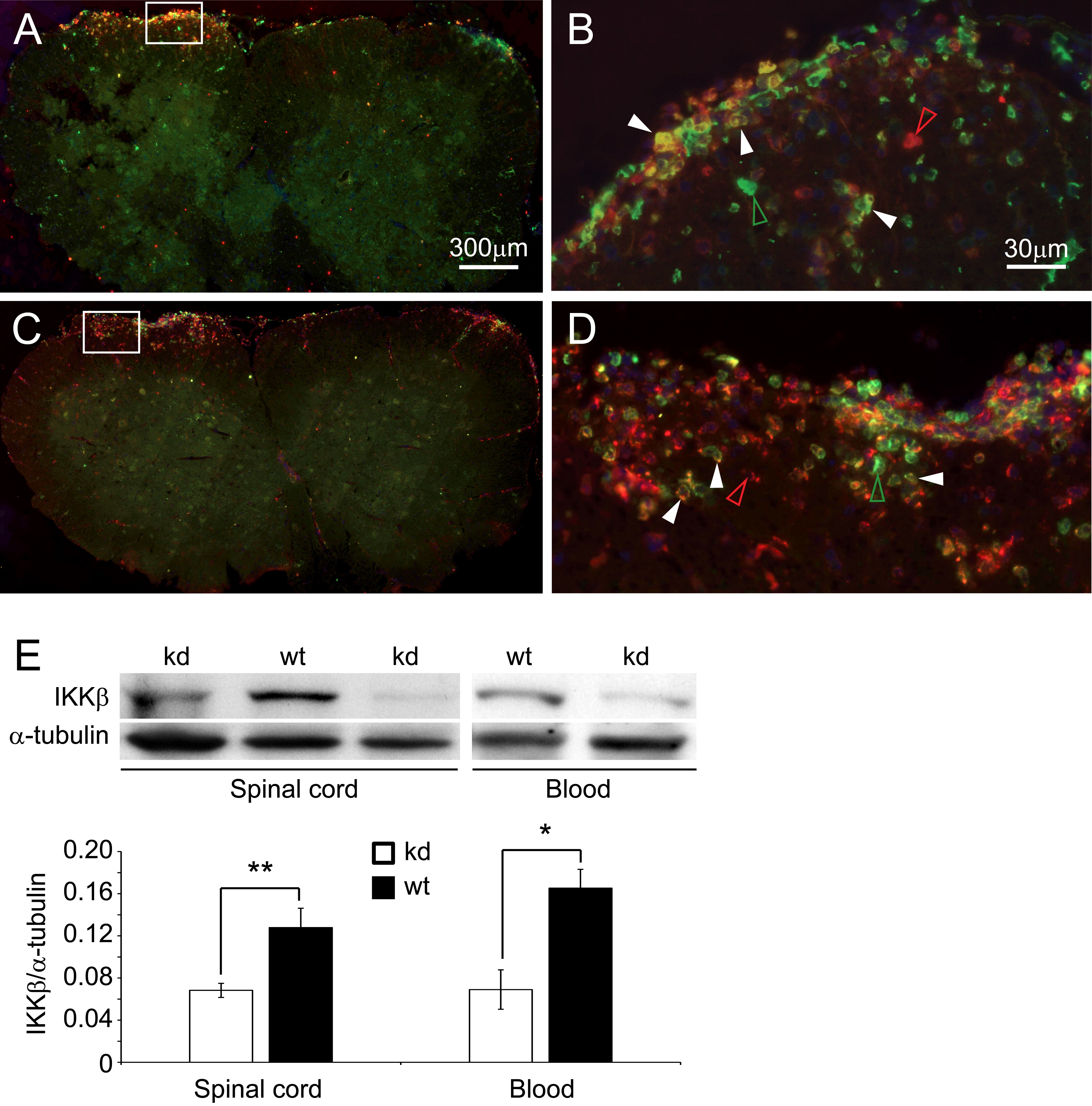
Immunocytochemistry/Immunofluorescence: Neutrophils Antibody (RM0028-3G23) [NBP2-12439] - Analysis of Neutrophils on paraffin embedded mouse transverse sections of spinal cord using anti- Neutrophils antibody. Image from verified customer review.![Immunohistochemistry-Paraffin: Neutrophils Antibody (RM0028-3G23) - Azide and BSA Free [NBP2-12439]](https://resources.rndsystems.com/images/products/Neutrophils-Antibody-RM0028-3G23-Immunohistochemistry-Paraffin-NBP2-12439-img0004.jpg "Immunohistochemistry-Paraffin: Neutrophils Antibody (RM0028-3G23) - Azide and BSA Free [NBP2-12439]")
Immunohistochemistry-Paraffin: Neutrophils Antibody (RM0028-3G23) - Azide and BSA Free [NBP2-12439]
Immunohistochemistry-Paraffin: Neutrophils Antibody (RM0028-3G23) [NBP2-12439] - Bouin's solution fixed and paraffin embedded mouse kidney section from anti-GBMmodel was subjected to immunohistochemistry staining (ABC) of neutrophils.Applications for Neutrophils Antibody (RM0028-3G23) - Azide and BSA Free
Application
Recommended Usage
Immunohistochemistry
1:100-300
Immunohistochemistry-Paraffin
1:100-1:300
Western Blot
1:500-1:1000
Reviewed Applications
Read 1 review rated 5 using NBP2-12439 in the following applications:
Formulation, Preparation, and Storage
Purification
Protein G purified
Reconstitution
Reconstitute with sterilized PBS to a final concentration of 0.5 mg/ml.
Formulation
Lyophilized from a 0.2 um filtered solution in PBS. 0.025 mg size is provided in liquid form, PBS
Format
Azide and BSA Free
Preservative
No Preservative
Concentration
LYOPH mg/ml
Shipping
The product is shipped with polar packs. Upon receipt, store it immediately at the temperature recommended below.
Stability & Storage
Store at -20 to -80C. Avoid freeze-thaw cycles.
Calculators
Product Documents for Neutrophils Antibody (RM0028-3G23) - Azide and BSA Free
Certificate of Analysis
To download a Certificate of Analysis, please enter a lot or batch number in the search box below.
Product Specific Notices for Neutrophils Antibody (RM0028-3G23) - Azide and BSA Free
This product is for research use only and is not approved for use in humans or in clinical diagnosis. Primary Antibodies are guaranteed for 1 year from date of receipt.
Citations for Neutrophils Antibody (RM0028-3G23) - Azide and BSA Free
 Powered by Bioz
Customer Reviews for Neutrophils Antibody (RM0028-3G23) - Azide and BSA Free (1)
5 out of 5
1 Customer Rating
Have you used Neutrophils Antibody (RM0028-3G23) - Azide and BSA Free?
Submit a review and receive an Amazon gift card!
$25/€18/£15/$25CAN/¥2500 Yen for a review with an image
$10/€7/£6/$10CAN/¥1110 Yen for a review without an image
Submit a review
Customer Images
Showing
1
-
1 of
1 review
Showing All
Filter By:
-
Application: ImmunofluorescenceSample Tested:Species: MouseVerified Customer | Posted 05/06/2016Infiltration of neutrophils in the EAE lesion site

There are no reviews that match your criteria.
Protocols
Find general support by application which include: protocols, troubleshooting, illustrated assays, videos and webinars.
- Antigen Retrieval Protocol (PIER)
- Antigen Retrieval for Frozen Sections Protocol
- Appropriate Fixation of IHC/ICC Samples
- Cellular Response to Hypoxia Protocols
- Chromogenic IHC Staining of Formalin-Fixed Paraffin-Embedded (FFPE) Tissue Protocol
- Chromogenic Immunohistochemistry Staining of Frozen Tissue
- ClariTSA™ Fluorophore Kits
- Detection & Visualization of Antibody Binding
- Fluorescent IHC Staining of Frozen Tissue Protocol
- Graphic Protocol for Heat-induced Epitope Retrieval
- Graphic Protocol for the Preparation and Fluorescent IHC Staining of Frozen Tissue Sections
- Graphic Protocol for the Preparation and Fluorescent IHC Staining of Paraffin-embedded Tissue Sections
- Graphic Protocol for the Preparation of Gelatin-coated Slides for Histological Tissue Sections
- ICC Cell Smear Protocol for Suspension Cells
- ICC Immunocytochemistry Protocol Videos
- ICC for Adherent Cells
- IHC Sample Preparation (Frozen sections vs Paraffin)
- Immunocytochemistry (ICC) Protocol
- Immunocytochemistry Troubleshooting
- Immunofluorescence of Organoids Embedded in Cultrex Basement Membrane Extract
- Immunofluorescent IHC Staining of Formalin-Fixed Paraffin-Embedded (FFPE) Tissue Protocol
- Immunohistochemistry (IHC) and Immunocytochemistry (ICC) Protocols
- Immunohistochemistry Frozen Troubleshooting
- Immunohistochemistry Paraffin Troubleshooting
- Preparing Samples for IHC/ICC Experiments
- Preventing Non-Specific Staining (Non-Specific Binding)
- Primary Antibody Selection & Optimization
- Protocol for Heat-Induced Epitope Retrieval (HIER)
- Protocol for Making a 4% Formaldehyde Solution in PBS
- Protocol for VisUCyte™ HRP Polymer Detection Reagent
- Protocol for the Fluorescent ICC Staining of Cell Smears - Graphic
- Protocol for the Fluorescent ICC Staining of Cultured Cells on Coverslips - Graphic
- Protocol for the Preparation & Fixation of Cells on Coverslips
- Protocol for the Preparation and Chromogenic IHC Staining of Frozen Tissue Sections
- Protocol for the Preparation and Chromogenic IHC Staining of Frozen Tissue Sections - Graphic
- Protocol for the Preparation and Chromogenic IHC Staining of Paraffin-embedded Tissue Sections
- Protocol for the Preparation and Chromogenic IHC Staining of Paraffin-embedded Tissue Sections - Graphic
- Protocol for the Preparation and Fluorescent ICC Staining of Cells on Coverslips
- Protocol for the Preparation and Fluorescent ICC Staining of Non-adherent Cells
- Protocol for the Preparation and Fluorescent ICC Staining of Stem Cells on Coverslips
- Protocol for the Preparation and Fluorescent IHC Staining of Frozen Tissue Sections
- Protocol for the Preparation and Fluorescent IHC Staining of Paraffin-embedded Tissue Sections
- Protocol for the Preparation of Gelatin-coated Slides for Histological Tissue Sections
- Protocol for the Preparation of a Cell Smear for Non-adherent Cell ICC - Graphic
- R&D Systems Quality Control Western Blot Protocol
- TUNEL and Active Caspase-3 Detection by IHC/ICC Protocol
- The Importance of IHC/ICC Controls
- Troubleshooting Guide: Immunohistochemistry
- Troubleshooting Guide: Western Blot Figures
- Western Blot Conditions
- Western Blot Protocol
- Western Blot Protocol for Cell Lysates
- Western Blot Troubleshooting
- Western Blot Troubleshooting Guide
- View all Protocols, Troubleshooting, Illustrated assays and Webinars
Loading...