NGFI-B alpha/Nur77/NR4A1 Antibody - BSA Free
Novus Biologicals | Catalog # NB100-56745
![Western Blot: NGFI-B alpha/Nur77/NR4A1 Antibody [NB100-56745]](https://resources.rndsystems.com/images/products/NGFI-B-alpha-Nur77-NR4A1-Antibody-Western-Blot-NB100-56745-img0005.jpg "Western Blot: NGFI-B alpha/Nur77/NR4A1 Antibody [NB100-56745]")
Key Product Details
Species Reactivity
Validated:
Cited:
Applications
Validated:
Cited:
Label
Antibody Source
Format
Product Specifications
Immunogen
Clonality
Host
Isotype
Scientific Data Images for NGFI-B alpha/Nur77/NR4A1 Antibody - BSA Free
Western Blot: NGFI-B alpha/Nur77/NR4A1 Antibody [NB100-56745]
NGFI-B-alpha-Nur77-NR4A1-Antibody-Western-Blot-NB100-56745-img0005.jpg![Immunohistochemistry-Paraffin: NGFI-B alpha/Nur77/NR4A1 Antibody [NB100-56745]](https://resources.rndsystems.com/images/products/NGFI-B-alpha-Nur77-NR4A1-Antibody-Immunohistochemistry-Paraffin-NB100-56745-img0007.jpg "Immunohistochemistry-Paraffin: NGFI-B alpha/Nur77/NR4A1 Antibody [NB100-56745]")
Immunohistochemistry-Paraffin: NGFI-B alpha/Nur77/NR4A1 Antibody [NB100-56745]
Immunohistochemistry-Paraffin: NGFI-B alpha/Nur77/NR4A1 Antibody [NB100-56745] - Analysis of a FFPE tissue section of mouse brain using 1:200 dilution of NGFI-B alpha/Nur77/NR4A1 antibody (NB100-56745). The staining was developed using HRP labeled anti-rabbit secondary antibody and DAB reagent, and nuclei of cells were counter-stained with hematoxylin.![Western Blot: NGFI-B alpha/Nur77/NR4A1 Antibody [NB100-56745]](https://resources.rndsystems.com/images/products/NGFI-B-alpha-Nur77-NR4A1-Antibody-Western-Blot-NB100-56745-img0003.jpg "Western Blot: NGFI-B alpha/Nur77/NR4A1 Antibody [NB100-56745]")
Western Blot: NGFI-B alpha/Nur77/NR4A1 Antibody [NB100-56745]
Western Blot: NGFI-B alpha/Nur77/NR4A1 Antibody [NB100-56745] - Analysis of Nur77 (Nak-1) in 15 ugs of NIH3T3 cell lysate using Nur77 antibody at 1:500 dilution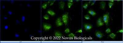

Western Blot: NGFI-B alpha/Nur77/NR4A1 Antibody [NB100-56745] -
nb100-56745_rabbit-polyclonal-ngfi-b-alpha-nur77-nr4a1-antibody-imgenex-img-528-2092023181155.jpg
Western Blot: NGFI-B alpha/Nur77/NR4A1 Antibody [NB100-56745] -
RNA-seq results were validated by Western blot analysis in WT and calpain-1 KO mice. Western blot images and quantifications showing the expression levels of HSPA1B, DNAJB1, IDE, ARC, PER2, PLA2G4E, and NR4A1 proteins in three independent replicates, respectively. Dot plots were used for the quantification of the expression levels of 7 proteins compared to actin control in WT and calpain-1 KO mice. Unpaired t-test in Prism 7 was used to calculate p values, *p < 0.05, **p < 0.01, ***p < 0.001.
NGFI-B alpha/Nur77/NR4A1 in U-2 OS Human Cell Line.
NGFI-B alpha/Nur77/NR4A1 was detected in immersion fixed U-2 OS human osteosarcoma cell line using Rabbit anti-NGFI-B alpha/Nur77/NR4A1 Antigen Affinity Purified Polyclonal Antibody conjugated to Alexa Fluor® 488 (Catalog # NB100-56745AF488) (green) at 10 µg/mL overnight at 4C. Cells were counterstained with DAPI (blue). Cells were imaged using a 100X objective and digitally deconvolved.Applications for NGFI-B alpha/Nur77/NR4A1 Antibody - BSA Free
Chromatin Immunoprecipitation
Chromatin Immunoprecipitation (ChIP)
Immunocytochemistry/ Immunofluorescence
Immunohistochemistry
Immunohistochemistry-Paraffin
Immunoprecipitation
Western Blot
Reviewed Applications
Read 3 reviews rated 4.7 using NB100-56745 in the following applications:
Formulation, Preparation, and Storage
Purification
Formulation
Format
Preservative
Concentration
Shipping
Stability & Storage
Background: NGFI-B alpha/Nur77/NR4A1
Long Name
Alternate Names
Gene Symbol
UniProt
Additional NGFI-B alpha/Nur77/NR4A1 Products
Product Documents for NGFI-B alpha/Nur77/NR4A1 Antibody - BSA Free
Certificate of Analysis
To download a Certificate of Analysis, please enter a lot or batch number in the search box below.
Product Specific Notices for NGFI-B alpha/Nur77/NR4A1 Antibody - BSA Free
This product is for research use only and is not approved for use in humans or in clinical diagnosis. Primary Antibodies are guaranteed for 1 year from date of receipt.
Related Research Areas
Citations for NGFI-B alpha/Nur77/NR4A1 Antibody - BSA Free
 Powered by Bioz
Customer Reviews for NGFI-B alpha/Nur77/NR4A1 Antibody - BSA Free (3)
Have you used NGFI-B alpha/Nur77/NR4A1 Antibody - BSA Free?
Submit a review and receive an Amazon gift card!
$25/€18/£15/$25CAN/¥2500 Yen for a review with an image
$10/€7/£6/$10CAN/¥1110 Yen for a review without an image
Submit a review
Customer Images


-
Application: Western BlotSample Tested: G361 human melanoma cell lineSpecies: HumanVerified Customer | Posted 04/04/2020

-
Application: Western BlotSample Tested: 4T1 cells whole cell lysate and Hepa 1-6 mouse hepatoma cell lineSpecies: MouseVerified Customer | Posted 12/19/2018
-
Application: Western BlotSample Tested: MDA MB 231 cells and A549 cellsSpecies: HumanVerified Customer | Posted 10/17/2018
There are no reviews that match your criteria.
Protocols
View specific protocols for NGFI-B alpha/Nur77/NR4A1 Antibody - BSA Free (NB100-56745):
Culture cells to appropriate density in 35 mm culture dishes or 6-well plates.
1. Remove culture medium and wash the cells briefly in PBS. Add 10% formalin to the dish and fix at room temperature for 10 minutes.
2. Remove the formalin and wash the cells in PBS.
3. Permeablize the cells with 0.1% Triton X100 or other suitable detergent for 10 min.
4. Remove the permeablization buffer and wash three times for 10 minutes each in PBS. Be sure to not let the specimen dry out.
5. To block nonspecific antibody binding, incubate in 10% normal goat serum from 1 hour to overnight at room temperature.
6. Add primary antibody at appropriate dilution and incubate overnight at 4C.
7. Remove primary antibody and replace with PBS. Wash three times for 10 minutes each.
8. Add secondary antibody at appropriate dilution. Incubate for 1 hour at room temperature.
9. Remove secondary antibody and replace with PBS. Wash three times for 10 minutes each.
10. Counter stain DNA with DAPi if required.
Bring slides to a boil in 10 mM sodium citrate buffer (pH 6.0) then maintain at a sub-boiling temperature for 10 minutes. Cool slides on bench-top for 30 minutes (keep slides in the sodium citrate buffer all the time).
Staining:
1. Wash sections in deionized water three times for 5 minutes each.
2. Wash sections in PBS for 5 minutes.
3. Block each section with 100-400 ul blocking solution (1% BSA in PBS) for 1 hour at room temperature.
4. Remove blocking solution and add 100-400 ul diluted primary antibody. Incubate overnight at 4 C.
5. Remove antibody solution and wash sections in wash buffer three times for 5 minutes each.
6. Add 100-400 ul HRP polymer conjugated secondary antibody. Incubate 30 minutes at room temperature.
7. Wash sections three times in wash buffer for 5 minutes each.
8. Add 100-400 ul DAB substrate to each section and monitor staining closely.
9. As soon as the sections develop, immerse slides in deionized water.
10. Counterstain sections in hematoxylin.
11. Wash sections in deionized water two times for 5 minutes each.
12. Dehydrate sections.
13. Mount coverslips.
1. Perform SDS-PAGE on samples to be analyzed, loading 10-25 ug of total protein per lane.
2. Transfer proteins to PVDF membrane according to the instructions provided by the manufacturer of the membrane and transfer apparatus.
3. Stain the membrane with Ponceau S (or similar product) to assess transfer success, and mark molecular weight standards where appropriate.
4. Rinse the blot TBS -0.05% Tween 20 (TBST).
5. Block the membrane in 5% Non-fat milk in TBST (blocking buffer) for at least 1 hour.
6. Wash the membrane in TBST three times for 10 minutes each.
7. Dilute primary antibody in blocking buffer and incubate overnight at 4C with gentle rocking.
8. Wash the membrane in TBST three times for 10 minutes each.
9. Incubate the membrane in diluted HRP conjugated secondary antibody in blocking buffer (as per manufacturer's instructions) for 1 hour at room temperature.
10. Wash the blot in TBST three times for 10 minutes each (this step can be repeated as required to reduce background).
11. Apply the detection reagent of choice in accordance with the manufacturers instructions.
Find general support by application which include: protocols, troubleshooting, illustrated assays, videos and webinars.
- Antigen Retrieval Protocol (PIER)
- Antigen Retrieval for Frozen Sections Protocol
- Appropriate Fixation of IHC/ICC Samples
- Cellular Response to Hypoxia Protocols
- ChIP Protocol Video
- Chromatin Immunoprecipitation (ChIP) Protocol
- Chromatin Immunoprecipitation Protocol
- Chromogenic IHC Staining of Formalin-Fixed Paraffin-Embedded (FFPE) Tissue Protocol
- Chromogenic Immunohistochemistry Staining of Frozen Tissue
- ClariTSA™ Fluorophore Kits
- Detection & Visualization of Antibody Binding
- Fluorescent IHC Staining of Frozen Tissue Protocol
- Graphic Protocol for Heat-induced Epitope Retrieval
- Graphic Protocol for the Preparation and Fluorescent IHC Staining of Frozen Tissue Sections
- Graphic Protocol for the Preparation and Fluorescent IHC Staining of Paraffin-embedded Tissue Sections
- Graphic Protocol for the Preparation of Gelatin-coated Slides for Histological Tissue Sections
- ICC Cell Smear Protocol for Suspension Cells
- ICC Immunocytochemistry Protocol Videos
- ICC for Adherent Cells
- IHC Sample Preparation (Frozen sections vs Paraffin)
- Immunocytochemistry (ICC) Protocol
- Immunocytochemistry Troubleshooting
- Immunofluorescence of Organoids Embedded in Cultrex Basement Membrane Extract
- Immunofluorescent IHC Staining of Formalin-Fixed Paraffin-Embedded (FFPE) Tissue Protocol
- Immunohistochemistry (IHC) and Immunocytochemistry (ICC) Protocols
- Immunohistochemistry Frozen Troubleshooting
- Immunohistochemistry Paraffin Troubleshooting
- Immunoprecipitation Protocol
- Preparing Samples for IHC/ICC Experiments
- Preventing Non-Specific Staining (Non-Specific Binding)
- Primary Antibody Selection & Optimization
- Protocol for Heat-Induced Epitope Retrieval (HIER)
- Protocol for Making a 4% Formaldehyde Solution in PBS
- Protocol for VisUCyte™ HRP Polymer Detection Reagent
- Protocol for the Fluorescent ICC Staining of Cell Smears - Graphic
- Protocol for the Fluorescent ICC Staining of Cultured Cells on Coverslips - Graphic
- Protocol for the Preparation & Fixation of Cells on Coverslips
- Protocol for the Preparation and Chromogenic IHC Staining of Frozen Tissue Sections
- Protocol for the Preparation and Chromogenic IHC Staining of Frozen Tissue Sections - Graphic
- Protocol for the Preparation and Chromogenic IHC Staining of Paraffin-embedded Tissue Sections
- Protocol for the Preparation and Chromogenic IHC Staining of Paraffin-embedded Tissue Sections - Graphic
- Protocol for the Preparation and Fluorescent ICC Staining of Cells on Coverslips
- Protocol for the Preparation and Fluorescent ICC Staining of Non-adherent Cells
- Protocol for the Preparation and Fluorescent ICC Staining of Stem Cells on Coverslips
- Protocol for the Preparation and Fluorescent IHC Staining of Frozen Tissue Sections
- Protocol for the Preparation and Fluorescent IHC Staining of Paraffin-embedded Tissue Sections
- Protocol for the Preparation of Gelatin-coated Slides for Histological Tissue Sections
- Protocol for the Preparation of a Cell Smear for Non-adherent Cell ICC - Graphic
- R&D Systems Quality Control Western Blot Protocol
- TUNEL and Active Caspase-3 Detection by IHC/ICC Protocol
- The Importance of IHC/ICC Controls
- Troubleshooting Guide: Immunohistochemistry
- Troubleshooting Guide: Western Blot Figures
- Western Blot Conditions
- Western Blot Protocol
- Western Blot Protocol for Cell Lysates
- Western Blot Troubleshooting
- Western Blot Troubleshooting Guide
- View all Protocols, Troubleshooting, Illustrated assays and Webinars
FAQs for NGFI-B alpha/Nur77/NR4A1 Antibody - BSA Free
-
Q: We have a customer who would like to use NB100-56745 in ChIP experiments. However, they would like to see the data that suggests this antibody could be used in ChIP, which we do not have. Could you pass along that data? Thanks!
A: Product NB100-56745 is a new product added to our catalog from our recent acquisition with Imgenex. They had verified the use of ChIP with this product from the following publication: p21 expression is induced by activation of nuclear nerve growth factor-induced Ba (Nur77) in pancreatic cancer cells. Lee S-O, S Chintharlapalli, S Liu, S Papineni, SD Cho, K Yoon, S Safe. Mol Cancer Res 7:1169-1178 (2009). IF: Fig 2 (Panc1 cells) WB: Figs 3C and 4B (Panc1 or L3.6pl cells transfected with scrambled or Nur77 siRNA); Figs 6A, 6D (Panc1 cells) ChIP: Fig 5 (Panc1 cells) Note: Nur77 was detected in the nuclear, but not the cytoplasmic fraction of Panc1 cells by WB (Fig 6D). Note: The Nur77 IMG-528 antibody was Nur77 siRNA transfected validated by western blot. Nur77 siRNA, but not scrambled siRNA reduced the intensity of the WB signal in PANC1 and L3.6pi cells (Figs 3C, 4B).