NMDAR1 Antibody (R1JHL)
Novus Biologicals | Catalog # NB300-118
![Western Blot: NMDAR1 Antibody (R1JHL)Azide and BSA Free [NB300-118]](https://resources.rndsystems.com/images/products/NMDAR1-Antibody-R1JHL-Western-Blot-NB300-118-img0001.jpg "Western Blot: NMDAR1 Antibody (R1JHL)Azide and BSA Free [NB300-118]")
Loading...
Key Product Details
Species Reactivity
Validated:
Human, Mouse, Rat
Cited:
Human, Mouse, Rat
Applications
Validated:
Immunohistochemistry, Immunohistochemistry-Paraffin, Immunohistochemistry-Frozen, Western Blot, Immunocytochemistry/ Immunofluorescence
Cited:
Immunohistochemistry-Paraffin, Immunohistochemistry-Frozen, Western Blot, Flow Cytometry, Immunocytochemistry/ Immunofluorescence
Label
Unconjugated
Antibody Source
Monoclonal Mouse IgG Clone # R1JHL
Loading...
Product Specifications
Immunogen
Fusion protein containing amino acids 1-564 of the NMDAR1 subunit. Accession # P35439
Reactivity Notes
Human reactivity reported in scientific literature (PMID: 20414717). Please note that this antibody is reactive to Mouse and derived from the same host, Mouse. Mouse-On-Mouse blocking reagent may be needed for IHC and ICC experiments to reduce high background signal. You can find these reagents under catalog numbers PK-2200-NB and MP-2400-NB. Please contact Technical Support if you have any questions.
Marker
Neuronal Marker
Specificity
Specific for endogenous levels of the ~120 kDa NR1 subunit of the NMDA receptor.
Clonality
Monoclonal
Host
Mouse
Isotype
IgG
Theoretical MW
120 kDa.
Disclaimer note: The observed molecular weight of the protein may vary from the listed predicted molecular weight due to post translational modifications, post translation cleavages, relative charges, and other experimental factors.
Disclaimer note: The observed molecular weight of the protein may vary from the listed predicted molecular weight due to post translational modifications, post translation cleavages, relative charges, and other experimental factors.
Description
Recommended that the undiluted antibody be aliquoted into smaller working volumes (10-30 uL/vial depending on usage).
Scientific Data Images for NMDAR1 Antibody (R1JHL)
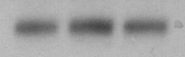
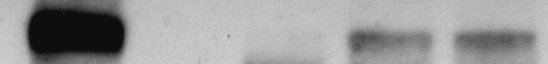
Western Blot: NMDAR1 Antibody (R1JHL)Azide and BSA Free [NB300-118]
Western Blot: NMDAR1 Antibody (R1JHL) [NB300-118] - 10 ug of rat hippocampal lysate showing specific immunolabeling of the ~120 kDa NR1 subunit of the NMDA receptor.![Western Blot: NMDAR1 Antibody (R1JHL)Azide and BSA Free [NB300-118]](https://resources.rndsystems.com/images/products/NMDAR1-Antibody-R1JHL-Western-Blot-NB300-118-img0005.jpg "Western Blot: NMDAR1 Antibody (R1JHL)Azide and BSA Free [NB300-118]")
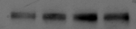
Western Blot: NMDAR1 Antibody (R1JHL)Azide and BSA Free [NB300-118]
NMDAR1-Antibody-R1JHL-Western-Blot-NB300-118-img0005.jpg![Immunocytochemistry/ Immunofluorescence: NMDAR1 Antibody (R1JHL) - Azide and BSA Free [NB300-118]](https://resources.rndsystems.com/images/products/NMDAR1-Antibody-R1JHL-Immunocytochemistry-Immunofluorescence-NB300-118-img0003.jpg "Immunocytochemistry/ Immunofluorescence: NMDAR1 Antibody (R1JHL) - Azide and BSA Free [NB300-118]")
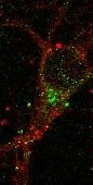
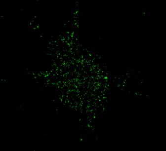
Immunocytochemistry/ Immunofluorescence: NMDAR1 Antibody (R1JHL) - Azide and BSA Free [NB300-118]
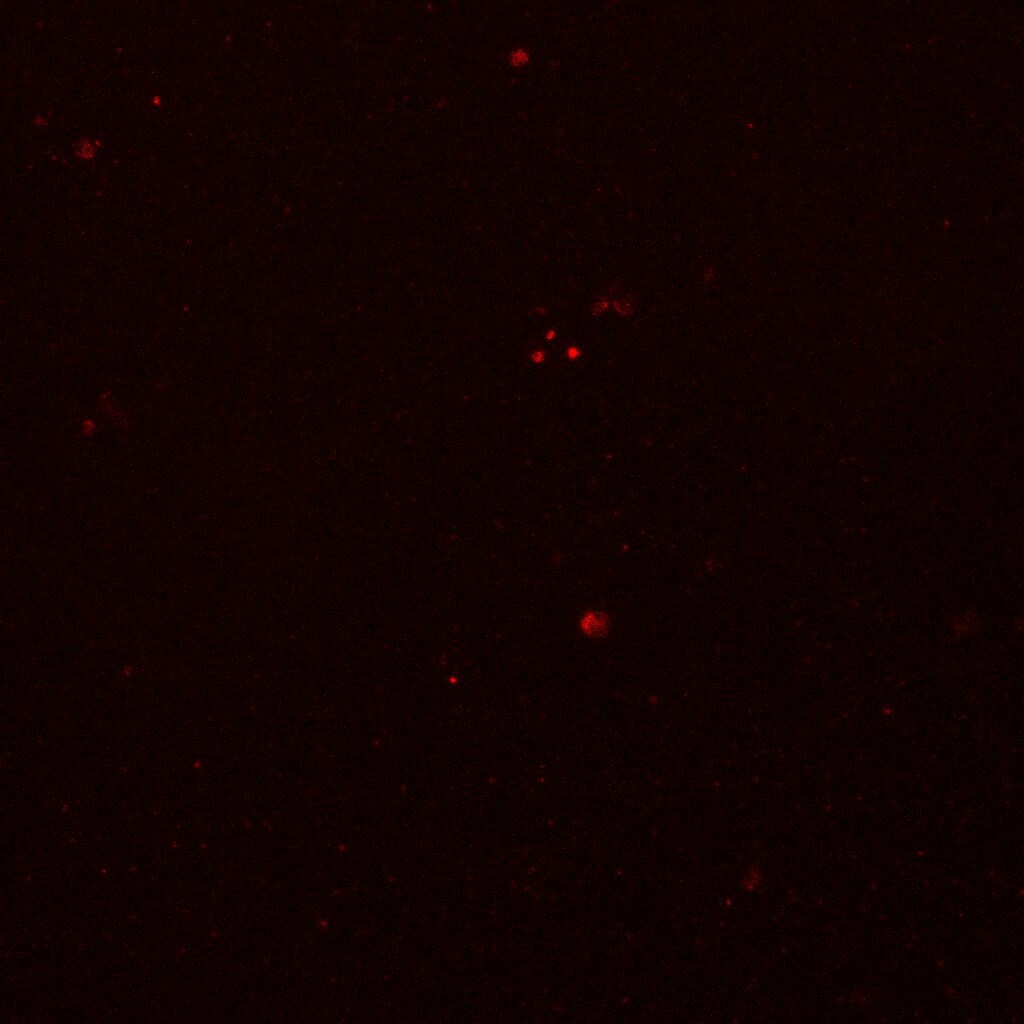
Immunocytochemistry/Immunofluorescence: NMDAR1 Antibody (R1JHL) [NB300-118] - NR1 surface staining of mouse corical neruons. This image was submitted via customer Review.![Immunocytochemistry/ Immunofluorescence: NMDAR1 Antibody (R1JHL) - Azide and BSA Free [NB300-118]](https://resources.rndsystems.com/images/products/NMDAR1-Antibody-R1JHL-Immunocytochemistry-Immunofluorescence-NB300-118-img0002.jpg "Immunocytochemistry/ Immunofluorescence: NMDAR1 Antibody (R1JHL) - Azide and BSA Free [NB300-118]")
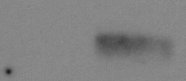
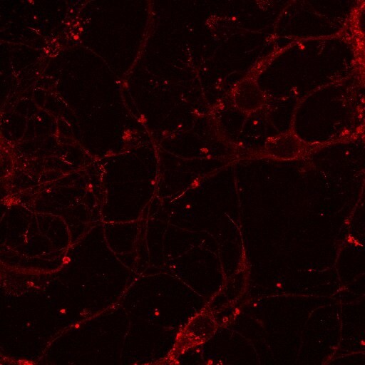
Immunocytochemistry/ Immunofluorescence: NMDAR1 Antibody (R1JHL) - Azide and BSA Free [NB300-118]
Immunocytochemistry/Immunofluorescence: NMDAR1 Antibody (R1JHL) [NB300-118] - Mouse neuron stained with anti-NMDAR1 antibody. Image from verified customer review.![Immunocytochemistry/ Immunofluorescence: NMDAR1 Antibody (R1JHL) - Azide and BSA Free [NB300-118]](https://resources.rndsystems.com/images/products/NMDAR1-Antibody-R1JHL-Immunocytochemistry-Immunofluorescence-NB300-118-img0004.jpg "Immunocytochemistry/ Immunofluorescence: NMDAR1 Antibody (R1JHL) - Azide and BSA Free [NB300-118]")
Immunocytochemistry/ Immunofluorescence: NMDAR1 Antibody (R1JHL) - Azide and BSA Free [NB300-118]
Immunocytochemistry/Immunofluorescence: NMDAR1 Antibody (R1JHL) [NB300-118] - Mixed rat cortical cultures (21 DIV) were labelled with anti-NMDAR1 (1:200) and anti-mouse CF568 (1:250). The antibody labels small punctae along the neuronal processes. No distinct features such as spines are visible. This image was submitted via customer Review. - Azide and BSA Free [NB300-118] -")
Immunocytochemistry/ Immunofluorescence: NMDAR1 Antibody (R1JHL) - Azide and BSA Free [NB300-118] -
Immunocytochemistry/ Immunofluorescence: NMDAR1 Antibody (R1JHL) - Azide and BSA Free [NB300-118] - Notch1 colocalizes postsynaptically with Reelin signaling components. (A) Representative IEM images from hippocampal slices using an antibody specific for Notch1 show that gold particles are localized at postsynaptic as well as presynaptic membrane terminals. (B) Bar graph summarizing the counting of gold particles on the length of presynaptic & postsynaptic membranes indicates that the majority of the Notch1 gold particles are localized postsynaptically (5*10−3 ± 0.8*10−3 vs. 2*10−3 ± 0.4*10−3, n = 3 mice; Student’s t-test, p < 0.01). (C) Fluorescent immunolabeling on 14 days primary neuronal WT cultures shows colocalization of Notch1 & ApoER2 in soma & processes of pyramidal neurons. Both Notch1 & ApoER2 are similarly co-expressed in NMDAR1 positive puncta (R = 0.92 ± 0.03 & R = 0.86 ± 0.08, respectively; Student’s t-test, p = 0.23). (C′) Close up of a dendrite displaying Notch1, ApoER2 & NMDAR1 expression & “zoom in” captions of puncta showing clustering of the three receptors in teal. (D) Fluorescent immunolabeling on primary neuronal cultures shows that Notch1 & Dab1 localize in the same pyramidal neuron’s soma & processes labeled by phalloidin (R = 0.87 ± 0.06 & R = 0.74 ± 0.07 respectively; Student’s t-test, p = 0.5). (D′) Close up of a dendrite with Notch1, Dab1 & F-actin labeling & “zoom in” captions of dendritic puncta showing clustering of Notch1, Dab1 & F-actin in teal. **p < 0.01. Error bars are SEM & scale bar in (A) is 200 nm for all IEM panels, in (C,D) 50 μm & in (C′,D′) 10 μm & 500 nm in zoom in captions. Image collected & cropped by CiteAb from the following publication (https://pubmed.ncbi.nlm.nih.gov/26635527), licensed under a CC-BY license. Not internally tested by Novus Biologicals.Applications for NMDAR1 Antibody (R1JHL)
Application
Recommended Usage
Immunohistochemistry-Frozen
1 ug per ml
Western Blot
1:1000
Application Notes
Use in ICC/IF and IHC-F reported in scientific literature (PMID: 20414717). Use in IHC-P reported in scientific literature (PMID 26635527).
Reviewed Applications
Read 14 reviews rated 4.2 using NB300-118 in the following applications:
Formulation, Preparation, and Storage
Purification
Tissue culture supernatant
Reconstitution
Reconstitute with 50 ul PBS to desired concentration.
Formulation
Lyophilized
Preservative
No Preservative
Concentration
This product is unpurified. The exact concentration of antibody is not quantifiable.
Shipping
The product is shipped with polar packs. Upon receipt, store it immediately at the temperature recommended below.
Stability & Storage
Store at -20C. Avoid freeze-thaw cycles.
Calculators
Background: NMDAR1
Alternate Names
glutamate [NMDA] receptor subunit zeta-1, glutamate receptor, ionotropic, N-methyl D-aspartate 1, NMDA1, NMDAR1glutamate [NMDA] receptor subunit zeta 1, NMD-R1, N-methyl-D-aspartate receptor channel, subunit zeta-1, N-methyl-D-aspartate receptor subunit NR1, NR1
Entrez Gene IDs
24408 (Rat)
Gene Symbol
GRIN1
UniProt
Additional NMDAR1 Products
Product Documents for NMDAR1 Antibody (R1JHL)
Certificate of Analysis
To download a Certificate of Analysis, please enter a lot or batch number in the search box below.
Product Specific Notices for NMDAR1 Antibody (R1JHL)
This product is for research use only and is not approved for use in humans or in clinical diagnosis. Primary Antibodies are guaranteed for 1 year from date of receipt.
Citations for NMDAR1 Antibody (R1JHL)
 Powered by Bioz
Customer Reviews for NMDAR1 Antibody (R1JHL) (14)
4.2 out of 5
14 Customer Ratings
Have you used NMDAR1 Antibody (R1JHL)?
Submit a review and receive an Amazon gift card!
$25/€18/£15/$25CAN/¥2500 Yen for a review with an image
$10/€7/£6/$10CAN/¥1110 Yen for a review without an image
Submit a review
Customer Images

Showing
1
-
5 of
14 reviews
Showing All
Filter By:
-
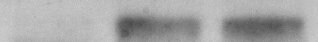
Application: Western BlotSample Tested: Mouse brainSpecies: MouseVerified Customer | Posted 01/31/2018Brain NR1

-
Application: ImmunocytochemistrySample Tested: Cultured hippocampal neuronsSpecies: MouseVerified Customer | Posted 08/04/2017
-
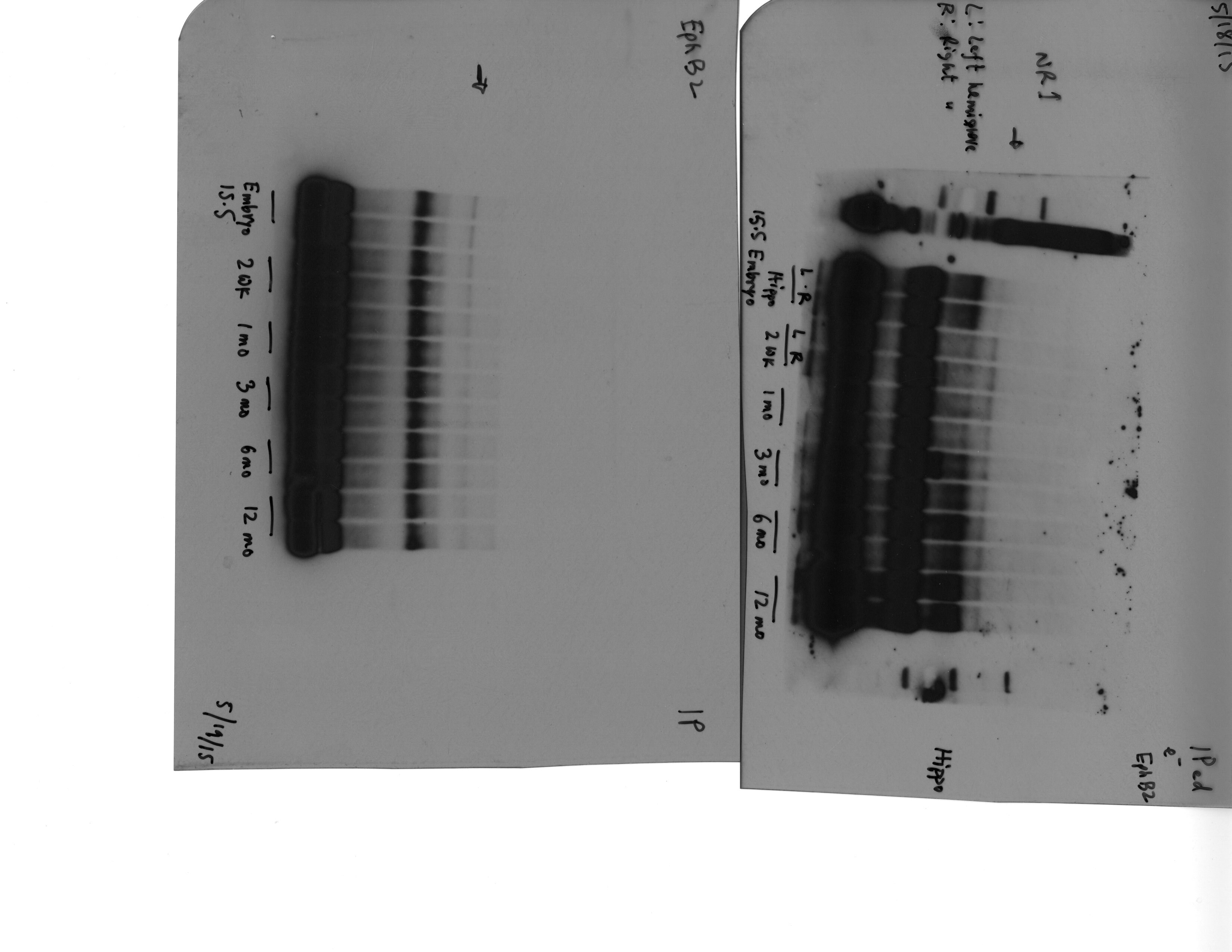
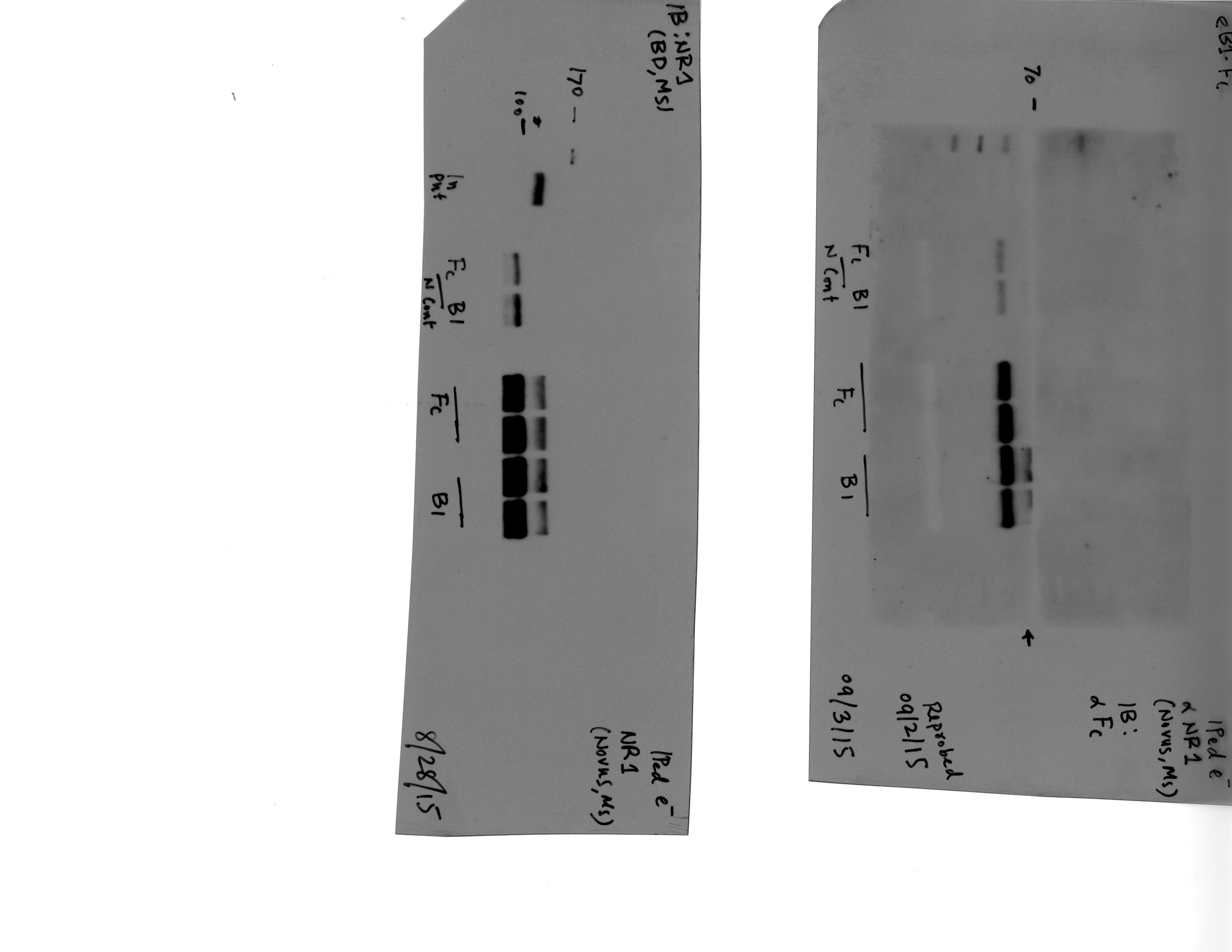
Application: ImmunoprecipitationSample Tested: Mice Brain LysateSpecies: MouseVerified Customer | Posted 08/04/2017
-
Application: Western BlotSample Tested: 293TSpecies: HumanVerified Customer | Posted 08/04/2017
-
Application: ImmunoprecipitationSample Tested: brain and spinal cordSpecies: MouseVerified Customer | Posted 07/14/2017
-
Application: ImmunocytochemistrySample Tested: Cortical neuronsSpecies: MouseVerified Customer | Posted 07/14/2017NR1 surface staining
-
Application: Western BlotSample Tested: brain and spinal cordSpecies: HumanVerified Customer | Posted 07/14/2017Human brain NR1
-
Application: ImmunocytochemistrySample Tested: rat cortical culture (3 weeks old)Species: RatVerified Customer | Posted 06/27/2017Mixed rat cortical cultures (21 DIV) were labelled with anti-NMDAR1 (1:200) and anti-mouse CF568 (1:250). The antibody labels small punctae along the neuronal processes. No distinct features such as spines are visible.Fixation Solution and Conditions: 4% paraformaldehyde in PBS, 15 minutes RT Blocking Solution & Duration: 3% BSA in PBS with 0.2% saponin, 30 minutes RT Primary Antibody Diluent and Dilutions Tested: 1:200 in 1% BSA in PBS with 0.2% saponin, over night 4°C Secondary Antibody Manufacturer, Host Species, Dilution, & Diluent: Thermo donkey anti-mouse CF568, 1:250 in PBS with 0.2% saponin, 1 hour, RT
-
Application: ImmunofluorescenceSample Tested: Mouse neuronsSpecies: MouseVerified Customer | Posted 11/26/2015
-
Application: ImmunocytochemistrySample Tested: Mouse neuronsSpecies: MouseVerified Customer | Posted 11/26/2015
-
Application: ImmunoprecipitationSample Tested: Mouse Brain TissueSpecies: MouseVerified Customer | Posted 10/16/2015Co-IP_NR1 and EphB2
-
Application: ImmunoprecipitationSample Tested: Mouse neuronal culturesSpecies: MouseVerified Customer | Posted 10/15/2015Co-IP
-
Application: ImmunoprecipitationSample Tested: Mouse neuronsSpecies: MouseVerified Customer | Posted 10/15/2015CoIP
-
Application: ImmunofluorescenceSample Tested: NR1 plasmid transfected HEK293T cellsSpecies: HumanVerified Customer | Posted 09/30/2014
There are no reviews that match your criteria.
Protocols
Find general support by application which include: protocols, troubleshooting, illustrated assays, videos and webinars.
- Antigen Retrieval Protocol (PIER)
- Antigen Retrieval for Frozen Sections Protocol
- Appropriate Fixation of IHC/ICC Samples
- Cellular Response to Hypoxia Protocols
- Chromogenic IHC Staining of Formalin-Fixed Paraffin-Embedded (FFPE) Tissue Protocol
- Chromogenic Immunohistochemistry Staining of Frozen Tissue
- ClariTSA™ Fluorophore Kits
- Detection & Visualization of Antibody Binding
- Fluorescent IHC Staining of Frozen Tissue Protocol
- Graphic Protocol for Heat-induced Epitope Retrieval
- Graphic Protocol for the Preparation and Fluorescent IHC Staining of Frozen Tissue Sections
- Graphic Protocol for the Preparation and Fluorescent IHC Staining of Paraffin-embedded Tissue Sections
- Graphic Protocol for the Preparation of Gelatin-coated Slides for Histological Tissue Sections
- ICC Cell Smear Protocol for Suspension Cells
- ICC Immunocytochemistry Protocol Videos
- ICC for Adherent Cells
- IHC Sample Preparation (Frozen sections vs Paraffin)
- Immunocytochemistry (ICC) Protocol
- Immunocytochemistry Troubleshooting
- Immunofluorescence of Organoids Embedded in Cultrex Basement Membrane Extract
- Immunofluorescent IHC Staining of Formalin-Fixed Paraffin-Embedded (FFPE) Tissue Protocol
- Immunohistochemistry (IHC) and Immunocytochemistry (ICC) Protocols
- Immunohistochemistry Frozen Troubleshooting
- Immunohistochemistry Paraffin Troubleshooting
- Preparing Samples for IHC/ICC Experiments
- Preventing Non-Specific Staining (Non-Specific Binding)
- Primary Antibody Selection & Optimization
- Protocol for Heat-Induced Epitope Retrieval (HIER)
- Protocol for Making a 4% Formaldehyde Solution in PBS
- Protocol for VisUCyte™ HRP Polymer Detection Reagent
- Protocol for the Fluorescent ICC Staining of Cell Smears - Graphic
- Protocol for the Fluorescent ICC Staining of Cultured Cells on Coverslips - Graphic
- Protocol for the Preparation & Fixation of Cells on Coverslips
- Protocol for the Preparation and Chromogenic IHC Staining of Frozen Tissue Sections
- Protocol for the Preparation and Chromogenic IHC Staining of Frozen Tissue Sections - Graphic
- Protocol for the Preparation and Chromogenic IHC Staining of Paraffin-embedded Tissue Sections
- Protocol for the Preparation and Chromogenic IHC Staining of Paraffin-embedded Tissue Sections - Graphic
- Protocol for the Preparation and Fluorescent ICC Staining of Cells on Coverslips
- Protocol for the Preparation and Fluorescent ICC Staining of Non-adherent Cells
- Protocol for the Preparation and Fluorescent ICC Staining of Stem Cells on Coverslips
- Protocol for the Preparation and Fluorescent IHC Staining of Frozen Tissue Sections
- Protocol for the Preparation and Fluorescent IHC Staining of Paraffin-embedded Tissue Sections
- Protocol for the Preparation of Gelatin-coated Slides for Histological Tissue Sections
- Protocol for the Preparation of a Cell Smear for Non-adherent Cell ICC - Graphic
- R&D Systems Quality Control Western Blot Protocol
- TUNEL and Active Caspase-3 Detection by IHC/ICC Protocol
- The Importance of IHC/ICC Controls
- Troubleshooting Guide: Immunohistochemistry
- Troubleshooting Guide: Western Blot Figures
- Western Blot Conditions
- Western Blot Protocol
- Western Blot Protocol for Cell Lysates
- Western Blot Troubleshooting
- Western Blot Troubleshooting Guide
- View all Protocols, Troubleshooting, Illustrated assays and Webinars
Loading...