NPM1 Antibody - BSA Free
Novus Biologicals | Catalog # NB110-61646
![Western Blot: NPM1 AntibodyBSA Free [NB110-61646]](https://resources.rndsystems.com/images/products/Nucleophosmin-Antibody-Western-Blot-NB110-61646-img0012.jpg "Western Blot: NPM1 AntibodyBSA Free [NB110-61646]")
Key Product Details
Species Reactivity
Validated:
Cited:
Applications
Validated:
Cited:
Label
Antibody Source
Format
Product Specifications
Immunogen
Reactivity Notes
Localization
Specificity
Clonality
Host
Isotype
Theoretical MW
Disclaimer note: The observed molecular weight of the protein may vary from the listed predicted molecular weight due to post translational modifications, post translation cleavages, relative charges, and other experimental factors.
Scientific Data Images for NPM1 Antibody - BSA Free
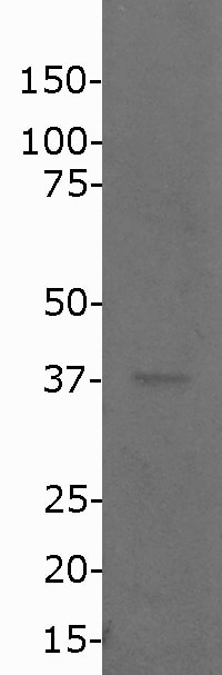
Western Blot: NPM1 AntibodyBSA Free [NB110-61646]
Western Blot: Nucleophosmin Antibody [NB110-61646] - Detection of mutant Nucleophosmin in OCI-AML3 lysates.![Immunocytochemistry/ Immunofluorescence: NPM1 Antibody - BSA Free [NB110-61646]](https://resources.rndsystems.com/images/products/Nucleophosmin-Antibody-Immunocytochemistry-Immunofluorescence-NB110-61646-img0011.jpg "Immunocytochemistry/ Immunofluorescence: NPM1 Antibody - BSA Free [NB110-61646]")
Immunocytochemistry/ Immunofluorescence: NPM1 Antibody - BSA Free [NB110-61646]
Immunocytochemistry/Immunofluorescence: Nucleophosmin Antibody [NB110-61646] - Nucleophosmin localization by immunofluorescence in HL-60 cells (negative control).![Immunocytochemistry/ Immunofluorescence: NPM1 Antibody - BSA Free [NB110-61646]](https://resources.rndsystems.com/images/products/Nucleophosmin-Antibody-Immunocytochemistry-Immunofluorescence-NB110-61646-img0010.jpg "Immunocytochemistry/ Immunofluorescence: NPM1 Antibody - BSA Free [NB110-61646]")
Immunocytochemistry/ Immunofluorescence: NPM1 Antibody - BSA Free [NB110-61646]
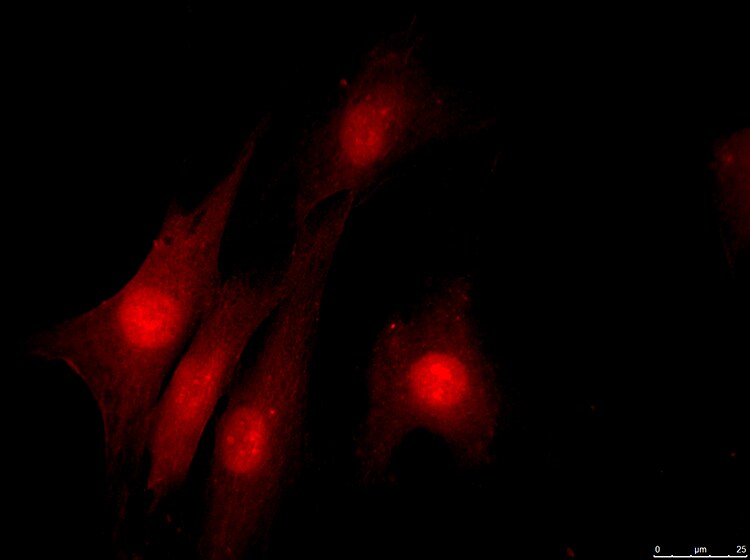
Immunocytochemistry/Immunofluorescence: Nucleophosmin Antibody [NB110-61646] - Nucleophosmin localization by immunofluorescence in OCI-AML3 cells.Applications for NPM1 Antibody - BSA Free
Immunocytochemistry/ Immunofluorescence
Immunohistochemistry
Immunohistochemistry-Paraffin
Immunoprecipitation
Western Blot
Reviewed Applications
Read 2 reviews rated 3 using NB110-61646 in the following applications:
Formulation, Preparation, and Storage
Purification
Formulation
Format
Preservative
Concentration
Shipping
Stability & Storage
Background: NPM1
Long Name
Alternate Names
Gene Symbol
Additional NPM1 Products
Product Documents for NPM1 Antibody - BSA Free
Certificate of Analysis
To download a Certificate of Analysis, please enter a lot or batch number in the search box below.
Product Specific Notices for NPM1 Antibody - BSA Free
This product is for research use only and is not approved for use in humans or in clinical diagnosis. Primary Antibodies are guaranteed for 1 year from date of receipt.
Related Research Areas
Citations for NPM1 Antibody - BSA Free
 Powered by Bioz
Customer Reviews for NPM1 Antibody - BSA Free (2)
Have you used NPM1 Antibody - BSA Free?
Submit a review and receive an Amazon gift card!
$25/€18/£15/$25CAN/¥2500 Yen for a review with an image
$10/€7/£6/$10CAN/¥1110 Yen for a review without an image
Submit a review
Customer Images
-
Application: ImmunocytochemistrySample Tested: fibroblastsSpecies: HumanVerified Customer | Posted 03/16/2018There was strong signal, but the signal was strong in the both the nucleus and the cytoplasm. 1:100 dilution was used, so perhaps using more diluted antibody would help.

-
Application: Western BlotSample Tested: Human lymphocytesSpecies: HumanVerified Customer | Posted 01/18/20181:500 16 hours 4C in 5% milk-TBS/T 1 minute exposure, 25ug protein.
There are no reviews that match your criteria.
Protocols
View specific protocols for NPM1 Antibody - BSA Free (NB110-61646):
Western Blot
1. Run 50 ug of cytoplasmic fraction into each lane of an 12% SDS-PAGE.
2. Block the membrane.
3. Dilute the primary anti-mNPM1 (NB110-61646) in 1% NFDM and incubate for 2 hours at room temperature.
4. Wash the membrane.
5. Dilute the sedondary anti-rabbit IgG, conjugated to HRP in 1% NFDM and incubate for 1 hour at room temperature.
6. Develop the blot.
Find general support by application which include: protocols, troubleshooting, illustrated assays, videos and webinars.
- Antigen Retrieval Protocol (PIER)
- Antigen Retrieval for Frozen Sections Protocol
- Appropriate Fixation of IHC/ICC Samples
- Cellular Response to Hypoxia Protocols
- Chromogenic IHC Staining of Formalin-Fixed Paraffin-Embedded (FFPE) Tissue Protocol
- Chromogenic Immunohistochemistry Staining of Frozen Tissue
- ClariTSA™ Fluorophore Kits
- Detection & Visualization of Antibody Binding
- Fluorescent IHC Staining of Frozen Tissue Protocol
- Graphic Protocol for Heat-induced Epitope Retrieval
- Graphic Protocol for the Preparation and Fluorescent IHC Staining of Frozen Tissue Sections
- Graphic Protocol for the Preparation and Fluorescent IHC Staining of Paraffin-embedded Tissue Sections
- Graphic Protocol for the Preparation of Gelatin-coated Slides for Histological Tissue Sections
- ICC Cell Smear Protocol for Suspension Cells
- ICC Immunocytochemistry Protocol Videos
- ICC for Adherent Cells
- IHC Sample Preparation (Frozen sections vs Paraffin)
- Immunocytochemistry (ICC) Protocol
- Immunocytochemistry Troubleshooting
- Immunofluorescence of Organoids Embedded in Cultrex Basement Membrane Extract
- Immunofluorescent IHC Staining of Formalin-Fixed Paraffin-Embedded (FFPE) Tissue Protocol
- Immunohistochemistry (IHC) and Immunocytochemistry (ICC) Protocols
- Immunohistochemistry Frozen Troubleshooting
- Immunohistochemistry Paraffin Troubleshooting
- Immunoprecipitation Protocol
- Preparing Samples for IHC/ICC Experiments
- Preventing Non-Specific Staining (Non-Specific Binding)
- Primary Antibody Selection & Optimization
- Protocol for Heat-Induced Epitope Retrieval (HIER)
- Protocol for Making a 4% Formaldehyde Solution in PBS
- Protocol for VisUCyte™ HRP Polymer Detection Reagent
- Protocol for the Fluorescent ICC Staining of Cell Smears - Graphic
- Protocol for the Fluorescent ICC Staining of Cultured Cells on Coverslips - Graphic
- Protocol for the Preparation & Fixation of Cells on Coverslips
- Protocol for the Preparation and Chromogenic IHC Staining of Frozen Tissue Sections
- Protocol for the Preparation and Chromogenic IHC Staining of Frozen Tissue Sections - Graphic
- Protocol for the Preparation and Chromogenic IHC Staining of Paraffin-embedded Tissue Sections
- Protocol for the Preparation and Chromogenic IHC Staining of Paraffin-embedded Tissue Sections - Graphic
- Protocol for the Preparation and Fluorescent ICC Staining of Cells on Coverslips
- Protocol for the Preparation and Fluorescent ICC Staining of Non-adherent Cells
- Protocol for the Preparation and Fluorescent ICC Staining of Stem Cells on Coverslips
- Protocol for the Preparation and Fluorescent IHC Staining of Frozen Tissue Sections
- Protocol for the Preparation and Fluorescent IHC Staining of Paraffin-embedded Tissue Sections
- Protocol for the Preparation of Gelatin-coated Slides for Histological Tissue Sections
- Protocol for the Preparation of a Cell Smear for Non-adherent Cell ICC - Graphic
- R&D Systems Quality Control Western Blot Protocol
- TUNEL and Active Caspase-3 Detection by IHC/ICC Protocol
- The Importance of IHC/ICC Controls
- Troubleshooting Guide: Immunohistochemistry
- Troubleshooting Guide: Western Blot Figures
- Western Blot Conditions
- Western Blot Protocol
- Western Blot Protocol for Cell Lysates
- Western Blot Troubleshooting
- Western Blot Troubleshooting Guide
- View all Protocols, Troubleshooting, Illustrated assays and Webinars
FAQs for NPM1 Antibody - BSA Free
-
Q: Does this antibody require antigen retrieval? If so, what antigen retrieval method do you recommend?
A: For the validation of our Nucleophosmin antibody (NB110-61646), we did not use any customized protocol and in our routine protocol, we use Sodium Citrate buffer pH 6.0 based antigen retrieval method. Please note that formalin fixation as well as paraffin embedding crosslinks the proteins which decreases staining signal by causing conformational changes on the antigens. The inclusion/conditions of antigen retrieval depends primarily upon for how long the samples were in the fixative solution. If the fixation time for samples was more than 4 hours, we would recommend performing Sodium Citrate buffer ph 6.0 based antigen retrieval.