Nucleolin Antibody (NCL/7014R)
Novus Biologicals | Catalog # NBP3-11037
Recombinant Monoclonal Antibody
Key Product Details
Species Reactivity
Human, Bovine (Negative), Mouse (Negative), Rat (Negative)
Applications
Immunohistochemistry, Immunohistochemistry-Paraffin, Western Blot, Flow Cytometry, Immunocytochemistry/ Immunofluorescence
Label
Unconjugated
Antibody Source
Recombinant Monoclonal Rabbit IgG Kappa Clone # NCL/7014R
Loading...
Product Specifications
Immunogen
Recombinant full-length human Nucleolin protein (Uniprot: P19338)
Reactivity Notes
Does not react with Mouse, Feline, Bovine, Canine or Equine.
Localization
Nucleolus
Marker
Marker of Human Cells
Specificity
Recognizes a protein of ~76kDa, which is identified as Nucleolin (NCL). It is the major nucleolar phosphoprotein of growing eukaryotic cells. NCL is located mainly in dense fibrillar regions of the nucleolus. It is found associated with intranucleolar chromatin and pre-ribosomal particles. Human NCL gene consists of 14 exons with 13 introns and spans approximately 11kb. It induces chromatin decondensation by binding to histone H1. It is thought to play a role in pre-rRNA transcription and ribosome assembly. This monoclonal antibody can be used to stain the nucleoli in cell or tissue preparations and can be used as a marker of the nucleoli in subcellular fractions. It produces a speckled pattern in the nuclei of cells of normal and malignant cells and may be used to stain the nucleoli of cells in fixed or frozen tissue sections. It can be used with paraformaldehyde fixed frozen tissue or cell preparations and formalin fixed, paraffin-embedded tissue sections.
Clonality
Monoclonal
Host
Rabbit
Isotype
IgG Kappa
Theoretical MW
76 kDa.
Disclaimer note: The observed molecular weight of the protein may vary from the listed predicted molecular weight due to post translational modifications, post translation cleavages, relative charges, and other experimental factors.
Disclaimer note: The observed molecular weight of the protein may vary from the listed predicted molecular weight due to post translational modifications, post translation cleavages, relative charges, and other experimental factors.
Description
200ug/ml of antibody purified from Bioreactor Concentrate by Protein A or G. Prepared in 10 mM PBS with 0.05% BSA & 0.05% azide. Also available WITHOUT BSA & azide at 1.0 mg/ml. (NBP3-11038)
Antibody with azide - store at 2 to 8C. Antibody without azide - store at -20 to -80 C.
Antibody with azide - store at 2 to 8C. Antibody without azide - store at -20 to -80 C.
Scientific Data Images for Nucleolin Antibody (NCL/7014R)
![Western Blot: Nucleolin Antibody (NCL/7014R) [NBP3-11037]](https://resources.rndsystems.com/images/products/Nucleolin-Antibody-NCL-7014R-Western-Blot-NBP3-11037-img0003.jpg "Western Blot: Nucleolin Antibody (NCL/7014R) [NBP3-11037]")
Western Blot: Nucleolin Antibody (NCL/7014R) [NBP3-11037]
Western Blot: Nucleolin Antibody (NCL/7014R) [NBP3-11037] - Western blot analysis of Jurkat cell lysate using Nucleolin antibody (NCL/7014R).![Immunohistochemistry-Paraffin: Nucleolin Antibody (NCL/7014R) [NBP3-11037]](https://resources.rndsystems.com/images/products/Nucleolin-Antibody-NCL-7014R-Immunohistochemistry-Paraffin-NBP3-11037-img0010.jpg "Immunohistochemistry-Paraffin: Nucleolin Antibody (NCL/7014R) [NBP3-11037]")
Immunohistochemistry-Paraffin: Nucleolin Antibody (NCL/7014R) [NBP3-11037]
Immunohistochemistry-Paraffin: Nucleolin Antibody (NCL/7014R) [NBP3-11037] - Formalin-fixed, paraffin-embedded cow heart stained with Nucleolin Antibody (NCL/7014R) at 2ug/ml. HIER: Tris/EDTA, pH9.0, 45min. HRP-polymer, 30min. DAB, 5min.![Immunohistochemistry-Paraffin: Nucleolin Antibody (NCL/7014R) [NBP3-11037]](https://resources.rndsystems.com/images/products/Nucleolin-Antibody-NCL-7014R-Immunohistochemistry-Paraffin-NBP3-11037-img0001.jpg "Immunohistochemistry-Paraffin: Nucleolin Antibody (NCL/7014R) [NBP3-11037]")
Immunohistochemistry-Paraffin: Nucleolin Antibody (NCL/7014R) [NBP3-11037]
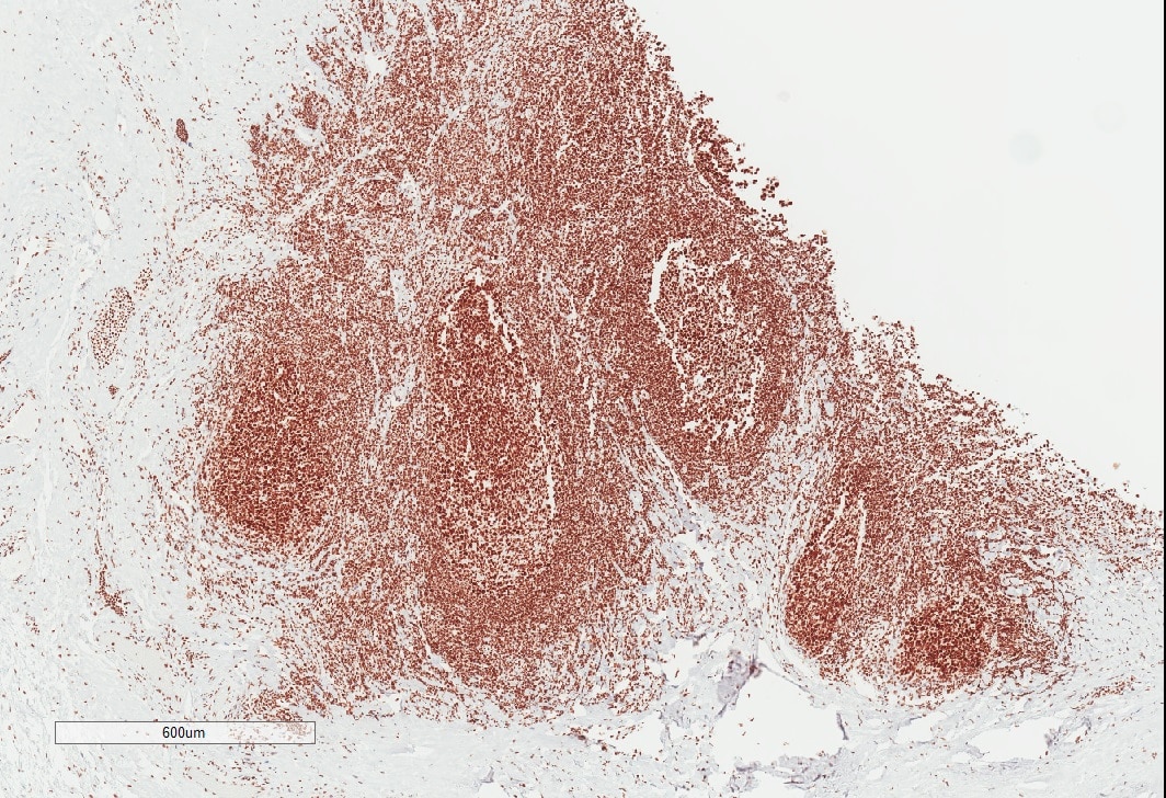
Immunohistochemistry-Paraffin: Nucleolin Antibody (NCL/7014R) [NBP3-11037] - Human tonsil stained with Nucleolin Antibody. IHC-P image submitted by a verified customer review.![Immunohistochemistry-Paraffin: Nucleolin Antibody (NCL/7014R) [NBP3-11037]](https://resources.rndsystems.com/images/products/Nucleolin-Antibody-NCL-7014R-Immunohistochemistry-Paraffin-NBP3-11037-img0002.jpg "Immunohistochemistry-Paraffin: Nucleolin Antibody (NCL/7014R) [NBP3-11037]")
Immunohistochemistry-Paraffin: Nucleolin Antibody (NCL/7014R) [NBP3-11037]
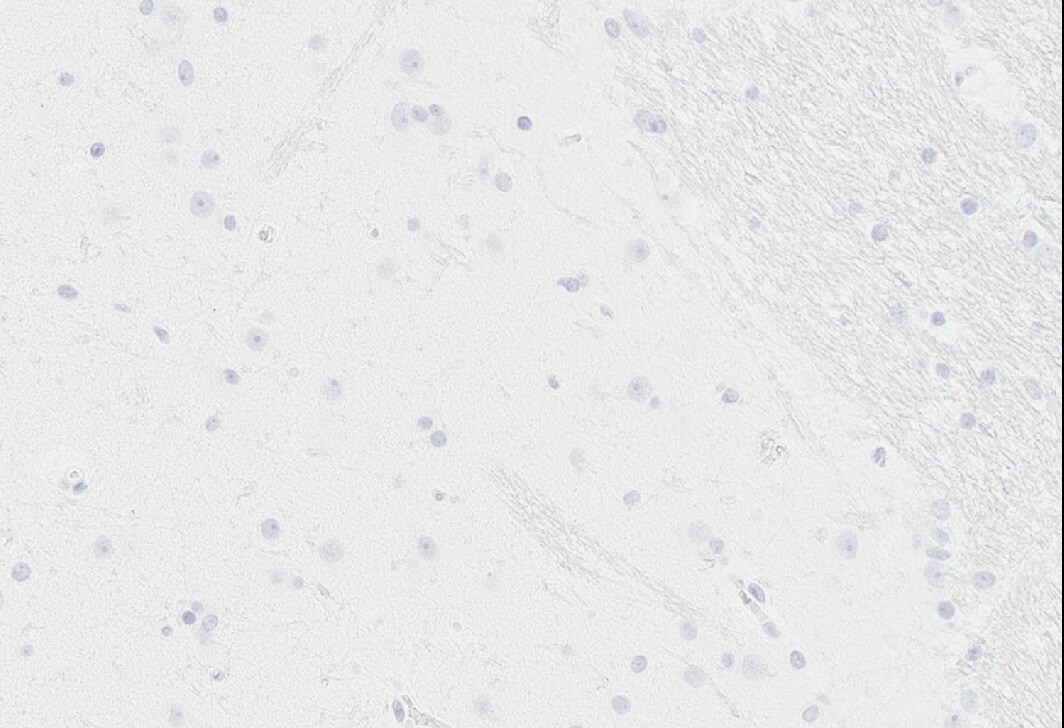
Immunohistochemistry-Paraffin: Nucleolin Antibody (NCL/7014R) [NBP3-11037] - Analysis of human cells administered in pig brain using Nucleolin antibody. Antibody did NOT stain porcine tissue allowing the presence of human cells to be determined. Primary antibody dilution 1:100. Image from verified customer review.![Immunohistochemistry-Paraffin: Nucleolin Antibody (NCL/7014R) [NBP3-11037]](https://resources.rndsystems.com/images/products/Nucleolin-Antibody-NCL-7014R-Immunohistochemistry-Paraffin-NBP3-11037-img0004.jpg "Immunohistochemistry-Paraffin: Nucleolin Antibody (NCL/7014R) [NBP3-11037]")
Immunohistochemistry-Paraffin: Nucleolin Antibody (NCL/7014R) [NBP3-11037]
Immunohistochemistry-Paraffin: Nucleolin Antibody (NCL/7014R) [NBP3-11037] - Formalin-fixed, paraffin-embedded human salivary gland stained with Nucleolin Antibody (NCL/7014R). HIER: Tris/EDTA, pH9.0, 45min. HRP-polymer, 30min. DAB, 5min.![Immunohistochemistry-Paraffin: Nucleolin Antibody (NCL/7014R) [NBP3-11037]](https://resources.rndsystems.com/images/products/Nucleolin-Antibody-NCL-7014R-Immunohistochemistry-Paraffin-NBP3-11037-img0005.jpg "Immunohistochemistry-Paraffin: Nucleolin Antibody (NCL/7014R) [NBP3-11037]")
Immunohistochemistry-Paraffin: Nucleolin Antibody (NCL/7014R) [NBP3-11037]
Immunohistochemistry-Paraffin: Nucleolin Antibody (NCL/7014R) [NBP3-11037] - Formalin-fixed, paraffin-embedded human kidney stained with Nucleolin Antibody (NCL/7014R).![Immunohistochemistry-Paraffin: Nucleolin Antibody (NCL/7014R) [NBP3-11037]](https://resources.rndsystems.com/images/products/Nucleolin-Antibody-NCL-7014R-Immunohistochemistry-Paraffin-NBP3-11037-img0006.jpg "Immunohistochemistry-Paraffin: Nucleolin Antibody (NCL/7014R) [NBP3-11037]")
Immunohistochemistry-Paraffin: Nucleolin Antibody (NCL/7014R) [NBP3-11037]
Immunohistochemistry-Paraffin: Nucleolin Antibody (NCL/7014R) [NBP3-11037] - Formalin-fixed, paraffin-embedded human kidney stained with Nucleolin Antibody (NCL/7014R).![Immunohistochemistry-Paraffin: Nucleolin Antibody (NCL/7014R) [NBP3-11037]](https://resources.rndsystems.com/images/products/Nucleolin-Antibody-NCL-7014R-Immunohistochemistry-Paraffin-NBP3-11037-img0007.jpg "Immunohistochemistry-Paraffin: Nucleolin Antibody (NCL/7014R) [NBP3-11037]")
Immunohistochemistry-Paraffin: Nucleolin Antibody (NCL/7014R) [NBP3-11037]
Immunohistochemistry-Paraffin: Nucleolin Antibody (NCL/7014R) [NBP3-11037] - Formalin-fixed, paraffin-embedded cat liver stained with Nucleolin Antibody (NCL/7014R) at 2ug/ml. HIER: Tris/EDTA, pH9.0, 45min. HRP-polymer, 30min. DAB, 5min.![Immunohistochemistry-Paraffin: Nucleolin Antibody (NCL/7014R) [NBP3-11037]](https://resources.rndsystems.com/images/products/Nucleolin-Antibody-NCL-7014R-Immunohistochemistry-Paraffin-NBP3-11037-img0008.jpg "Immunohistochemistry-Paraffin: Nucleolin Antibody (NCL/7014R) [NBP3-11037]")
Immunohistochemistry-Paraffin: Nucleolin Antibody (NCL/7014R) [NBP3-11037]
Immunohistochemistry-Paraffin: Nucleolin Antibody (NCL/7014R) [NBP3-11037] - Formalin-fixed, paraffin-embedded horse lung stained with Nucleolin Antibody (NCL/7014R) at 2ug/ml. HIER: Tris/EDTA, pH9.0, 45min. HRP-polymer, 30min. DAB, 5min.![Immunohistochemistry-Paraffin: Nucleolin Antibody (NCL/7014R) [NBP3-11037]](https://resources.rndsystems.com/images/products/Nucleolin-Antibody-NCL-7014R-Immunohistochemistry-Paraffin-NBP3-11037-img0009.jpg "Immunohistochemistry-Paraffin: Nucleolin Antibody (NCL/7014R) [NBP3-11037]")
Immunohistochemistry-Paraffin: Nucleolin Antibody (NCL/7014R) [NBP3-11037]
Immunohistochemistry-Paraffin: Nucleolin Antibody (NCL/7014R) [NBP3-11037] - Formalin-fixed, paraffin-embedded mouse stomach stained with Nucleolin Antibody (NCL/7014R) at 2ug/ml. HIER: Tris/EDTA, pH9.0, 45min. HRP-polymer, 30min. DAB, 5min.")
Nucleolin Antibody (NCL/7014R)
Formalin-fixed, paraffin-embedded human colon stained with Nucleolin antibody (NCL/7014R) at 2ug/ml. HIER: Tris/EDTA, pH9.0, 45min. Secondary: HRP-polymer, 30min. DAB, 5min.Applications for Nucleolin Antibody (NCL/7014R)
Application
Recommended Usage
Flow Cytometry
1-2 ug/million cells
Immunocytochemistry/ Immunofluorescence
1-2 ug/ml
Immunohistochemistry
1-2ug/ml
Immunohistochemistry-Paraffin
1-2 ug/ml
Western Blot
1-2 ug/ml
Application Notes
Immunohistochemistry (Formalin-fixed): 1-2ug/ml for 30 minutes at RT. Staining of formalin-fixed tissues requires heating tissue sections in 10mM Tris with 1mM EDTA, pH 9.0, for 45 min at 95C followed by cooling at RT for 20 minutes Optimal dilution for a specific application should be determined.
Reviewed Applications
Read 2 reviews rated 5 using NBP3-11037 in the following applications:
Flow Cytometry Panel Builder
Bio-Techne Knows Flow Cytometry
Save time and reduce costly mistakes by quickly finding compatible reagents using the Panel Builder Tool.
Advanced Features
- Spectra Viewer - Custom analysis of spectra from multiple fluorochromes
- Spillover Popups - Visualize the spectra of individual fluorochromes
- Antigen Density Selector - Match fluorochrome brightness with antigen density

Formulation, Preparation, and Storage
Purification
Protein A or G purified
Formulation
10 mM PBS with 0.05% BSA
Preservative
0.05% Sodium Azide
Concentration
0.2 mg/ml
Shipping
The product is shipped with polar packs. Upon receipt, store it immediately at the temperature recommended below.
Stability & Storage
Store at 4C.
Background: Nucleolin
Alternate Names
C23, FLJ45706, FLJ59041, nucleolin, Protein C23
Gene Symbol
NCL
UniProt
Additional Nucleolin Products
Product Documents for Nucleolin Antibody (NCL/7014R)
Certificate of Analysis
To download a Certificate of Analysis, please enter a lot or batch number in the search box below.
Product Specific Notices for Nucleolin Antibody (NCL/7014R)
This product is for research use only and is not approved for use in humans or in clinical diagnosis. Primary Antibodies are guaranteed for 1 year from date of receipt.
Customer Reviews for Nucleolin Antibody (NCL/7014R) (2)
5 out of 5
2 Customer Ratings
Have you used Nucleolin Antibody (NCL/7014R)?
Submit a review and receive an Amazon gift card!
$25/€18/£15/$25CAN/¥2500 Yen for a review with an image
$10/€7/£6/$10CAN/¥1110 Yen for a review without an image
Submit a review
Customer Images
Showing
1
-
2 of
2 reviews
Showing All
Filter By:
-
Application: Immunohistochemistry-ParaffinSample Tested: brain and spinal cordSpecies: PigVerified Customer | Posted 02/01/2022Nucleolin 1:100 on pig brainWe were looking for human cells that were administered in pig brain. We needed an antibody that did not stain porcine tissue. This antibody did not stain pig and helped us determine the presence of human cells.
 Bio-Techne ResponseThis review was submitted through the legacy Novus Innovators Program, reflecting a new species or application tested on a primary antibody.
 -
Application: Immunohistochemistry-ParaffinSample Tested: human tonsilSpecies: HumanVerified Customer | Posted 06/03/2021Human tonsil stained with Nucleolin AntibodyCitrate pH6 retrieval at 110C, antibody dilution 1:100/30 min RT, anti-rabbit polymer detection
There are no reviews that match your criteria.
Protocols
Find general support by application which include: protocols, troubleshooting, illustrated assays, videos and webinars.
- 7-Amino Actinomycin D (7-AAD) Cell Viability Flow Cytometry Protocol
- Antigen Retrieval Protocol (PIER)
- Antigen Retrieval for Frozen Sections Protocol
- Appropriate Fixation of IHC/ICC Samples
- Cellular Response to Hypoxia Protocols
- Chromogenic IHC Staining of Formalin-Fixed Paraffin-Embedded (FFPE) Tissue Protocol
- Chromogenic Immunohistochemistry Staining of Frozen Tissue
- ClariTSA™ Fluorophore Kits
- Detection & Visualization of Antibody Binding
- Extracellular Membrane Flow Cytometry Protocol
- Flow Cytometry Protocol for Cell Surface Markers
- Flow Cytometry Protocol for Staining Membrane Associated Proteins
- Flow Cytometry Staining Protocols
- Flow Cytometry Troubleshooting Guide
- Fluorescent IHC Staining of Frozen Tissue Protocol
- Graphic Protocol for Heat-induced Epitope Retrieval
- Graphic Protocol for the Preparation and Fluorescent IHC Staining of Frozen Tissue Sections
- Graphic Protocol for the Preparation and Fluorescent IHC Staining of Paraffin-embedded Tissue Sections
- Graphic Protocol for the Preparation of Gelatin-coated Slides for Histological Tissue Sections
- ICC Cell Smear Protocol for Suspension Cells
- ICC Immunocytochemistry Protocol Videos
- ICC for Adherent Cells
- IHC Sample Preparation (Frozen sections vs Paraffin)
- Immunocytochemistry (ICC) Protocol
- Immunocytochemistry Troubleshooting
- Immunofluorescence of Organoids Embedded in Cultrex Basement Membrane Extract
- Immunofluorescent IHC Staining of Formalin-Fixed Paraffin-Embedded (FFPE) Tissue Protocol
- Immunohistochemistry (IHC) and Immunocytochemistry (ICC) Protocols
- Immunohistochemistry Frozen Troubleshooting
- Immunohistochemistry Paraffin Troubleshooting
- Intracellular Flow Cytometry Protocol Using Alcohol (Methanol)
- Intracellular Flow Cytometry Protocol Using Detergents
- Intracellular Nuclear Staining Flow Cytometry Protocol Using Detergents
- Intracellular Staining Flow Cytometry Protocol Using Alcohol Permeabilization
- Intracellular Staining Flow Cytometry Protocol Using Detergents to Permeabilize Cells
- Preparing Samples for IHC/ICC Experiments
- Preventing Non-Specific Staining (Non-Specific Binding)
- Primary Antibody Selection & Optimization
- Propidium Iodide Cell Viability Flow Cytometry Protocol
- Protocol for Heat-Induced Epitope Retrieval (HIER)
- Protocol for Liperfluo
- Protocol for Making a 4% Formaldehyde Solution in PBS
- Protocol for VisUCyte™ HRP Polymer Detection Reagent
- Protocol for the Characterization of Human Th22 Cells
- Protocol for the Characterization of Human Th9 Cells
- Protocol for the Fluorescent ICC Staining of Cell Smears - Graphic
- Protocol for the Fluorescent ICC Staining of Cultured Cells on Coverslips - Graphic
- Protocol for the Preparation & Fixation of Cells on Coverslips
- Protocol for the Preparation and Chromogenic IHC Staining of Frozen Tissue Sections
- Protocol for the Preparation and Chromogenic IHC Staining of Frozen Tissue Sections - Graphic
- Protocol for the Preparation and Chromogenic IHC Staining of Paraffin-embedded Tissue Sections
- Protocol for the Preparation and Chromogenic IHC Staining of Paraffin-embedded Tissue Sections - Graphic
- Protocol for the Preparation and Fluorescent ICC Staining of Cells on Coverslips
- Protocol for the Preparation and Fluorescent ICC Staining of Non-adherent Cells
- Protocol for the Preparation and Fluorescent ICC Staining of Stem Cells on Coverslips
- Protocol for the Preparation and Fluorescent IHC Staining of Frozen Tissue Sections
- Protocol for the Preparation and Fluorescent IHC Staining of Paraffin-embedded Tissue Sections
- Protocol for the Preparation of Gelatin-coated Slides for Histological Tissue Sections
- Protocol for the Preparation of a Cell Smear for Non-adherent Cell ICC - Graphic
- Protocol: Annexin V and PI Staining by Flow Cytometry
- Protocol: Annexin V and PI Staining for Apoptosis by Flow Cytometry
- R&D Systems Quality Control Western Blot Protocol
- TUNEL and Active Caspase-3 Detection by IHC/ICC Protocol
- The Importance of IHC/ICC Controls
- Troubleshooting Guide: Fluorokine Flow Cytometry Kits
- Troubleshooting Guide: Immunohistochemistry
- Troubleshooting Guide: Western Blot Figures
- Western Blot Conditions
- Western Blot Protocol
- Western Blot Protocol for Cell Lysates
- Western Blot Troubleshooting
- Western Blot Troubleshooting Guide
- View all Protocols, Troubleshooting, Illustrated assays and Webinars
Loading...