![Western Blot: Nup153 Antibody [NB100-93329]](https://resources.rndsystems.com/images/products/Nup153-Antibody-Western-Blot-NB100-93329-img0004.jpg "Western Blot: Nup153 Antibody [NB100-93329]")
![Western Blot: Nup153 Antibody [NB100-93329]](https://resources.rndsystems.com/images/products/Nup153-Antibody-Western-Blot-NB100-93329-img0005.jpg "Western Blot: Nup153 Antibody [NB100-93329]")
Loading...
Key Product Details
Validated by
Independent Antibodies
Species Reactivity
Validated:
Human
Cited:
Human
Applications
Validated:
Knockout Validated, Western Blot, Immunocytochemistry/ Immunofluorescence, Immunoprecipitation
Cited:
Western Blot, Immunocytochemistry/ Immunofluorescence
Label
Unconjugated
Antibody Source
Polyclonal Rabbit IgG
Loading...
Product Specifications
Immunogen
The immunogen recognized by this antibody maps to a region between residue 50 and 100 of human nucleoporin 153kDa using the numbering given in entry NP_005115.2 (GeneID 9972).
Clonality
Polyclonal
Host
Rabbit
Isotype
IgG
Scientific Data Images for Nup153 Antibody
Western Blot: Nup153 Antibody [NB100-93329]
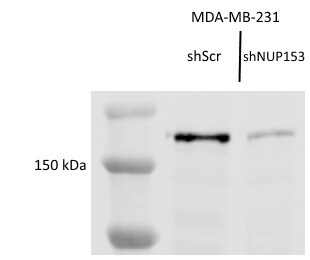
Western Blot: Nup153 Antibody [NB100-93329] - Knockdown of NUP153 in MDA-MB-231 breast cancer cells. Lane 1: ladder. Lane 2: Scramble Control Lane 3: NUP153 knockdown. Substantially reduced protein detection in knockdown sample and not in scramble control reflect antibody's unique specificity for Nup153. Image submitted by a verified customer review.![Immunocytochemistry/ Immunofluorescence: Nup153 Antibody [NB100-93329]](https://resources.rndsystems.com/images/products/Nup153-Antibody-Immunocytochemistry-Immunofluorescence-NB100-93329-img0003.jpg "Immunocytochemistry/ Immunofluorescence: Nup153 Antibody [NB100-93329]")
Immunocytochemistry/ Immunofluorescence: Nup153 Antibody [NB100-93329]
Immunocytochemistry/Immunofluorescence: Nup153 Antibody [NB100-93329] - Formaldehyde-fixed asynchronous HeLa cells. Antibody: Affinity purified rabbit anti-NUP153 used at a dilution of 1:200 (1ug/ml). Detection: Red-fluorescent goat anti-rabbit IgG highly cross adsorbed Antibody used at a dilution of 1:100.Applications for Nup153 Antibody
Application
Recommended Usage
Immunocytochemistry/ Immunofluorescence
1:100 to 1:500
Immunoprecipitation
2-5 ug/mg lysate
Western Blot
1:2000-1:10000
Application Notes
Formaldehyde fixation is recommended. Permeabilization with Triton-X 100 is recommended for formaldehyde-fixed cells. Nup153 antibody validated for WB from a verified customer review.
Reviewed Applications
Read 2 reviews rated 4.5 using NB100-93329 in the following applications:
Formulation, Preparation, and Storage
Purification
Immunogen affinity purified
Formulation
TBS and 0.1% BSA
Preservative
0.09% Sodium Azide
Concentration
0.2 mg/ml
Shipping
The product is shipped with polar packs. Upon receipt, store it immediately at the temperature recommended below.
Stability & Storage
Store at 4C. Do not freeze.
Background: Nup153
Alternate Names
CG4453, Dmel\CG4453, dmNup153, dNup153, HNUP153, N153, nuclear pore complex protein hnup153,153 kDa nucleoporin, nuclear pore complex protein Nup153, nucleoporin 153kD, nucleoporin 153kDa, Nucleoporin Nup153, Nup 153, NUP153, Nup153 Nucleoporin 153
Entrez Gene IDs
9972 (Human)
Gene Symbol
NUP153
UniProt
Additional Nup153 Products
Product Documents for Nup153 Antibody
Certificate of Analysis
To download a Certificate of Analysis, please enter a lot or batch number in the search box below.
Product Specific Notices for Nup153 Antibody
This product is for research use only and is not approved for use in humans or in clinical diagnosis. Primary Antibodies are guaranteed for 1 year from date of receipt.
Citations for Nup153 Antibody
 Powered by Bioz
Customer Reviews for Nup153 Antibody (2)
4.5 out of 5
2 Customer Ratings
Have you used Nup153 Antibody?
Submit a review and receive an Amazon gift card!
$25/€18/£15/$25CAN/¥2500 Yen for a review with an image
$10/€7/£6/$10CAN/¥1110 Yen for a review without an image
Submit a review
Customer Images
Showing
1
-
2 of
2 reviews
Showing All
Filter By:
-
Application: Western BlotSample Tested: MDA-MB-231 nuclear extractsSpecies: HumanVerified Customer | Posted 01/31/2018Knockdown of NUP153 in MDA-MB-231 breast cancer cells.Knockdown of NUP153 in MDA-MB-231 breast cancer cells. Lane 1: ladder. Lane 2: Scramble Control Lane 3: NUP153 knockdown. Substantially reduced protein detection in knockdown sample and not in scramble control reflect antibody's unique specificity for Nup153. This image was submitted by customer review.

-
Application: Western BlotSample Tested: See PMID 23791529Species: HumanVerified Customer | Posted 12/23/2014
There are no reviews that match your criteria.
Protocols
Find general support by application which include: protocols, troubleshooting, illustrated assays, videos and webinars.
- Appropriate Fixation of IHC/ICC Samples
- Cellular Response to Hypoxia Protocols
- ClariTSA™ Fluorophore Kits
- Detection & Visualization of Antibody Binding
- ICC Cell Smear Protocol for Suspension Cells
- ICC Immunocytochemistry Protocol Videos
- ICC for Adherent Cells
- Immunocytochemistry (ICC) Protocol
- Immunocytochemistry Troubleshooting
- Immunofluorescence of Organoids Embedded in Cultrex Basement Membrane Extract
- Immunohistochemistry (IHC) and Immunocytochemistry (ICC) Protocols
- Immunoprecipitation Protocol
- Preparing Samples for IHC/ICC Experiments
- Preventing Non-Specific Staining (Non-Specific Binding)
- Primary Antibody Selection & Optimization
- Protocol for VisUCyte™ HRP Polymer Detection Reagent
- Protocol for the Fluorescent ICC Staining of Cell Smears - Graphic
- Protocol for the Fluorescent ICC Staining of Cultured Cells on Coverslips - Graphic
- Protocol for the Preparation and Fluorescent ICC Staining of Cells on Coverslips
- Protocol for the Preparation and Fluorescent ICC Staining of Non-adherent Cells
- Protocol for the Preparation and Fluorescent ICC Staining of Stem Cells on Coverslips
- Protocol for the Preparation of a Cell Smear for Non-adherent Cell ICC - Graphic
- R&D Systems Quality Control Western Blot Protocol
- TUNEL and Active Caspase-3 Detection by IHC/ICC Protocol
- The Importance of IHC/ICC Controls
- Troubleshooting Guide: Western Blot Figures
- Western Blot Conditions
- Western Blot Protocol
- Western Blot Protocol for Cell Lysates
- Western Blot Troubleshooting
- Western Blot Troubleshooting Guide
- View all Protocols, Troubleshooting, Illustrated assays and Webinars
Loading...