Olig2 Antibody (064) - Azide and BSA Free
Novus Biologicals | Catalog # NBP2-89201
Recombinant Monoclonal Antibody
![Western Blot: Olig2 Antibody (064) [NBP2-89201]](https://resources.rndsystems.com/images/products/Olig2-Antibody-064-Western-Blot-NBP2-89201-img0003.jpg "Western Blot: Olig2 Antibody (064) [NBP2-89201]")
Key Product Details
Species Reactivity
Validated:
Human
Cited:
Mouse
Applications
Validated:
Immunohistochemistry, Immunohistochemistry-Paraffin, Western Blot, Flow Cytometry
Cited:
Immunohistochemistry-Paraffin, Immunocytochemistry/ Immunofluorescence
Label
Unconjugated
Antibody Source
Recombinant Monoclonal Rabbit IgG Clone # 064 expressed in HEK293
Format
Azide and BSA Free
Loading...
Product Specifications
Immunogen
This antibody was obtained from a rabbit immunized with purified, synthetic peptide corresponding to the N-terminus of the Human Olig2.
Clonality
Monoclonal
Host
Rabbit
Isotype
IgG
Description
This antibody can be stored at 2C-8C for one month without detectable loss of activity. Antibody products are stable for twelve months from date of receipt when stored at -20C to -80C. Avoid repeated freeze-thaw cycles.
Scientific Data Images for Olig2 Antibody (064) - Azide and BSA Free
Western Blot: Olig2 Antibody (064) [NBP2-89201]
Western Blot: Olig2 Antibody (064) [NBP2-89201] - A: Jurkat Whole Cell Lysate Lysates/proteins at 30 ug per lane. SecondaryGoat Anti-Rabbit IgG H&L (Dylight800) at 1/10000 dilution. Developed using the Odyssey technique. Performed under reducing conditions. Predicted band size:32 kDa Observed band size:37 kDa![Immunohistochemistry-Paraffin: Olig2 Antibody (064) [NBP2-89201]](https://resources.rndsystems.com/images/products/Olig2-Antibody-064-Immunohistochemistry-Paraffin-NBP2-89201-img0002.jpg "Immunohistochemistry-Paraffin: Olig2 Antibody (064) [NBP2-89201]")
Immunohistochemistry-Paraffin: Olig2 Antibody (064) [NBP2-89201]
Immunohistochemistry-Paraffin: Olig2 Antibody (064) [NBP2-89201] - Staining of human olig-2 in human brain with rabbit monoclonal antibody (1:200).![Immunohistochemistry-Paraffin: Olig2 Antibody (064) [NBP2-89201]](https://resources.rndsystems.com/images/products/Olig2-Antibody-064-Immunohistochemistry-Paraffin-NBP2-89201-img0001.jpg "Immunohistochemistry-Paraffin: Olig2 Antibody (064) [NBP2-89201]")
Immunohistochemistry-Paraffin: Olig2 Antibody (064) [NBP2-89201]
Immunohistochemistry-Paraffin: Olig2 Antibody (064) [NBP2-89201] - Staining of human olig-2 in human glioma with rabbit monoclonal antibody (1:200). [NBP2-89201] -")
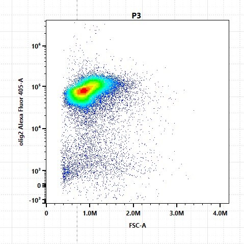
Flow Cytometry: Rabbit Monoclonal Olig2 Antibody (064) [NBP2-89201] -
Flow Cytometry: Rabbit Monoclonal Olig2 Antibody (064) [NBP2-89201] - Mouse brain cells fixed and stained for Olig2 protein: 1:100. AF405 conjugated version of antibody used (Catalog # NBP2-89201AF405). Image from a verified customer review. [NBP2-89201] -")
Flow Cytometry: Rabbit Monoclonal Olig2 Antibody (064) [NBP2-89201] -
Flow Cytometry: Rabbit Monoclonal Olig2 Antibody (064) [NBP2-89201] - Rat brain cells fixed and stained with Olig2. AF405 conjugated version of antibody used (Catalog # NBP2-89201AF405). Image from a verified customer review.Applications for Olig2 Antibody (064) - Azide and BSA Free
Application
Recommended Usage
Flow Cytometry
Validated for Flow Cytometry from a verified customer review.
Immunohistochemistry-Paraffin
1:100-1:500
Western Blot
1:500-1:2000
Reviewed Applications
Read 1 review rated 5 using NBP2-89201 in the following applications:
Flow Cytometry Panel Builder
Bio-Techne Knows Flow Cytometry
Save time and reduce costly mistakes by quickly finding compatible reagents using the Panel Builder Tool.
Advanced Features
- Spectra Viewer - Custom analysis of spectra from multiple fluorochromes
- Spillover Popups - Visualize the spectra of individual fluorochromes
- Antigen Density Selector - Match fluorochrome brightness with antigen density

Formulation, Preparation, and Storage
Purification
Protein A purified
Formulation
0.2 um filtered solution in PBS
Format
Azide and BSA Free
Preservative
No Preservative
Concentration
Please see the vial label for concentration. If unlisted please contact technical services.
Shipping
The product is shipped with polar packs. Upon receipt, store it immediately at the temperature recommended below.
Stability & Storage
Store at 4C short term. Aliquot and store at -20C long term. Avoid freeze-thaw cycles.
Background: Olig2
Long Name
Oligodendrocyte Lineage Transcription Factor 2
Alternate Names
BHLHB1, PRKCBP2, RACK17
Gene Symbol
OLIG2
Additional Olig2 Products
Product Documents for Olig2 Antibody (064) - Azide and BSA Free
Certificate of Analysis
To download a Certificate of Analysis, please enter a lot or batch number in the search box below.
Product Specific Notices for Olig2 Antibody (064) - Azide and BSA Free
This product is for research use only and is not approved for use in humans or in clinical diagnosis. Primary Antibodies are guaranteed for 1 year from date of receipt.
Citations for Olig2 Antibody (064) - Azide and BSA Free
 Powered by Bioz
Customer Reviews for Olig2 Antibody (064) - Azide and BSA Free (1)
5 out of 5
1 Customer Rating
Have you used Olig2 Antibody (064) - Azide and BSA Free?
Submit a review and receive an Amazon gift card!
$25/€18/£15/$25CAN/¥2500 Yen for a review with an image
$10/€7/£6/$10CAN/¥1110 Yen for a review without an image
Submit a review
Customer Images
Showing
1
-
1 of
1 review
Showing All
Filter By:
-
Application: Flow CytometrySample Tested: Adult mouse brainSpecies: MouseVerified Customer | Posted 04/02/2024mouse brain cells fixed and stained for olig2 protein : 1:100 NBP2-89201AF405 (Alexa fluor 405)
 Bio-Techne ResponseThis review was submitted through the legacy Novus Innovators Program, reflecting a new species or application tested on a primary antibody.
There are no reviews that match your criteria.
Protocols
Find general support by application which include: protocols, troubleshooting, illustrated assays, videos and webinars.
- 7-Amino Actinomycin D (7-AAD) Cell Viability Flow Cytometry Protocol
- Antigen Retrieval Protocol (PIER)
- Antigen Retrieval for Frozen Sections Protocol
- Appropriate Fixation of IHC/ICC Samples
- Cellular Response to Hypoxia Protocols
- Chromogenic IHC Staining of Formalin-Fixed Paraffin-Embedded (FFPE) Tissue Protocol
- Chromogenic Immunohistochemistry Staining of Frozen Tissue
- ClariTSA™ Fluorophore Kits
- Detection & Visualization of Antibody Binding
- Extracellular Membrane Flow Cytometry Protocol
- Flow Cytometry Protocol for Cell Surface Markers
- Flow Cytometry Protocol for Staining Membrane Associated Proteins
- Flow Cytometry Staining Protocols
- Flow Cytometry Troubleshooting Guide
- Fluorescent IHC Staining of Frozen Tissue Protocol
- Graphic Protocol for Heat-induced Epitope Retrieval
- Graphic Protocol for the Preparation and Fluorescent IHC Staining of Frozen Tissue Sections
- Graphic Protocol for the Preparation and Fluorescent IHC Staining of Paraffin-embedded Tissue Sections
- Graphic Protocol for the Preparation of Gelatin-coated Slides for Histological Tissue Sections
- IHC Sample Preparation (Frozen sections vs Paraffin)
- Immunofluorescent IHC Staining of Formalin-Fixed Paraffin-Embedded (FFPE) Tissue Protocol
- Immunohistochemistry (IHC) and Immunocytochemistry (ICC) Protocols
- Immunohistochemistry Frozen Troubleshooting
- Immunohistochemistry Paraffin Troubleshooting
- Intracellular Flow Cytometry Protocol Using Alcohol (Methanol)
- Intracellular Flow Cytometry Protocol Using Detergents
- Intracellular Nuclear Staining Flow Cytometry Protocol Using Detergents
- Intracellular Staining Flow Cytometry Protocol Using Alcohol Permeabilization
- Intracellular Staining Flow Cytometry Protocol Using Detergents to Permeabilize Cells
- Preparing Samples for IHC/ICC Experiments
- Preventing Non-Specific Staining (Non-Specific Binding)
- Primary Antibody Selection & Optimization
- Propidium Iodide Cell Viability Flow Cytometry Protocol
- Protocol for Heat-Induced Epitope Retrieval (HIER)
- Protocol for Liperfluo
- Protocol for Making a 4% Formaldehyde Solution in PBS
- Protocol for VisUCyte™ HRP Polymer Detection Reagent
- Protocol for the Characterization of Human Th22 Cells
- Protocol for the Characterization of Human Th9 Cells
- Protocol for the Preparation & Fixation of Cells on Coverslips
- Protocol for the Preparation and Chromogenic IHC Staining of Frozen Tissue Sections
- Protocol for the Preparation and Chromogenic IHC Staining of Frozen Tissue Sections - Graphic
- Protocol for the Preparation and Chromogenic IHC Staining of Paraffin-embedded Tissue Sections
- Protocol for the Preparation and Chromogenic IHC Staining of Paraffin-embedded Tissue Sections - Graphic
- Protocol for the Preparation and Fluorescent IHC Staining of Frozen Tissue Sections
- Protocol for the Preparation and Fluorescent IHC Staining of Paraffin-embedded Tissue Sections
- Protocol for the Preparation of Gelatin-coated Slides for Histological Tissue Sections
- Protocol: Annexin V and PI Staining by Flow Cytometry
- Protocol: Annexin V and PI Staining for Apoptosis by Flow Cytometry
- R&D Systems Quality Control Western Blot Protocol
- TUNEL and Active Caspase-3 Detection by IHC/ICC Protocol
- The Importance of IHC/ICC Controls
- Troubleshooting Guide: Fluorokine Flow Cytometry Kits
- Troubleshooting Guide: Immunohistochemistry
- Troubleshooting Guide: Western Blot Figures
- Western Blot Conditions
- Western Blot Protocol
- Western Blot Protocol for Cell Lysates
- Western Blot Troubleshooting
- Western Blot Troubleshooting Guide
- View all Protocols, Troubleshooting, Illustrated assays and Webinars
Loading...
Associated Pathways
