Oligodendrocytes are myelinating cells in the central nervous system (CNS) and form the myelin sheath of axons to support rapid nerve conduction. Oligodendrocyte Marker O4 is an antigen on the surface of oligodendrocyte progenitors (1, 2). It has been commonly used as the earliest recognized marker specific for the oligodendroglial lineage (3-8).
Oligodendrocyte Marker O4 APC-conjugated Antibody
R&D Systems | Catalog # FAB1326A

Key Product Details
Species Reactivity
Validated:
Human, Mouse, Rat, Chicken
Cited:
Mouse
Applications
Validated:
Flow Cytometry
Cited:
Flow Cytometry
Label
Allophycocyanin (Excitation = 620-650 nm, Emission = 660-670 nm)
Antibody Source
Monoclonal Mouse IgM Clone # O4
Loading...
Product Specifications
Immunogen
Bovine brain corpus callosum white matter
Specificity
Detects human, mouse, rat, and chicken Oligodendrocyte Marker O4.
Clonality
Monoclonal
Host
Mouse
Isotype
IgM
Scientific Data Images for Oligodendrocyte Marker O4 APC-conjugated Antibody
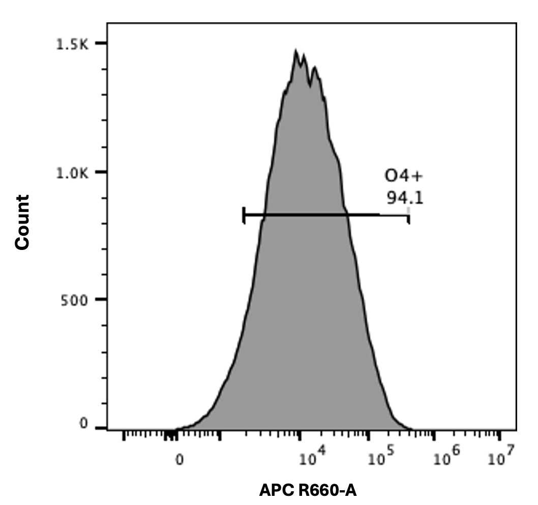
Detection of Oligodendrocyte Marker O4 in Differentiated Rat Cortical Stem Cells by Flow Cytometry.
Differentiated Rat Cortical Stem Cells (Catalog # NSC001) were stained with Mouse Anti-Human/Mouse/ Rat/Chicken Oligodendrocyte Marker O4 APC-conjugated Monoclonal Antibody (Catalog # FAB1326A, filled histogram) or isotype control antibody (Catalog # IC015A, open histogram). View our protocol for Staining Membrane-associated Proteins.Applications for Oligodendrocyte Marker O4 APC-conjugated Antibody
Application
Recommended Usage
Flow Cytometry
10 µL/106 cells
Sample: Differentiated Rat Cortical Stem Cells (Catalog # NSC001)
Sample: Differentiated Rat Cortical Stem Cells (Catalog # NSC001)
Reviewed Applications
Read 2 reviews rated 4.5 using FAB1326A in the following applications:
Spectra Viewer
Plan Your Experiments
Use our spectra viewer to interactively plan your experiments, assessing multiplexing options. View the excitation and emission spectra for our fluorescent dye range and other commonly used dyes.
Spectra Viewer
Flow Cytometry Panel Builder
Bio-Techne Knows Flow Cytometry
Save time and reduce costly mistakes by quickly finding compatible reagents using the Panel Builder Tool.
Advanced Features
- Spectra Viewer - Custom analysis of spectra from multiple fluorochromes
- Spillover Popups - Visualize the spectra of individual fluorochromes
- Antigen Density Selector - Match fluorochrome brightness with antigen density

Formulation, Preparation, and Storage
Purification
IgM-specific Affinity-purified from hybridoma culture supernatant
Formulation
Supplied in a saline solution containing BSA and Sodium Azide.
Shipping
The product is shipped with polar packs. Upon receipt, store it immediately at the temperature recommended below.
Stability & Storage
Protect from light. Do not freeze.
- 12 months from date of receipt, 2 to 8 °C as supplied.
Background: Oligodendrocyte Marker O4
References
- Schachner, M. et al. (1981) Dev. Biol. 83:328.
- Bansal, R. et al. (1989) J. Neurosci. Res. 24:548.
- Bansal, R. and Pfeiffer, S.E. (1989) Proc. Natl. Acad. Sci. USA 86:6181.
- Gard, A. et al. (1995) Dev. Biol. 167:596.
- Reynolds, R. and Hardy, R. (1997) J. Neurosci. Res. 47:455.
- Ono, K. et al. (1997) J. Neurosci. Res. 48:212.
- Pang, Y. et al. (2000) J. Neurosci. Res. 62:510.
- Cai, Z. et al. (2001) Brain Res. 898:126.
Additional Oligodendrocyte Marker O4 Products
Product Documents for Oligodendrocyte Marker O4 APC-conjugated Antibody
Certificate of Analysis
To download a Certificate of Analysis, please enter a lot or batch number in the search box below.
Note: Certificate of Analysis not available for kit components.
Product Specific Notices for Oligodendrocyte Marker O4 APC-conjugated Antibody
For research use only
Related Research Areas
Citations for Oligodendrocyte Marker O4 APC-conjugated Antibody
 Powered by Bioz
Customer Reviews for Oligodendrocyte Marker O4 APC-conjugated Antibody (2)
4.5 out of 5
2 Customer Ratings
Have you used Oligodendrocyte Marker O4 APC-conjugated Antibody?
Submit a review and receive an Amazon gift card!
$25/€18/£15/$25CAN/¥2500 Yen for a review with an image
$10/€7/£6/$10CAN/¥1110 Yen for a review without an image
Submit a review
Customer Images
Showing
1
-
2 of
2 reviews
Showing All
Filter By:
-
Application: Flow CytometrySample Tested: Brain tissueSpecies: HumanVerified Customer | Posted 08/26/2025

-
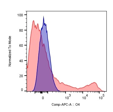
Application: Flow CytometrySample Tested: brain and spinal cordSpecies: MouseVerified Customer | Posted 02/07/2019CNS cells of EAE mice stained for O4. Viable non-stained cells (blue) and stained cells (red).Experimental autoimmune encephalomyelitis was induced in C57BL6/J mice, and cells from central nervous system (brain+spinal cord) were isolated when the animals were at the peak of the disease. Cells were stained for MOG, O4, GalC and AN2, plus for viability to exclude dead cells. MOG antibody was biotinylated, so cells were subsequently incubated with streptavidin-BV786 to stain MOG+ cells. Viable cells were gated and O4 staining is showed in non-stained cells (blue histogram) and in stained cells (red histogram).
There are no reviews that match your criteria.
Protocols
Find general support by application which include: protocols, troubleshooting, illustrated assays, videos and webinars.
- 7-Amino Actinomycin D (7-AAD) Cell Viability Flow Cytometry Protocol
- Extracellular Membrane Flow Cytometry Protocol
- Flow Cytometry Protocol for Cell Surface Markers
- Flow Cytometry Protocol for Staining Membrane Associated Proteins
- Flow Cytometry Staining Protocols
- Flow Cytometry Troubleshooting Guide
- Intracellular Flow Cytometry Protocol Using Alcohol (Methanol)
- Intracellular Flow Cytometry Protocol Using Detergents
- Intracellular Nuclear Staining Flow Cytometry Protocol Using Detergents
- Intracellular Staining Flow Cytometry Protocol Using Alcohol Permeabilization
- Intracellular Staining Flow Cytometry Protocol Using Detergents to Permeabilize Cells
- Propidium Iodide Cell Viability Flow Cytometry Protocol
- Protocol for Liperfluo
- Protocol for the Characterization of Human Th22 Cells
- Protocol for the Characterization of Human Th9 Cells
- Protocol: Annexin V and PI Staining by Flow Cytometry
- Protocol: Annexin V and PI Staining for Apoptosis by Flow Cytometry
- Troubleshooting Guide: Fluorokine Flow Cytometry Kits
- View all Protocols, Troubleshooting, Illustrated assays and Webinars
Loading...