Oligodendrocytes are myelinating cells in the central nervous system (CNS) and form the myelin sheath of axons to support rapid nerve conduction. Oligodendrocyte Marker O4 is an antigen on the surface of oligodendrocyte progenitors (1, 2). It has been commonly used as the earliest recognized marker specific for the oligodendroglial lineage (3-8).
Best Seller
Oligodendrocyte Marker O4 Antibody (O4)
R&D Systems | Catalog # MAB1326

Key Product Details
Species Reactivity
Validated:
Human, Mouse, Rat, Chicken
Cited:
Human, Mouse, Rat, Porcine, Avian - Chicken, Rabbit, Salmon, Transgenic Mouse
Applications
Validated:
Flow Cytometry, Immunocytochemistry, CyTOF-ready
Cited:
Immunohistochemistry, Immunohistochemistry-Paraffin, Immunohistochemistry-Frozen, Western Blot, Neutralization, Flow Cytometry, Immunofluorescence, Immunocytochemistry, Cell Culture, Immunopanning
Label
Unconjugated
Antibody Source
Monoclonal Mouse IgM Clone # O4
Loading...
Product Specifications
Immunogen
Bovine brain corpus callosum white matter
Specificity
Detects human, mouse, rat, and chicken Oligodendrocyte Marker O4.
Clonality
Monoclonal
Host
Mouse
Isotype
IgM
Scientific Data Images for Oligodendrocyte Marker O4 Antibody (O4)
Detection of Oligodendrocyte Marker O4 in Differentiated Rat Cortical Stem Cells by Flow Cytometry.
Rat cortical stem cells (Catalog # NSC001) either differentiated (filled histogram) or undifferentiated (open histogram) were stained with Mouse Anti-Human/Mouse/Rat/Chicken Oligodendrocyte Marker O4 Monoclonal Antibody (Catalog # MAB1326), followed by PE-conjugated Anti-Mouse IgM Secondary Antibody (Catalog # F0116). View our protocol for Staining Membrane-associated Proteins.
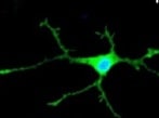
Oligodendrocyte Marker O4 in Differentiated Rat Cortical Stem Cells.
Oligodendrocyte Marker O4 was detected in immersion fixed differentiated rat cortical stem cells using 1 µg/mL Mouse Anti-Human/Mouse/ Rat/Chicken Oligodendrocyte Marker O4 Monoclonal Antibody (Catalog # MAB1326) for 3 hours at room temperature. Cells were stained (red). View our protocol for Fluorescent ICC Staining of Cells on Coverslips.
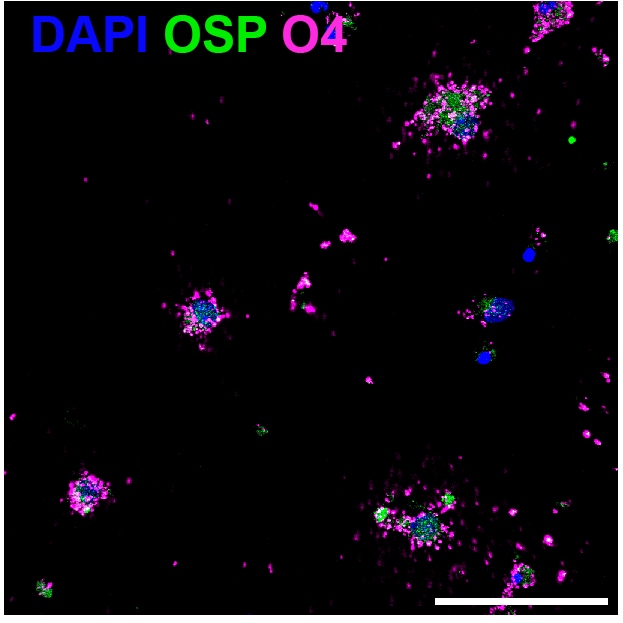
Olig2 and Oligodendrocyte Marker O4 in Rat Cortical Stem Cells.
Olig2 and Oligodendrocyte Marker O4 were detected in 7 day differentiated rat cortical stem cells using 10 µg/mL Human Olig2 Antigen Affinity-purified Polyclonal Antibody (Catalog # AF2418) and 10 µg/mL Mouse Anti-Human/Mouse/Rat/Chicken O4 Monoclonal Antibody (Catalog # MAB1326). Cells were incubated with primary antibodies for 3 hours at room temperature. Cells were stained for Olig2 using the NorthernLights™ 637-conjugated Anti-Goat IgG Secondary Antibody (red; Catalog # NL002), and stained for O4 using an anti-mouse IgM secondary antibody (pseudo-stained green). View our protocol for Fluorescent ICC Staining of Cells on Coverslips.
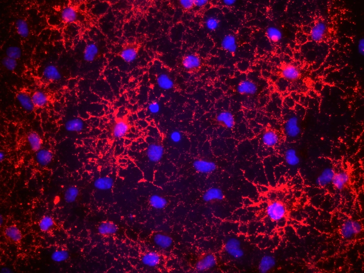
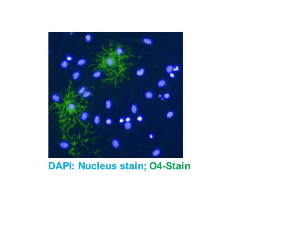
Oligodendrocyte Marker O4 in Rat Cortical Stem Cells.
Oligodendrocyte Marker O4 was detected in immersion fixed 7 day differentiated rat cortical stem cells using 1 µg/mL Mouse Anti-Human/Mouse/ Rat/Chicken Oligodendrocyte Marker O4 Monoclonal Antibody (Catalog # MAB1326) for 3 hours at room temperature. Cells were stained (red) and counterstained with DAPI (blue). View our protocol for Fluorescent ICC Staining of Cells on Coverslips.Applications for Oligodendrocyte Marker O4 Antibody (O4)
Application
Recommended Usage
CyTOF-ready
Ready to be labeled using established conjugation methods. No BSA or other carrier proteins that could interfere with conjugation.
Flow Cytometry
0.25 µg/106 cells
Sample: Differentiated rat cortical stem cells
Sample: Differentiated rat cortical stem cells
Immunocytochemistry
1-10 µg/mL
Sample: Immersion fixed 7 day differentiated rat cortical stem cells
Sample: Immersion fixed 7 day differentiated rat cortical stem cells
Reviewed Applications
Read 7 reviews rated 4.7 using MAB1326 in the following applications:
Flow Cytometry Panel Builder
Bio-Techne Knows Flow Cytometry
Save time and reduce costly mistakes by quickly finding compatible reagents using the Panel Builder Tool.
Advanced Features
- Spectra Viewer - Custom analysis of spectra from multiple fluorochromes
- Spillover Popups - Visualize the spectra of individual fluorochromes
- Antigen Density Selector - Match fluorochrome brightness with antigen density

Formulation, Preparation, and Storage
Purification
IgM-specific Affinity-purified from hybridoma culture supernatant
Reconstitution
Reconstitute at 0.5 mg/mL in sterile PBS. For liquid material, refer to CoA for concentration.
Loading...
Formulation
Lyophilized from a 0.2 μm filtered solution in PBS with Trehalose. *Small pack size (SP) is supplied either lyophilized or as a 0.2 µm filtered solution in PBS.
Shipping
Lyophilized product is shipped at ambient temperature. Liquid small pack size (-SP) is shipped with polar packs. Upon receipt, store immediately at the temperature recommended below.
Stability & Storage
Use a manual defrost freezer and avoid repeated freeze-thaw cycles.
- 12 months from date of receipt, -20 to -70 °C as supplied.
- 1 month, 2 to 8 °C under sterile conditions after reconstitution.
- 6 months, -20 to -70 °C under sterile conditions after reconstitution.
Calculators
Background: Oligodendrocyte Marker O4
References
- Schachner, M. et al. (1981) Dev. Biol. 83:328.
- Bansal, R. et al. (1989) J. Neurosci. Res. 24:548.
- Bansal, R. and Pfeiffer, S.E. (1989) Proc. Natl. Acad. Sci. USA 86:6181.
- Gard, A. et al. (1995) Dev. Biol. 167:596.
- Reynolds, R. and Hardy, R. (1997) J. Neurosci. Res. 47:455.
- Ono, K. et al. (1997) J. Neurosci. Res. 48:212.
- Pang, Y. et al. (2000) J. Neurosci. Res. 62:510.
- Cai, Z. et al. (2001) Brain Res. 898:126.
Additional Oligodendrocyte Marker O4 Products
Product Documents for Oligodendrocyte Marker O4 Antibody (O4)
Certificate of Analysis
To download a Certificate of Analysis, please enter a lot or batch number in the search box below.
Note: Certificate of Analysis not available for kit components.
Product Specific Notices for Oligodendrocyte Marker O4 Antibody (O4)
For research use only
Related Research Areas
Citations for Oligodendrocyte Marker O4 Antibody (O4)
 Powered by Bioz
Customer Reviews for Oligodendrocyte Marker O4 Antibody (O4) (7)
4.7 out of 5
7 Customer Ratings
Have you used Oligodendrocyte Marker O4 Antibody (O4)?
Submit a review and receive an Amazon gift card!
$25/€18/£15/$25CAN/¥2500 Yen for a review with an image
$10/€7/£6/$10CAN/¥1110 Yen for a review without an image
Submit a review
Customer Images
Showing
1
-
5 of
7 reviews
Showing All
Filter By:
-
Application: Immunocytochemistry/ImmunofluorescenceSample Tested: Brain (cerebral cortex)Species: RatVerified Customer | Posted 11/03/2023Rat cortical oligodendrocyte culture. Excelent staining, showing a punctate pattern. The O4 positive cells were also positive for the pan-oligodendrocyte marker OSP.

-
Application: Immunocytochemistry/ImmunofluorescenceSample Tested: Glial cellsSpecies: MouseVerified Customer | Posted 12/06/2021
-
Application: ImmunocytochemistrySample Tested: OligodendrocytesSpecies: RatVerified Customer | Posted 07/16/2019ICC without detergents
-
Application: Immunocytochemistry/ImmunofluorescenceSample Tested: Adult brainSpecies: MouseVerified Customer | Posted 04/16/2019
-
Application: Immunocytochemistry/ImmunofluorescenceSample Tested: IPS2 induced pluripotent stem cellsSpecies: MouseVerified Customer | Posted 01/25/2018
-
Application: Immunocytochemistry/ImmunofluorescenceSample Tested: Neural progenitor cellsSpecies: MouseVerified Customer | Posted 12/15/2016Our research focuses on how neural stem cells make the choice to differentiate into the various adult lineages such as neurons, astrocytes, and oligodendrocytes. We utilize the O4 oligodendrocyte marker as a tool to label differentiated oligodendrocytes and assess oligogenesis in various microenvironments.
-
Application: ImmunofluorescenceSample Tested: Primary oligodendrocyte culturesSpecies: MouseVerified Customer | Posted 12/19/2014
There are no reviews that match your criteria.
Protocols
Find general support by application which include: protocols, troubleshooting, illustrated assays, videos and webinars.
- 7-Amino Actinomycin D (7-AAD) Cell Viability Flow Cytometry Protocol
- Appropriate Fixation of IHC/ICC Samples
- Cellular Response to Hypoxia Protocols
- ClariTSA™ Fluorophore Kits
- Detection & Visualization of Antibody Binding
- Extracellular Membrane Flow Cytometry Protocol
- Flow Cytometry Protocol for Cell Surface Markers
- Flow Cytometry Protocol for Staining Membrane Associated Proteins
- Flow Cytometry Staining Protocols
- Flow Cytometry Troubleshooting Guide
- ICC Cell Smear Protocol for Suspension Cells
- ICC Immunocytochemistry Protocol Videos
- ICC for Adherent Cells
- Immunocytochemistry (ICC) Protocol
- Immunocytochemistry Troubleshooting
- Immunofluorescence of Organoids Embedded in Cultrex Basement Membrane Extract
- Immunohistochemistry (IHC) and Immunocytochemistry (ICC) Protocols
- Intracellular Flow Cytometry Protocol Using Alcohol (Methanol)
- Intracellular Flow Cytometry Protocol Using Detergents
- Intracellular Nuclear Staining Flow Cytometry Protocol Using Detergents
- Intracellular Staining Flow Cytometry Protocol Using Alcohol Permeabilization
- Intracellular Staining Flow Cytometry Protocol Using Detergents to Permeabilize Cells
- Preparing Samples for IHC/ICC Experiments
- Preventing Non-Specific Staining (Non-Specific Binding)
- Primary Antibody Selection & Optimization
- Propidium Iodide Cell Viability Flow Cytometry Protocol
- Protocol for Liperfluo
- Protocol for VisUCyte™ HRP Polymer Detection Reagent
- Protocol for the Characterization of Human Th22 Cells
- Protocol for the Characterization of Human Th9 Cells
- Protocol for the Fluorescent ICC Staining of Cell Smears - Graphic
- Protocol for the Fluorescent ICC Staining of Cultured Cells on Coverslips - Graphic
- Protocol for the Preparation and Fluorescent ICC Staining of Cells on Coverslips
- Protocol for the Preparation and Fluorescent ICC Staining of Non-adherent Cells
- Protocol for the Preparation and Fluorescent ICC Staining of Stem Cells on Coverslips
- Protocol for the Preparation of a Cell Smear for Non-adherent Cell ICC - Graphic
- Protocol: Annexin V and PI Staining by Flow Cytometry
- Protocol: Annexin V and PI Staining for Apoptosis by Flow Cytometry
- TUNEL and Active Caspase-3 Detection by IHC/ICC Protocol
- The Importance of IHC/ICC Controls
- Troubleshooting Guide: Fluorokine Flow Cytometry Kits
- View all Protocols, Troubleshooting, Illustrated assays and Webinars
FAQs for Oligodendrocyte Marker O4 Antibody (O4)
Showing
1
-
1 of
1 FAQ
Showing All
-
Q: What is the immunogen of Human/Mouse/Rat/Chicken Oligodendrocyte Marker O4 Antibody, Catalog # MAB1326?
A: The immunogen of Catalog # MAB1326 is bovine brain corpus callosum white matter. It is a natural source of Oligodendrocyte Marker O4, not a recombinant protein or synthetic peptide.
Loading...