OLLAS Epitope Tag Antibody (L2) - BSA Free
Novus Biologicals | Catalog # NBP1-06713
Key Product Details
Species Reactivity
Validated:
Cited:
Applications
Validated:
Cited:
Label
Antibody Source
Format
Product Specifications
Immunogen
Clonality
Host
Isotype
Scientific Data Images for OLLAS Epitope Tag Antibody (L2) - BSA Free
![Western Blot: OLLAS Epitope Tag Antibody (L2)BSA Free [NBP1-06713]](https://resources.rndsystems.com/images/products/OLLAS-Epitope-Tag-Antibody-L2-Western-Blot-NBP1-06713-img0009.jpg "Western Blot: OLLAS Epitope Tag Antibody (L2)BSA Free [NBP1-06713]")
Western Blot: OLLAS Epitope Tag Antibody (L2)BSA Free [NBP1-06713]
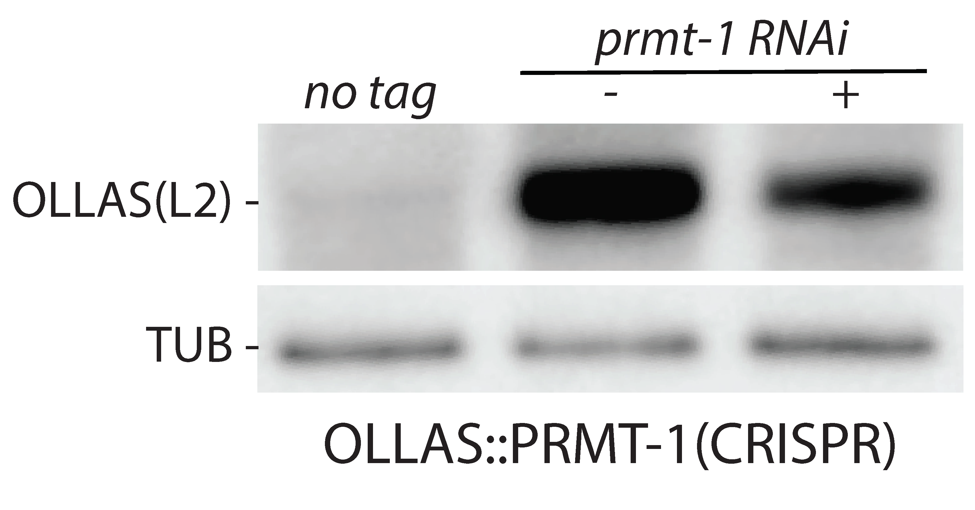
Western Blot: OLLAS Epitope Tag Antibody (L2) [NBP1-06713] - C. elegans whole nematode. OLLAS::PRMT-1 Western Blot using NBP1-06713SS (1:1000). Western blot image submitted by a verified customer review.![Immunocytochemistry/ Immunofluorescence: OLLAS Epitope Tag Antibody (L2) - BSA Free [NBP1-06713]](https://resources.rndsystems.com/images/products/OLLAS-Epitope-Tag-Antibody-L2-Immunocytochemistry-Immunofluorescence-NBP1-06713-img0006.jpg "Immunocytochemistry/ Immunofluorescence: OLLAS Epitope Tag Antibody (L2) - BSA Free [NBP1-06713]")
Immunocytochemistry/ Immunofluorescence: OLLAS Epitope Tag Antibody (L2) - BSA Free [NBP1-06713]
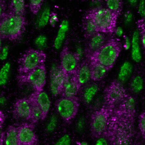
Immunocytochemistry/Immunofluorescence: OLLAS Epitope Tag Antibody (L2) [NBP1-06713] - Oncohistone incorporation patterns induce changes in nuclear PRC2 distribution and sterility phenotypes. Live cell imaging of GFP-tagged MES-2/EZH2 (the catalytic subunit of C. elegans PRC2), and immunofluorescence of H3K27me3 and H3.3/oncohistone (tagged with OLLAS Epitope Tag) in pachytene nuclei of H3-like K27M oncohistone (H3-like mut) worms. Scale bars represent 5 um. Chromosome X was identified by depletion of H3.3 and H3K4me3 staining shown in Supplementary Fig. 3, and is marked with an asterisk. Image collected and cropped by CiteAb from the following publication (https://www.nature.com/articles/s41467-019-10404-9) licensed under a CC-BY license Image using the Biotin format of this antibody.![Immunohistochemistry: OLLAS Epitope Tag Antibody (L2) - BSA Free [NBP1-06713]](https://resources.rndsystems.com/images/products/OLLAS-Epitope-Tag-Antibody-L2-BSA-Free-Immunohistochemistry-NBP1-06713-img0010.jpg "Immunohistochemistry: OLLAS Epitope Tag Antibody (L2) - BSA Free [NBP1-06713]")
Immunohistochemistry: OLLAS Epitope Tag Antibody (L2) - BSA Free [NBP1-06713]
OLLAS-Epitope-Tag-Antibody-L2-BSA-Free-Immunohistochemistry-NBP1-06713-img0010.jpg![Western Blot: OLLAS Epitope Tag Antibody (L2)BSA Free [NBP1-06713]](https://resources.rndsystems.com/images/products/OLLAS-Epitope-Tag-Antibody-L2-Western-Blot-NBP1-06713-img0001.jpg "Western Blot: OLLAS Epitope Tag Antibody (L2)BSA Free [NBP1-06713]")
Western Blot: OLLAS Epitope Tag Antibody (L2)BSA Free [NBP1-06713]
Western Blot: OLLAS Epitope Tag Antibody (L2) [NBP1-06713] - Comparison of binding sensitivity of Novus Biologicals' monoclonal antibodies to OLLAS (NBP1-06713), DYKDDDDK (NBP1-06712) and the FLAG-M2 monoclonal antibody from Sigma-Aldrich. FLAG (TM) and ANTI-FLAG (TM) are registered trademarks of Sigma-Aldrich Biotechnology LP and Sigma-Aldrich Co.![Western Blot: OLLAS Epitope Tag Antibody (L2)BSA Free [NBP1-06713]](https://resources.rndsystems.com/images/products/OLLAS-Epitope-Tag-Antibody-L2-Western-Blot-NBP1-06713-img0003.jpg "Western Blot: OLLAS Epitope Tag Antibody (L2)BSA Free [NBP1-06713]")
Western Blot: OLLAS Epitope Tag Antibody (L2)BSA Free [NBP1-06713]
Western Blot: OLLAS Epitope Tag Antibody (L2) [NBP1-06713] - OLLAS tagged SFO-p41 protein.![Immunohistochemistry Whole-Mount: OLLAS Epitope Tag Antibody (L2) - BSA Free [NBP1-06713]](https://resources.rndsystems.com/images/products/OLLAS-Epitope-Tag-Antibody-L2-Immunohistochemistry-Whole-Mount-NBP1-06713-img0005.jpg "Immunohistochemistry Whole-Mount: OLLAS Epitope Tag Antibody (L2) - BSA Free [NBP1-06713]")
Immunohistochemistry Whole-Mount: OLLAS Epitope Tag Antibody (L2) - BSA Free [NBP1-06713]
Immunohistochemistry Whole-Mount: OLLAS Epitope Tag Antibody (L2) [NBP1-06713] - Drosophila adult midgut expressing an OLLAS-tagged protein stained with OLLAS Epitope Tag Antibody (magenta). Nuclei are shown in green. IHC image submitted by a verified customer review.![Immunohistochemistry-Frozen: OLLAS Epitope Tag Antibody (L2) - BSA Free [NBP1-06713]](https://resources.rndsystems.com/images/products/OLLAS-Epitope-Tag-Antibody-L2-Immunohistochemistry-Frozen-NBP1-06713-img0004.jpg "Immunohistochemistry-Frozen: OLLAS Epitope Tag Antibody (L2) - BSA Free [NBP1-06713]")
Immunohistochemistry-Frozen: OLLAS Epitope Tag Antibody (L2) - BSA Free [NBP1-06713]
Immunohistochemistry-Frozen: OLLAS Epitope Tag Antibody (L2) [NBP1-06713] - Vector Map of pCMV-SD OLLAS![Immunohistochemistry-Paraffin: OLLAS Epitope Tag Antibody (L2) - BSA Free [NBP1-06713]](https://resources.rndsystems.com/images/products/OLLAS-Epitope-Tag-Antibody-L2-Immunohistochemistry-Paraffin-NBP1-06713-img0008.jpg "Immunohistochemistry-Paraffin: OLLAS Epitope Tag Antibody (L2) - BSA Free [NBP1-06713]")
Immunohistochemistry-Paraffin: OLLAS Epitope Tag Antibody (L2) - BSA Free [NBP1-06713]
OLLAS-Epitope-Tag-Antibody-L2-Immunohistochemistry-Paraffin-NBP1-06713-img0008.jpg![Immunohistochemistry-Paraffin: OLLAS Epitope Tag Antibody (L2) - BSA Free [NBP1-06713]](https://resources.rndsystems.com/images/products/OLLAS-Epitope-Tag-Antibody-L2-Immunohistochemistry-Paraffin-NBP1-06713-img0007.jpg "Immunohistochemistry-Paraffin: OLLAS Epitope Tag Antibody (L2) - BSA Free [NBP1-06713]")
Immunohistochemistry-Paraffin: OLLAS Epitope Tag Antibody (L2) - BSA Free [NBP1-06713]
OLLAS-Epitope-Tag-Antibody-L2-Immunohistochemistry-Paraffin-NBP1-06713-img0007.jpg - BSA Free [NBP1-06713] -")
Western Blot: OLLAS Epitope Tag Antibody (L2) - BSA Free [NBP1-06713] -
Western Blot: OLLAS Epitope Tag Antibody (L2) - BSA Free [NBP1-06713] - Utilization of pMVP for the creation of unique PDX1 vectors. pENTR plasmids containing cDNA for human PDX1 with or without a stop codon (i.e. open) were recombined with pMVP components to generate an assortment of PDX1-expressing (A) adenovirus & (C) expression plasmid vectors. (B) Immunoblot blot analysis of INS1 832/13 cell lysates harvested 48h after treatment with crude adenovirus lysates. For the epitope tagged conditions, note the appearance of the endogenous (lower) & overexpressed (upper) PDX1 bands (top blot). For eGFP blot, the use of P2A adds 23 amino acids to the N-terminal protein (i.e. eGFP). Immunoblot analysis of HEK293 cell lysates 24 h after transfection of expression vectors encoding PDX1 with C-terminal (D) epitope tags, (E) eGFP reporter or fusion, (G) & mCherry reporter or fusion. (F, H) Fluorescence microscopy of the eGFP & mCherry containing conditions analyzed in panels E & G. Visible bands resulting from ‘Uncleaved’ P2A products (red circle •) & degradation products (*) are labeled. Image collected & cropped by CiteAb from the following publication (https://pubmed.ncbi.nlm.nih.gov/30590691), licensed under a CC-BY license. Not internally tested by Novus Biologicals. - BSA Free [NBP1-06713] -")
Immunocytochemistry/ Immunofluorescence: OLLAS Epitope Tag Antibody (L2) - BSA Free [NBP1-06713] -
MES proteins are expressed in nos-1nos-2 embryonic PGCs.Transcriptome comparison between PGCs isolated from wild-type and mes-2(RNAi) L1 larvae. (A) Volcano plot showing log2 fold change of gene expression between mes-2(RNAi) and wild-type L1 PGCs. The numbers of genes whose expression were up or downregulated in mes-2(RNAi) compared to wild-type L1 PGCs are indicated. Dashed lines mark the significance cutoff of q = 0.05 above which genes were counted as misexpressed. (B) Bar graph showing chromosomal distribution of mes-2(RNAi) upregulated genes. Asterisks indicate significantly more genes than expected (hypergeometric test, p-value<0.001 [**]). (C) Top: Photomicrograph of live embryo expressing GFP tagged MES-2 in wild- type and nos-1(gv5)nos-2(ax3103) embryos. Middle: Photomicrograph of fixed wild-type and nos-1(gv5)nos-2(ax3103) embryos expressing OLLAS tagged MES-3. Bottom: Photomicrograph of fixed wild-type and nos-1(gv5)nos-2(RNAi) embryos stained with alpha -MES-4 antibody and K76 alpha -PGL-1 antibody. Images of 2-fold+ stage embryos were taken. Image collected and cropped by CiteAb from the following open publication (https://pubmed.ncbi.nlm.nih.gov/29111977), licensed under a CC-BY license. Not internally tested by Novus Biologicals. - BSA Free [NBP1-06713] -")
Immunocytochemistry/ Immunofluorescence: OLLAS Epitope Tag Antibody (L2) - BSA Free [NBP1-06713] -
Comparison of localization of the CD19::Nrx, nSyb::CD19 and CD19::sdc ligands into presynaptic sites in the ORNs targeting DA1.Different intracellular and transmembrane domains (from Nrx, nSyb, and sdc) were fused to CD19 and expressed into ORNs targeting the DA1 glomerulus using the CD19::Nrx (top panels), nSyb::CD19 (middle panels) and CD19::Sdc (bottom panels), with the R17H02 driver (in green). The brain samples were co-immunostained with antibodies against the OLLAS tag (present in the ligand) and against the pre-synaptic protein, BRP (in magenta). The ligand proteins are co-localized with or adjacent to BRP (arrows), demonstrating that all ligands are enriched at the presynaptic terminals of the ORNs, but CD19::Nrx was also expressed at strong levels in the axon shaft outside of the glomerulus (arrowheads in top panels). Scale bar = 20 μm. Image collected and cropped by CiteAb from the following open publication (https://pubmed.ncbi.nlm.nih.gov/29231171), licensed under a CC-BY license. Not internally tested by Novus Biologicals.Applications for OLLAS Epitope Tag Antibody (L2) - BSA Free
Chromatin Immunoprecipitation Sequencing
Immunoblotting
Immunocytochemistry/ Immunofluorescence
Immunohistochemistry
Immunohistochemistry-Frozen
Immunoprecipitation
Western Blot
Reviewed Applications
Read 2 reviews rated 4.5 using NBP1-06713 in the following applications:
Formulation, Preparation, and Storage
Purification
Formulation
Format
Preservative
Concentration
Shipping
Stability & Storage
Background: OLLAS Epitope Tag
Alternate Names
Additional OLLAS Epitope Tag Products
Product Documents for OLLAS Epitope Tag Antibody (L2) - BSA Free
Certificate of Analysis
To download a Certificate of Analysis, please enter a lot or batch number in the search box below.
Product Specific Notices for OLLAS Epitope Tag Antibody (L2) - BSA Free
This product is for research use only and is not approved for use in humans or in clinical diagnosis. Primary Antibodies are guaranteed for 1 year from date of receipt.
Citations for OLLAS Epitope Tag Antibody (L2) - BSA Free
 Powered by Bioz
Customer Reviews for OLLAS Epitope Tag Antibody (L2) - BSA Free (2)
Have you used OLLAS Epitope Tag Antibody (L2) - BSA Free?
Submit a review and receive an Amazon gift card!
$25/€18/£15/$25CAN/¥2500 Yen for a review with an image
$10/€7/£6/$10CAN/¥1110 Yen for a review without an image
Submit a review
Customer Images
-
Application: Western BlotSample Tested: whole C. elegans nematodeSpecies: C. elegansVerified Customer | Posted 06/26/2020OLLAS::PRMT-1 Western Blot using NBP1-06713SS (1:1000)

-
Application: Immunohistochemistry-Whole mountSample Tested: Drosophila adult midgutSpecies: DrosophilaVerified Customer | Posted 12/12/2016A Drosophila adult midgut expressing an OLLAS-tagged protein is stained with the anti-OLLAS antibody (magenta). Nuclei are shown in green.
There are no reviews that match your criteria.
Protocols
View specific protocols for OLLAS Epitope Tag Antibody (L2) - BSA Free (NBP1-06713):
Western Blot Protocol
1. Perform SDS-PAGE (4-12% MOPS) on samples to be analyzed, loading 5 ug of total protein per lane.
2. Transfer proteins to Nitrocellulose according to the instructions provided by the manufacturer of the transfer apparatus.
3. Rinse membrane with dH2O and then stain the blot using Ponceau S for 1-2 minutes to access the transfer of proteins onto the nitrocellulose membrane. Rinse the blot in water to remove excess stain and mark the lane locations and locations of molecular weight markers using a pencil.
4. Rinse the blot in TBS for approximately 5 minutes.
5. Block the membrane using 5% NFDM + 1% BSA in TBS + Tween, 1 hour at RT.
6. Rinse the membrane in dH2O and then wash the membrane in wash buffer [TBS + 0.1% Tween] 3 times for 10 minutes each.
7. Dilute the primary antibody (NBP1-06713) in blocking buffer and incubate 1 hour at room temperature.
8. Rinse the membrane in dH2O and then wash the membrane in wash buffer [TBS + 0.1% Tween] 3 times for 10 minutes each.
9. Dilute the appropriate secondary antibody in blocking buffer (as per manufacturers instructions) and incubate 1 hour at room temperature.
10. Wash the blot in wash buffer [TBS + 0.1% Tween] 3 times for 10 minutes each (this step can be repeated as required to reduce background).
11. Apply the detection reagent of choice in accordance with the manufacturers instructions (Pierce ECL).
Note: Tween-20 can be added to the blocking or antibody dilution buffer at a final concentration of 0.05-0.2%, provided it does not interfere with antibody-antigen binding.
Find general support by application which include: protocols, troubleshooting, illustrated assays, videos and webinars.
- Antigen Retrieval Protocol (PIER)
- Antigen Retrieval for Frozen Sections Protocol
- Appropriate Fixation of IHC/ICC Samples
- Cellular Response to Hypoxia Protocols
- Chromogenic IHC Staining of Formalin-Fixed Paraffin-Embedded (FFPE) Tissue Protocol
- Chromogenic Immunohistochemistry Staining of Frozen Tissue
- ClariTSA™ Fluorophore Kits
- Detection & Visualization of Antibody Binding
- Fluorescent IHC Staining of Frozen Tissue Protocol
- Graphic Protocol for Heat-induced Epitope Retrieval
- Graphic Protocol for the Preparation and Fluorescent IHC Staining of Frozen Tissue Sections
- Graphic Protocol for the Preparation and Fluorescent IHC Staining of Paraffin-embedded Tissue Sections
- Graphic Protocol for the Preparation of Gelatin-coated Slides for Histological Tissue Sections
- ICC Cell Smear Protocol for Suspension Cells
- ICC Immunocytochemistry Protocol Videos
- ICC for Adherent Cells
- IHC Sample Preparation (Frozen sections vs Paraffin)
- Immunocytochemistry (ICC) Protocol
- Immunocytochemistry Troubleshooting
- Immunofluorescence of Organoids Embedded in Cultrex Basement Membrane Extract
- Immunofluorescent IHC Staining of Formalin-Fixed Paraffin-Embedded (FFPE) Tissue Protocol
- Immunohistochemistry (IHC) and Immunocytochemistry (ICC) Protocols
- Immunohistochemistry Frozen Troubleshooting
- Immunohistochemistry Paraffin Troubleshooting
- Immunoprecipitation Protocol
- Preparing Samples for IHC/ICC Experiments
- Preventing Non-Specific Staining (Non-Specific Binding)
- Primary Antibody Selection & Optimization
- Protocol for Heat-Induced Epitope Retrieval (HIER)
- Protocol for Making a 4% Formaldehyde Solution in PBS
- Protocol for VisUCyte™ HRP Polymer Detection Reagent
- Protocol for the Fluorescent ICC Staining of Cell Smears - Graphic
- Protocol for the Fluorescent ICC Staining of Cultured Cells on Coverslips - Graphic
- Protocol for the Preparation & Fixation of Cells on Coverslips
- Protocol for the Preparation and Chromogenic IHC Staining of Frozen Tissue Sections
- Protocol for the Preparation and Chromogenic IHC Staining of Frozen Tissue Sections - Graphic
- Protocol for the Preparation and Chromogenic IHC Staining of Paraffin-embedded Tissue Sections
- Protocol for the Preparation and Chromogenic IHC Staining of Paraffin-embedded Tissue Sections - Graphic
- Protocol for the Preparation and Fluorescent ICC Staining of Cells on Coverslips
- Protocol for the Preparation and Fluorescent ICC Staining of Non-adherent Cells
- Protocol for the Preparation and Fluorescent ICC Staining of Stem Cells on Coverslips
- Protocol for the Preparation and Fluorescent IHC Staining of Frozen Tissue Sections
- Protocol for the Preparation and Fluorescent IHC Staining of Paraffin-embedded Tissue Sections
- Protocol for the Preparation of Gelatin-coated Slides for Histological Tissue Sections
- Protocol for the Preparation of a Cell Smear for Non-adherent Cell ICC - Graphic
- R&D Systems Quality Control Western Blot Protocol
- TUNEL and Active Caspase-3 Detection by IHC/ICC Protocol
- The Importance of IHC/ICC Controls
- Troubleshooting Guide: Immunohistochemistry
- Troubleshooting Guide: Western Blot Figures
- Western Blot Conditions
- Western Blot Protocol
- Western Blot Protocol for Cell Lysates
- Western Blot Troubleshooting
- Western Blot Troubleshooting Guide
- View all Protocols, Troubleshooting, Illustrated assays and Webinars
FAQs for OLLAS Epitope Tag Antibody (L2) - BSA Free
-
Q: Can you confirm if you measure the concentration of NBP1-06713 (anti-OLLAS epitope tag)?
A: Unfortunately, we do not measure the concentration of our OLLAS tag antibody, as the product is provided unpurified.
-
Q: I am interested in tagging proteins from E. coli and am wondering if ompF cross-reactivity is a problem?
A: The OLLAS tag is not predicted to cross-react with any portion of ompF.
-
Q: Can you confirm if you measure the concentration of NBP1-06713 (anti-OLLAS epitope tag)?
A: Unfortunately, we do not measure the concentration of our OLLAS tag antibody, as the product is provided unpurified.
-
Q: I am interested in tagging proteins from E. coli and am wondering if ompF cross-reactivity is a problem?
A: The OLLAS tag is not predicted to cross-react with any portion of ompF.