OPA1 Antibody (1284B) - BSA Free
Novus Biologicals | Catalog # NBP2-59770
Recombinant Monoclonal Antibody
Key Product Details
Species Reactivity
Validated:
Human, Mouse, Rat
Cited:
Mouse, Rat
Applications
Validated:
Immunohistochemistry, Immunohistochemistry-Paraffin, Western Blot, Immunocytochemistry/ Immunofluorescence
Cited:
Western Blot, IF/IHC
Label
Unconjugated
Antibody Source
Recombinant Monoclonal Rabbit IgG Clone # 1284B expressed in HEK293
Format
BSA Free
Loading...
Product Specifications
Immunogen
A synthetic peptide made to an internal region within residues 500-600 of human OPA1. [Swiss-Prot# O60313]
Clonality
Monoclonal
Host
Rabbit
Isotype
IgG
Scientific Data Images for OPA1 Antibody (1284B) - BSA Free
![Western Blot: OPA1 Antibody (1284B)BSA Free [NBP2-59770]](https://resources.rndsystems.com/images/products/OPA1-Antibody-1284B-Western-Blot-NBP2-59770-img0001.jpg "Western Blot: OPA1 Antibody (1284B)BSA Free [NBP2-59770]")
Western Blot: OPA1 Antibody (1284B)BSA Free [NBP2-59770]
Western Blot: OPA1 Antibody (1284B) [NBP2-59770] - Total protein from Mouse Neuro2A cells, Human HeLa cells and Rat PC12 cells was separated on a 7.5% gel by SDS-PAGE, transferred to PVDF membrane and blocked in 5% non-fat milk in TBST. The membrane was probed with 1.0 ug/ml anti-OPA1 in blocking buffer, and detected with an anti-mouse HRP secondary antibody using chemiluminescence.![Immunocytochemistry/ Immunofluorescence: OPA1 Antibody (1284B) - BSA Free [NBP2-59770]](https://resources.rndsystems.com/images/products/OPA1-Antibody-1284B-Immunocytochemistry-Immunofluorescence-NBP2-59770-img0022.jpg "Immunocytochemistry/ Immunofluorescence: OPA1 Antibody (1284B) - BSA Free [NBP2-59770]")
Immunocytochemistry/ Immunofluorescence: OPA1 Antibody (1284B) - BSA Free [NBP2-59770]
Immunocytochemistry/Immunofluorescence: OPA1 Antibody (1284B) [NBP2-59770] - A431 cells were fixed in 4% paraformaldehyde for 10 minutes and permeabilized in 0.05% Triton X-100 in PBS for 5 minutes. The cells were incubated with OPA1 Antibody [1284B] conjugated to Biotin (NBP2-59770B) at 5 ug/ml for 1 hour at room temperature then detected with Streptavidin conjugated to DyLight 550. Nuclei were counterstained with DAPI (Blue). Cells were imaged using a 40X objective.![Immunohistochemistry-Paraffin: OPA1 Antibody (1284B) - BSA Free [NBP2-59770]](https://resources.rndsystems.com/images/products/OPA1-Antibody-1284B-Immunohistochemistry-Paraffin-NBP2-59770-img0021.jpg "Immunohistochemistry-Paraffin: OPA1 Antibody (1284B) - BSA Free [NBP2-59770]")
Immunohistochemistry-Paraffin: OPA1 Antibody (1284B) - BSA Free [NBP2-59770]
Immunohistochemistry-Paraffin: OPA1 Antibody (1284B) [NBP2-59770] - IHC analysis of a formalin fixed paraffin embedded (FFPE) tissue section of mouse testes using OPA1 antibody at 1:1000 dilution. The primary antibody bound to OPA1 antigens in the tissue section was deteced using an HRP labeled secondary antibody and DAB reagent. Nuclei of the cells were counterstained with hematoxylin. This OPA1 antibody generated a diffused cytoplasmic staining of OPA1 protein in the tubular epithelial cells, the spermatids/spermatocytes and Leydig's cells.![Immunocytochemistry/ Immunofluorescence: OPA1 Antibody (1284B) - BSA Free [NBP2-59770]](https://resources.rndsystems.com/images/products/OPA1-Antibody-1284B-Immunocytochemistry-Immunofluorescence-NBP2-59770-img0002.jpg "Immunocytochemistry/ Immunofluorescence: OPA1 Antibody (1284B) - BSA Free [NBP2-59770]")
Immunocytochemistry/ Immunofluorescence: OPA1 Antibody (1284B) - BSA Free [NBP2-59770]
Immunocytochemistry/Immunofluorescence: OPA1 Antibody (1284B) [NBP2-59770] - HeLa cells were fixed for 10 minutes using 10% formalin and then permeabilized for 5 minutes using 1X TBS + 0.5% Triton X-100. The cells were incubated with anti-OPA1 at 5 ug/ml overnight at 4C and detected with an anti-rabbit DyLight 488 (Green) at a 1:500 dilution. Alpha tubulin (DM1A) NB100-690 was used as a co-stain at a 1:1000 dilution and detected with an anti-mouse DyLight 550 (Red) at a 1:500 dilution. Nuclei were counterstained with DAPI (Blue). Cells were imaged using a 40X objective.Applications for OPA1 Antibody (1284B) - BSA Free
Application
Recommended Usage
Immunocytochemistry/ Immunofluorescence
5 ug/ml
Immunohistochemistry
1:1000
Immunohistochemistry-Paraffin
1:1000
Western Blot
2 ug/ml
Reviewed Applications
Read 1 review rated 4 using NBP2-59770 in the following applications:
Formulation, Preparation, and Storage
Purification
Protein A or G purified
Formulation
PBS
Format
BSA Free
Preservative
0.02% Sodium Azide
Concentration
1.0 mg/ml
Shipping
The product is shipped with polar packs. Upon receipt, store it immediately at the temperature recommended below.
Stability & Storage
Store at 4C short term. Aliquot and store at -20C long term. Avoid freeze-thaw cycles.
Background: OPA1
Long Name
Optic Atrophy Protein 1
Alternate Names
BERHS, EC 3.6.5.5, LargeG, lilr3, MGM1, MTDPS14, NPG, NTG
Gene Symbol
OPA1
Additional OPA1 Products
Product Documents for OPA1 Antibody (1284B) - BSA Free
Certificate of Analysis
To download a Certificate of Analysis, please enter a lot or batch number in the search box below.
Product Specific Notices for OPA1 Antibody (1284B) - BSA Free
This product is for research use only and is not approved for use in humans or in clinical diagnosis. Primary Antibodies are guaranteed for 1 year from date of receipt.
Citations for OPA1 Antibody (1284B) - BSA Free
 Powered by Bioz
Customer Reviews for OPA1 Antibody (1284B) - BSA Free (1)
4 out of 5
1 Customer Rating
Have you used OPA1 Antibody (1284B) - BSA Free?
Submit a review and receive an Amazon gift card!
$25/€18/£15/$25CAN/¥2500 Yen for a review with an image
$10/€7/£6/$10CAN/¥1110 Yen for a review without an image
Submit a review
Customer Images
Showing
1
-
1 of
1 review
Showing All
Filter By:
-
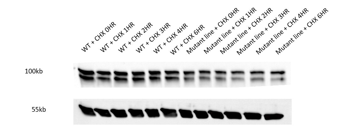
Application: Western BlotSample Tested: fibroblastsSpecies: HumanVerified Customer | Posted 02/01/2021Cyclohexamide chase of WT and Mutant fibroblast lines. OPA1 bands were seen at 120 and 80kDa. BIII loading control was used.SDS-PAGE run on a 8% Bis-Tris gel. Run in a biorad cassette system Loading gel - 100V then 120V to completion. Transfer 90minutes at 100V Blocked for 1 hour at RT with 5% Milk in PBS-T Primary: Stained overnight at 4oC with 1 in 1,000 OPA1 ab with 5% Milk in PBS-T Secondary: Rabbit anti-Goat. 1 in 30,000. Stained at RT for 1 hr with 5% Milk in PBS-T Loading control: 1 in 10,000 B-tubulin (Goat). Imaged using BioRad

There are no reviews that match your criteria.
Protocols
Find general support by application which include: protocols, troubleshooting, illustrated assays, videos and webinars.
- Antigen Retrieval Protocol (PIER)
- Antigen Retrieval for Frozen Sections Protocol
- Appropriate Fixation of IHC/ICC Samples
- Cellular Response to Hypoxia Protocols
- Chromogenic IHC Staining of Formalin-Fixed Paraffin-Embedded (FFPE) Tissue Protocol
- Chromogenic Immunohistochemistry Staining of Frozen Tissue
- ClariTSA™ Fluorophore Kits
- Detection & Visualization of Antibody Binding
- Fluorescent IHC Staining of Frozen Tissue Protocol
- Graphic Protocol for Heat-induced Epitope Retrieval
- Graphic Protocol for the Preparation and Fluorescent IHC Staining of Frozen Tissue Sections
- Graphic Protocol for the Preparation and Fluorescent IHC Staining of Paraffin-embedded Tissue Sections
- Graphic Protocol for the Preparation of Gelatin-coated Slides for Histological Tissue Sections
- ICC Cell Smear Protocol for Suspension Cells
- ICC Immunocytochemistry Protocol Videos
- ICC for Adherent Cells
- IHC Sample Preparation (Frozen sections vs Paraffin)
- Immunocytochemistry (ICC) Protocol
- Immunocytochemistry Troubleshooting
- Immunofluorescence of Organoids Embedded in Cultrex Basement Membrane Extract
- Immunofluorescent IHC Staining of Formalin-Fixed Paraffin-Embedded (FFPE) Tissue Protocol
- Immunohistochemistry (IHC) and Immunocytochemistry (ICC) Protocols
- Immunohistochemistry Frozen Troubleshooting
- Immunohistochemistry Paraffin Troubleshooting
- Preparing Samples for IHC/ICC Experiments
- Preventing Non-Specific Staining (Non-Specific Binding)
- Primary Antibody Selection & Optimization
- Protocol for Heat-Induced Epitope Retrieval (HIER)
- Protocol for Making a 4% Formaldehyde Solution in PBS
- Protocol for VisUCyte™ HRP Polymer Detection Reagent
- Protocol for the Fluorescent ICC Staining of Cell Smears - Graphic
- Protocol for the Fluorescent ICC Staining of Cultured Cells on Coverslips - Graphic
- Protocol for the Preparation & Fixation of Cells on Coverslips
- Protocol for the Preparation and Chromogenic IHC Staining of Frozen Tissue Sections
- Protocol for the Preparation and Chromogenic IHC Staining of Frozen Tissue Sections - Graphic
- Protocol for the Preparation and Chromogenic IHC Staining of Paraffin-embedded Tissue Sections
- Protocol for the Preparation and Chromogenic IHC Staining of Paraffin-embedded Tissue Sections - Graphic
- Protocol for the Preparation and Fluorescent ICC Staining of Cells on Coverslips
- Protocol for the Preparation and Fluorescent ICC Staining of Non-adherent Cells
- Protocol for the Preparation and Fluorescent ICC Staining of Stem Cells on Coverslips
- Protocol for the Preparation and Fluorescent IHC Staining of Frozen Tissue Sections
- Protocol for the Preparation and Fluorescent IHC Staining of Paraffin-embedded Tissue Sections
- Protocol for the Preparation of Gelatin-coated Slides for Histological Tissue Sections
- Protocol for the Preparation of a Cell Smear for Non-adherent Cell ICC - Graphic
- R&D Systems Quality Control Western Blot Protocol
- TUNEL and Active Caspase-3 Detection by IHC/ICC Protocol
- The Importance of IHC/ICC Controls
- Troubleshooting Guide: Immunohistochemistry
- Troubleshooting Guide: Western Blot Figures
- Western Blot Conditions
- Western Blot Protocol
- Western Blot Protocol for Cell Lysates
- Western Blot Troubleshooting
- Western Blot Troubleshooting Guide
- View all Protocols, Troubleshooting, Illustrated assays and Webinars
Loading...