OPA1 Antibody - BSA Free
Novus Biologicals | Catalog # NB110-55290
![Simple Western: OPA1 AntibodyBSA Free [NB110-55290]](https://resources.rndsystems.com/images/products/OPA1-Antibody-Simple-Western-NB110-55290-img0007.jpg "Simple Western: OPA1 AntibodyBSA Free [NB110-55290]")
Key Product Details
Species Reactivity
Validated:
Human, Mouse, Rat, Porcine, Chicken, Zebrafish
Cited:
Human, Mouse, Rat, Porcine, Fish - Danio rerio (Zebrafish)
Predicted:
Primate (100%). Backed by our 100% Guarantee.
Applications
Validated:
Immunohistochemistry, Immunohistochemistry-Paraffin, Western Blot, Immunoblotting, Immunocytochemistry/ Immunofluorescence, Simple Western
Cited:
Immunohistochemistry-Paraffin, Western Blot, Immunocytochemistry/ Immunofluorescence, Simple Western, IF/IHC
Label
Unconjugated
Antibody Source
Polyclonal Rabbit IgG
Format
BSA Free
Loading...
Product Specifications
Immunogen
A synthetic peptide made to an internal region within residues 500-600 of human OPA1. [Swiss-Prot: O60313]
Reactivity Notes
Zebrafish reactivity reported in scientific literature (PMID: 23516612, 26365306).
Localization
Mitochondrion; mitochondrial inner membrane; single-pass membrane protein. Mitochondrion; mitochondrial intermembrane space.
Clonality
Polyclonal
Host
Rabbit
Isotype
IgG
Theoretical MW
111 kDa.
Disclaimer note: The observed molecular weight of the protein may vary from the listed predicted molecular weight due to post translational modifications, post translation cleavages, relative charges, and other experimental factors.
Disclaimer note: The observed molecular weight of the protein may vary from the listed predicted molecular weight due to post translational modifications, post translation cleavages, relative charges, and other experimental factors.
Scientific Data Images for OPA1 Antibody - BSA Free
Simple Western: OPA1 AntibodyBSA Free [NB110-55290]
Simple Western: OPA1 Antibody [NB110-55290] - Lane view shows a specific band for OPA1 in 0.5 mg/ml of MEF lysate. This experiment was performed under reducing conditions using the 12-230 kDa separation system.![Western Blot: OPA1 AntibodyBSA Free [NB110-55290]](https://resources.rndsystems.com/images/products/OPA1-Antibody-Western-Blot-NB110-55290-img0010.jpg "Western Blot: OPA1 AntibodyBSA Free [NB110-55290]")
Western Blot: OPA1 AntibodyBSA Free [NB110-55290]
OPA1-Antibody-Western-Blot-NB110-55290-img0010.jpg![Western Blot: OPA1 AntibodyBSA Free [NB110-55290]](https://resources.rndsystems.com/images/products/OPA1-Antibody-Western-Blot-NB110-55290-img0009.jpg "Western Blot: OPA1 AntibodyBSA Free [NB110-55290]")
Western Blot: OPA1 AntibodyBSA Free [NB110-55290]
Western Blot: OPA1 Antibody [NB110-55290] - Total protein from Human HeLa cells, Mouse MEF cells and Rat Brain was separated on a 7.5% gel by SDS-PAGE, transferred to PVDF membrane and blocked in 5% non-fat milk in TBST. The membrane was probed with 2.0 ug/ml anti-OPA1 in 1% blocking buffer. Precision Plus Protein All Blue molecular weight markers (BioRad) were detected with 1 ug/ml Anti-Blue Marker antibody (NBP2-33376). The protein ladder was detected with an anti-mouse HRP secondary antibody and OPA1 with an anti-rabbit HRP secondary antibody using chemiluminescence.![Immunohistochemistry: OPA1 Antibody - BSA Free [NB110-55290]](https://resources.rndsystems.com/images/products/OPA1-Antibody-Immunohistochemistry-NB110-55290-img0005.jpg "Immunohistochemistry: OPA1 Antibody - BSA Free [NB110-55290]")
Immunohistochemistry: OPA1 Antibody - BSA Free [NB110-55290]
Immunohistochemistry: OPA1 Antibody [NB110-55290] - Staining in prostatic smooth muscle and glandular epithelium. Human Prostate 40X magnification.![Western Blot: OPA1 AntibodyBSA Free [NB110-55290]](https://resources.rndsystems.com/images/products/OPA1-Antibody-Western-Blot-NB110-55290-img0006.jpg "Western Blot: OPA1 AntibodyBSA Free [NB110-55290]")
Western Blot: OPA1 AntibodyBSA Free [NB110-55290]
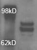
Western Blot: OPA1 Antibody [NB110-55290] - Detection of OPA1 protein in post-nuclear extracts of mouse embryonic fibroblasts.![Western Blot: OPA1 AntibodyBSA Free [NB110-55290]](https://resources.rndsystems.com/images/products/OPA1-Antibody-Western-Blot-NB110-55290-img0008.jpg "Western Blot: OPA1 AntibodyBSA Free [NB110-55290]")
Western Blot: OPA1 AntibodyBSA Free [NB110-55290]
Western Blot: OPA1 Antibody [NB110-55290] - Analysis of OPA1 in porcine retinal pigment epithelium lysate using anti-OPA1 antibody. Image from verified customer review.
Western Blot: OPA1 Antibody - BSA Free [NB110-55290] -
Western Blot: OPA1 Antibody - BSA Free [NB110-55290] - High content screening of the preadipocytes & beige cells, & detection of mitochondrial fission- & fusion-related proteins: (A) Representative high content screening images showing mitochondrial morphology in TG2+/+ & TG2−/− preadipocytes, scale bars represent 50 μm; (B) fractions of fragmented & tubular mitochondrial morphology in preadipocytes (%); (C) quantitative analysis of mitochondrial morphology in preadipocytes; (D) representative high content screening images showing mitochondrial morphology in TG2+/+ & TG2−/− beige cells; (E) fractions of fragmented & tubular mitochondrial morphology in beige cells (%); (F) quantitative analysis of mitochondrial morphology in beige cells, DAPI staining was used to determine the number of nuclei, the mitochondria were stained with Mito Tracker Deep Red either in the absence or presence of 10 μM antimycin A, Texas Red-X phalloidin was used to stain actin filaments for the detection of cell shapes (n = 3); (G–J) representative Western blot analyses & quantitative analyses of the mitochondrial fusion proteins (MFN2, OPA1) & mitochondrial fission proteins (DRP1, MFF) in preadipocytes & differentiated beige cells. beta -ACTIN was used as a loading control. Columns represent the mean values ± SD. Statistical analyses were performed using Student’s t-test. n = 5. * p < 0.05. Image collected & cropped by CiteAb from the following publication (https://pubmed.ncbi.nlm.nih.gov/35563567), licensed under a CC-BY license. Not internally tested by Novus Biologicals.Applications for OPA1 Antibody - BSA Free
Application
Recommended Usage
Immunoblotting
reported in scientific literature (PMID 25224038)
Immunocytochemistry/ Immunofluorescence
reported in scientific literature (PMID 34923139)
Immunohistochemistry
2.5 ug/ml
Immunohistochemistry-Paraffin
2.5 ug/ml
Simple Western
20 ug/ml
Western Blot
2 ug/ml
Application Notes
In Western blot, a band is seen at approx. 111 kDa. In Simple Western only 10 - 15 uL of the recommended dilution is used per data point.
See Simple Western Antibody Database for Simple Western validation: antibody dilution of 20 ug/mL
See Simple Western Antibody Database for Simple Western validation: antibody dilution of 20 ug/mL
Reviewed Applications
Read 2 reviews rated 5 using NB110-55290 in the following applications:
Formulation, Preparation, and Storage
Purification
Immunogen affinity purified
Formulation
PBS
Format
BSA Free
Preservative
0.02% Sodium Azide
Concentration
1 mg/ml
Shipping
The product is shipped with polar packs. Upon receipt, store it immediately at the temperature recommended below.
Stability & Storage
Aliquot and store at -20C or -80C. Avoid freeze-thaw cycles.
Background: OPA1
Long Name
Optic Atrophy Protein 1
Alternate Names
BERHS, EC 3.6.5.5, LargeG, lilr3, MGM1, MTDPS14, NPG, NTG
Gene Symbol
OPA1
Additional OPA1 Products
Product Documents for OPA1 Antibody - BSA Free
Certificate of Analysis
To download a Certificate of Analysis, please enter a lot or batch number in the search box below.
Product Specific Notices for OPA1 Antibody - BSA Free
This product is for research use only and is not approved for use in humans or in clinical diagnosis. Primary Antibodies are guaranteed for 1 year from date of receipt.
Citations for OPA1 Antibody - BSA Free
 Powered by Bioz
Customer Reviews for OPA1 Antibody - BSA Free (2)
5 out of 5
2 Customer Ratings
Have you used OPA1 Antibody - BSA Free?
Submit a review and receive an Amazon gift card!
$25/€18/£15/$25CAN/¥2500 Yen for a review with an image
$10/€7/£6/$10CAN/¥1110 Yen for a review without an image
Submit a review
Customer Images
Showing
1
-
2 of
2 reviews
Showing All
Filter By:
-
Application: Western BlotSample Tested: Retinal pigment epithelium lysateSpecies: OtherVerified Customer | Posted 06/04/2015Porcine OPA1

-
Application: Western BlotSample Tested: COS-1 cell, Sample Amount: 30 ugSpecies: OtherVerified Customer | Posted 01/19/2010
There are no reviews that match your criteria.
Protocols
View specific protocols for OPA1 Antibody - BSA Free (NB110-55290):
Western Blot Protocol
1. Perform SDS-PAGE (4-12%) on samples to be analyzed, loading 32 ug of total protein per lane.
2. Transfer proteins to Nitrocellulose according to the instructions provided by the manufacturer of the transfer
apparatus.
3. Rinse membrane with dH2O and then stain the blot using Ponceau S for 1-2 minutes to access the transfer of proteins onto the nitrocellulose membrane. Rinse the blot in water to remove excess stain and mark the lane locations and locations of molecular weight markers using a pencil.
4. Rinse the blot in TBS for approximately 5 minutes.
5. Block the membrane using 5% non-fat dry milk + 1% BSA in TBS, 1 hour at room temperature.
6. Rinse the membrane in dH2O and then wash the membrane in wash buffer [TBS + 0.1% Tween] 3 times for 10 minutes each.
7. Dilute the rabbit anti-Opa1 primary antibody (NB110-55290) in blocking buffer and incubate 2 hours at room temperature.
8. Rinse the membrane in dH2O and then wash the membrane in wash buffer [TBS + 0.1% Tween] 3 times for 10 minutes each.
9. Apply the diluted rabbit-IgG HRP-conjugated secondary antibody in blocking buffer (as per manufacturers
instructions) and incubate 1 hour at room temperature.
10. Wash the blot in wash buffer [TBS + 0.1% Tween] 3 times for 10 minutes each (this step can be repeated as required to reduce background).
11. Apply the detection reagent of choice in accordance with the manufacturers instructions (Pierce, ECL).
Note: Tween-20 can be added to the blocking or antibody diultion buffer at a final concentration of 0.05-0.2%, provided it does not interfere with antibody-antigen binding.
IHC-FFPE sections
I. Deparaffinization:
A. Treat slides with Xylene: 3 changes for 5 minutes each. Drain slides for 10 seconds between changes.
B. Treat slides with 100% Reagent Alcohol: 3 changes for 5 minutes each. Drain slides for 10 seconds between changes.
II. Quench Endogenous Peroxidase:
A.Place slides in peroxidase quenching solution: 15-30 minutes.
To Prepare 200 ml of Quenching Solution:
Add 3 ml of 30% Hydrogen Peroxide to 200 ml of Methanol.
Use within 4 hours of preparation
B. Place slides in distilled water: 2 changes for 2 minutes each.
III. Retrieve Epitopes:
A. Preheat Citrate Buffer. Place 200 ml of Citrate Buffer Working Solution into container, cover and place into steamer. Heat to 90-96 degrees Celcius.
B. Place rack of slides into hot Citrate Buffer for 20 minutes. Cover.
C.Carefully remove container with slides from steamer and cool on bench, uncovered, for 20 minutes.
D. Slowly add distilled water to further cool for 5 minutes.
E. Rinse slides with distilled water. 2 changes for 2 minutes each.
IV. Immunostaining Procedure:
A. Remove each slide from rack and circle tissue section with a hydrophobic barrier pen (e.g. Liquid Blocker-Super Pap-Pen).
B. Flood slide with Wash Solution. Do not allow tissue sections to dry for the rest of the procedure.
C. Drain wash solution and apply 4 drops of Blocking Reagent to each slide and incubate for 15 minutes.
D. Drain Blocking Reagent (do not wash off the Blocking Reagent), apply 200 ul of Primary Antibody solution to each slide, and incubate for 1 hour.
E. Wash slides with Wash Solution: 3 changes for 5 minutes each.
F. Drain wash solution, apply 4 drops of Secondary antibody to each slide and incubate for 1 hour.
G. Wash slides with Wash Solution: 3 changes for 5 minutes each.
H. Drain wash solution, apply 4 drops of DAB Substrate to each slide and develop for 5-10 minutes. Check development with microscope.
I. Wash slides with Wash Solution: 3 changes for 5 minutes each. Wash slides with Wash Solution: 3 changes for 5 minutes each
J. Drain wash solution, apply 4 drops of Hematoxylin to each slide and stain for 1-3 minutes. Increase time if darker counterstaining is desired.
K. Wash slides with Wash Solution: 2-3 changes for 2 minutes each.
L. Drain wash solution and apply 4 drops of Bluing Solution to each slide for 1-2 minutes.
M. Rinse slides in distilled water.
N. Soak slides in 70% reagent alcohol: 3 minutes with intermittent agitation.
O. Soak slides in 95% reagent alcohol: 2 changes for 3 minutes each with intermittent agitation.
P. Soak slides in 100% reagent alcohol: 3 changes for 3 minutes each with intermittent agitation. Drain slides for 10 seconds between each change.
Q. Soak slides in Xylene: 3 changes for 3 minutes each with intermittent agitation. Drain slides for 10 seconds between each change.
R. Apply 2-3 drops of non-aqueous mounting media to each slide and mount coverslip.
S. Lay slides on a flat surface to dry prior to viewing under microscope.
NOTES:
-Use treated slides (e.g. HistoBond) to assure adherence of FFPE sections to slide.
-Prior to deparaffinization, heat slides overnight in a 60 degrees Celcius oven.
-All steps in which Xylene is used should be performed in a fume hood.
-For Epitope Retrieval, a microwave or pressure cooker may be substituted for the steamer method. Adjust times as necessary depending on conditions.
-For the initial IHC run with a new primary antibody, test tissues with and without Epitope Retrieval. In some instances, Epitope Retrieval may not be necessary.
-200 ul is the recommended maximum volume to apply to a slide for full coverage. Using more than 200 ul may allow solutions to wick off the slide and create drying artifacts. For small tissue sections less than 200 ul may be used.
-5 minutes of development with DAB Substrate should be sufficient. Do not develop for more than 10 minutes. If 5 minutes of development causes background staining, further dilution of the primary antibody may be necessary.
-Hematoxylin should produce a light nuclear counterstain so as not to obscure the DAB staining. Counterstain for 1-1 1/2 minutes for nuclear antigens. Counterstain for 2-3 minutes for cytoplasmic and membranous antigens. If darker counterstaining is desired increase time (up to 10 minutes).
Find general support by application which include: protocols, troubleshooting, illustrated assays, videos and webinars.
- Antigen Retrieval Protocol (PIER)
- Antigen Retrieval for Frozen Sections Protocol
- Appropriate Fixation of IHC/ICC Samples
- Cellular Response to Hypoxia Protocols
- Chromogenic IHC Staining of Formalin-Fixed Paraffin-Embedded (FFPE) Tissue Protocol
- Chromogenic Immunohistochemistry Staining of Frozen Tissue
- ClariTSA™ Fluorophore Kits
- Detection & Visualization of Antibody Binding
- Fluorescent IHC Staining of Frozen Tissue Protocol
- Graphic Protocol for Heat-induced Epitope Retrieval
- Graphic Protocol for the Preparation and Fluorescent IHC Staining of Frozen Tissue Sections
- Graphic Protocol for the Preparation and Fluorescent IHC Staining of Paraffin-embedded Tissue Sections
- Graphic Protocol for the Preparation of Gelatin-coated Slides for Histological Tissue Sections
- ICC Cell Smear Protocol for Suspension Cells
- ICC Immunocytochemistry Protocol Videos
- ICC for Adherent Cells
- IHC Sample Preparation (Frozen sections vs Paraffin)
- Immunocytochemistry (ICC) Protocol
- Immunocytochemistry Troubleshooting
- Immunofluorescence of Organoids Embedded in Cultrex Basement Membrane Extract
- Immunofluorescent IHC Staining of Formalin-Fixed Paraffin-Embedded (FFPE) Tissue Protocol
- Immunohistochemistry (IHC) and Immunocytochemistry (ICC) Protocols
- Immunohistochemistry Frozen Troubleshooting
- Immunohistochemistry Paraffin Troubleshooting
- Preparing Samples for IHC/ICC Experiments
- Preventing Non-Specific Staining (Non-Specific Binding)
- Primary Antibody Selection & Optimization
- Protocol for Heat-Induced Epitope Retrieval (HIER)
- Protocol for Making a 4% Formaldehyde Solution in PBS
- Protocol for VisUCyte™ HRP Polymer Detection Reagent
- Protocol for the Fluorescent ICC Staining of Cell Smears - Graphic
- Protocol for the Fluorescent ICC Staining of Cultured Cells on Coverslips - Graphic
- Protocol for the Preparation & Fixation of Cells on Coverslips
- Protocol for the Preparation and Chromogenic IHC Staining of Frozen Tissue Sections
- Protocol for the Preparation and Chromogenic IHC Staining of Frozen Tissue Sections - Graphic
- Protocol for the Preparation and Chromogenic IHC Staining of Paraffin-embedded Tissue Sections
- Protocol for the Preparation and Chromogenic IHC Staining of Paraffin-embedded Tissue Sections - Graphic
- Protocol for the Preparation and Fluorescent ICC Staining of Cells on Coverslips
- Protocol for the Preparation and Fluorescent ICC Staining of Non-adherent Cells
- Protocol for the Preparation and Fluorescent ICC Staining of Stem Cells on Coverslips
- Protocol for the Preparation and Fluorescent IHC Staining of Frozen Tissue Sections
- Protocol for the Preparation and Fluorescent IHC Staining of Paraffin-embedded Tissue Sections
- Protocol for the Preparation of Gelatin-coated Slides for Histological Tissue Sections
- Protocol for the Preparation of a Cell Smear for Non-adherent Cell ICC - Graphic
- R&D Systems Quality Control Western Blot Protocol
- TUNEL and Active Caspase-3 Detection by IHC/ICC Protocol
- The Importance of IHC/ICC Controls
- Troubleshooting Guide: Immunohistochemistry
- Troubleshooting Guide: Western Blot Figures
- Western Blot Conditions
- Western Blot Protocol
- Western Blot Protocol for Cell Lysates
- Western Blot Troubleshooting
- Western Blot Troubleshooting Guide
- View all Protocols, Troubleshooting, Illustrated assays and Webinars
Loading...