p53 [p Ser392] Antibody (FP3.2 [FPS392]) - BSA Free
Novus Biologicals | Catalog # NB500-575
Key Product Details
Species Reactivity
Human
Applications
Immunohistochemistry, Immunohistochemistry-Paraffin, Western Blot
Label
Unconjugated
Antibody Source
Monoclonal Mouse IgG1 Clone # FP3.2 [FPS392]
Format
BSA Free
Loading...
Product Specifications
Immunogen
KLH-conjugated phosphopeptide RHKKLMFKTEGPDS[P]D, corresponding to amino acids 378-393 of human p53.
Modification
p Ser392
Specificity
The antibody FP3.2 [FPS392] reacts with human p53 tumour suppressor protein phosphorylated at CKII site (Ser 392).
Clonality
Monoclonal
Host
Mouse
Isotype
IgG1
Scientific Data Images for p53 [p Ser392] Antibody (FP3.2 [FPS392]) - BSA Free
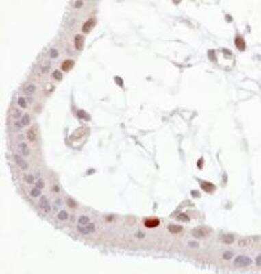
Immunohistochemistry: p53 [p Ser392] Antibody (FP3.2 [FPS392]) [NB500-575] - Wild-type p53 expressed in human trophoblast (paraffin-embedded sections). 1A - anti-p53 (total).
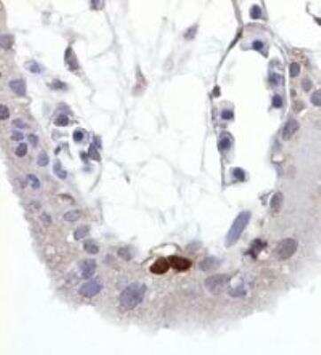
Immunohistochemistry: p53 [p Ser392] Antibody (FP3.2 [FPS392]) [NB500-575] - 1B anti-P53 (phospho Ser392) Note that some of total p53 positive nuclei are also FP3.2 (phospho p53) positive.
Applications for p53 [p Ser392] Antibody (FP3.2 [FPS392]) - BSA Free
Application
Recommended Usage
Immunohistochemistry
1:10-1:500
Immunohistochemistry-Paraffin
10 ug/ml
Western Blot
1 ug/ml
Application Notes
Immunohistochemistry-Paraffin sections - Staining technique: standard ABC technique (DAB+) Pretreatment: high temperature antigen retrieval (microwave, pressure cooker) in 10 mM citrate buffer pH 6.0 or 1 mM EDTA-NaOH buffer pH 8.0 Recommended dilution: 10 ug/ml Incubation: 1 hour at RT; or overnight at 4C
Formulation, Preparation, and Storage
Purification
Protein A purified
Formulation
Phosphate buffered saline (PBS), pH 7.4
Format
BSA Free
Preservative
15mM Sodium Azide
Concentration
1.0 mg/ml
Shipping
The product is shipped with polar packs. Upon receipt, store it immediately at the temperature recommended below.
Stability & Storage
Store at 4C. Do not freeze.
Background: p53
Alternate Names
BCC7, LFS1, TP53, TRP53
Gene Symbol
TP53
UniProt
Additional p53 Products
Product Documents for p53 [p Ser392] Antibody (FP3.2 [FPS392]) - BSA Free
Certificate of Analysis
To download a Certificate of Analysis, please enter a lot or batch number in the search box below.
Product Specific Notices for p53 [p Ser392] Antibody (FP3.2 [FPS392]) - BSA Free
This product is for research use only and is not approved for use in humans or in clinical diagnosis. Primary Antibodies are guaranteed for 1 year from date of receipt.
Customer Reviews for p53 [p Ser392] Antibody (FP3.2 [FPS392]) - BSA Free
There are currently no reviews for this product. Be the first to review p53 [p Ser392] Antibody (FP3.2 [FPS392]) - BSA Free and earn rewards!
Have you used p53 [p Ser392] Antibody (FP3.2 [FPS392]) - BSA Free?
Submit a review and receive an Amazon gift card!
$25/€18/£15/$25CAN/¥2500 Yen for a review with an image
$10/€7/£6/$10CAN/¥1110 Yen for a review without an image
Submit a review
Protocols
Find general support by application which include: protocols, troubleshooting, illustrated assays, videos and webinars.
- Antigen Retrieval Protocol (PIER)
- Antigen Retrieval for Frozen Sections Protocol
- Appropriate Fixation of IHC/ICC Samples
- Cellular Response to Hypoxia Protocols
- Chromogenic IHC Staining of Formalin-Fixed Paraffin-Embedded (FFPE) Tissue Protocol
- Chromogenic Immunohistochemistry Staining of Frozen Tissue
- ClariTSA™ Fluorophore Kits
- Detection & Visualization of Antibody Binding
- Fluorescent IHC Staining of Frozen Tissue Protocol
- Graphic Protocol for Heat-induced Epitope Retrieval
- Graphic Protocol for the Preparation and Fluorescent IHC Staining of Frozen Tissue Sections
- Graphic Protocol for the Preparation and Fluorescent IHC Staining of Paraffin-embedded Tissue Sections
- Graphic Protocol for the Preparation of Gelatin-coated Slides for Histological Tissue Sections
- IHC Sample Preparation (Frozen sections vs Paraffin)
- Immunofluorescent IHC Staining of Formalin-Fixed Paraffin-Embedded (FFPE) Tissue Protocol
- Immunohistochemistry (IHC) and Immunocytochemistry (ICC) Protocols
- Immunohistochemistry Frozen Troubleshooting
- Immunohistochemistry Paraffin Troubleshooting
- Preparing Samples for IHC/ICC Experiments
- Preventing Non-Specific Staining (Non-Specific Binding)
- Primary Antibody Selection & Optimization
- Protocol for Heat-Induced Epitope Retrieval (HIER)
- Protocol for Making a 4% Formaldehyde Solution in PBS
- Protocol for VisUCyte™ HRP Polymer Detection Reagent
- Protocol for the Preparation & Fixation of Cells on Coverslips
- Protocol for the Preparation and Chromogenic IHC Staining of Frozen Tissue Sections
- Protocol for the Preparation and Chromogenic IHC Staining of Frozen Tissue Sections - Graphic
- Protocol for the Preparation and Chromogenic IHC Staining of Paraffin-embedded Tissue Sections
- Protocol for the Preparation and Chromogenic IHC Staining of Paraffin-embedded Tissue Sections - Graphic
- Protocol for the Preparation and Fluorescent IHC Staining of Frozen Tissue Sections
- Protocol for the Preparation and Fluorescent IHC Staining of Paraffin-embedded Tissue Sections
- Protocol for the Preparation of Gelatin-coated Slides for Histological Tissue Sections
- R&D Systems Quality Control Western Blot Protocol
- TUNEL and Active Caspase-3 Detection by IHC/ICC Protocol
- The Importance of IHC/ICC Controls
- Troubleshooting Guide: Immunohistochemistry
- Troubleshooting Guide: Western Blot Figures
- Western Blot Conditions
- Western Blot Protocol
- Western Blot Protocol for Cell Lysates
- Western Blot Troubleshooting
- Western Blot Troubleshooting Guide
- View all Protocols, Troubleshooting, Illustrated assays and Webinars
Loading...
Associated Pathways
 Apoptosis Signaling Pathway
 MAPK Signaling: Oxidative Stress Pathway
 mTOR Signaling Pathway
