Parkin Antibody (1H4) - Azide and BSA Free
Novus Biologicals | Catalog # H00005071-M01
![Western Blot: Parkin Antibody (1H4) [H00005071-M01]](https://resources.rndsystems.com/images/products/Parkin-Antibody-1H4-Western-Blot-H00005071-M01-img0008.jpg "Western Blot: Parkin Antibody (1H4) [H00005071-M01]")
Loading...
Key Product Details
Species Reactivity
Human, Rat
Applications
Western Blot, ELISA, Immunocytochemistry/ Immunofluorescence
Label
Unconjugated
Antibody Source
Monoclonal Mouse IgG3 Kappa Clone # 1H4
Format
Azide and BSA Free
Loading...
Product Specifications
Immunogen
PARK2 (AAH22014, 288 a.a. ~ 387 a.a) partial recombinant protein with GST tag. MW of the GST tag alone is 26 KDa. PCVGTGDTVVLRGALGGFRRGVAGCPNSLIKELHHFRILGEEQYNRYQQYGAEECVLQMGGVLCPRPGCGAGLLPEPDQRKVTCEGGNGLGCGYGQRRTK
Specificity
PARK2 - Parkinson disease (autosomal recessive, juvenile) 2, parkin
Clonality
Monoclonal
Host
Mouse
Isotype
IgG3 Kappa
Description
Quality control test: Antibody Reactive Against Recombinant Protein.
Scientific Data Images for Parkin Antibody (1H4) - Azide and BSA Free
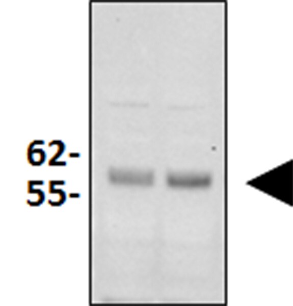
Western Blot: Parkin Antibody (1H4) [H00005071-M01]
Western Blot: Parkin Antibody (1H4) [H00005071-M01] - Parkin monoclonal antibody (M01), clone 1H4. Western Blot analysis of Parkin expression in PC-12.![Immunocytochemistry/ Immunofluorescence: Parkin Antibody (1H4) [H00005071-M01]](https://resources.rndsystems.com/images/products/Parkin-Antibody-1H4-Immunocytochemistry-Immunofluorescence-H00005071-M01-img0004.jpg "Immunocytochemistry/ Immunofluorescence: Parkin Antibody (1H4) [H00005071-M01]")
Immunocytochemistry/ Immunofluorescence: Parkin Antibody (1H4) [H00005071-M01]
Immunocytochemistry/Immunofluorescence: Parkin Antibody (1H4) [H00005071-M01] - Analysis of monoclonal antibody to PARK2 on HeLa cell. Antibody concentration 10 ug/ml.![Western Blot: Parkin Antibody (1H4) [H00005071-M01]](https://resources.rndsystems.com/images/products/Parkin-Antibody-1H4-Western-Blot-H00005071-M01-img0006.jpg "Western Blot: Parkin Antibody (1H4) [H00005071-M01]")
Western Blot: Parkin Antibody (1H4) [H00005071-M01]
Western Blot: Parkin Antibody (1H4) [H00005071-M01] - PARK2 monoclonal antibody (M01), clone 1H4 Analysis of PARK2 expression in Jurkat.![ELISA: Parkin Antibody (1H4) [H00005071-M01]](https://resources.rndsystems.com/images/products/Parkin-Antibody-1H4-ELISA-H00005071-M01-img0007.jpg "ELISA: Parkin Antibody (1H4) [H00005071-M01]")
ELISA: Parkin Antibody (1H4) [H00005071-M01]
ELISA: Parkin Antibody (1H4) [H00005071-M01] - Detection limit for recombinant GST tagged PARK2 is 0.1 ng/ml as a capture antibody.Applications for Parkin Antibody (1H4) - Azide and BSA Free
Application
Recommended Usage
ELISA
Optimal dilutions of this antibody should be experimentally determined.
Immunocytochemistry/ Immunofluorescence
Optimal dilutions of this antibody should be experimentally determined.
Western Blot
Optimal dilutions of this antibody should be experimentally determined.
Application Notes
Antibody reactivity against cell lysate and recombinant protein for WB. It has also been used for IF and ELISA.
Reviewed Applications
Read 2 reviews rated 3.5 using H00005071-M01 in the following applications:
Formulation, Preparation, and Storage
Purification
IgG purified
Formulation
In 1x PBS, pH 7.4
Format
Azide and BSA Free
Preservative
No Preservative
Concentration
Concentrations vary lot to lot. See vial label for concentration. If unlisted please contact technical services.
Shipping
The product is shipped with polar packs. Upon receipt, store it immediately at the temperature recommended below.
Stability & Storage
Aliquot and store at -20C or -80C. Avoid freeze-thaw cycles.
Background: Parkin
Long Name
Parkinson Disease [autosomal recessive, juvenile] 2, Parkin [PARK2], transcript variant 1
Alternate Names
AR-JP, PARK2, PDJ, PRKN
Entrez Gene IDs
5071 (Human)
Gene Symbol
PRKN
OMIM
211980 (Human)
UniProt
Additional Parkin Products
Product Documents for Parkin Antibody (1H4) - Azide and BSA Free
Certificate of Analysis
To download a Certificate of Analysis, please enter a lot or batch number in the search box below.
Product Specific Notices for Parkin Antibody (1H4) - Azide and BSA Free
This product is produced by and distributed for Abnova, a company based in Taiwan.
This product is for research use only and is not approved for use in humans or in clinical diagnosis. Primary Antibodies are guaranteed for 1 year from date of receipt.
Related Research Areas
Citations for Parkin Antibody (1H4) - Azide and BSA Free
 Powered by Bioz
Customer Reviews for Parkin Antibody (1H4) - Azide and BSA Free (2)
3.5 out of 5
2 Customer Ratings
Have you used Parkin Antibody (1H4) - Azide and BSA Free?
Submit a review and receive an Amazon gift card!
$25/€18/£15/$25CAN/¥2500 Yen for a review with an image
$10/€7/£6/$10CAN/¥1110 Yen for a review without an image
Submit a review
Customer Images
Showing
1
-
2 of
2 reviews
Showing All
Filter By:
-
Application: Western BlotSample Tested: Mouse skeletal muscle homogenateSpecies: MouseVerified Customer | Posted 09/26/2017Western Blot: Parkin Antibody (1H4) [H00005071-M01] - Western blot analysisi was performed in mouse skeletal muscle proteins, separated by 4-12% SDS-PAGE. Proteins were transferred on nitrocellulose membrane and blocked in 5% non-fat milk for 1h at room temperature. Anti Parkin antibody was used 1:1000

-
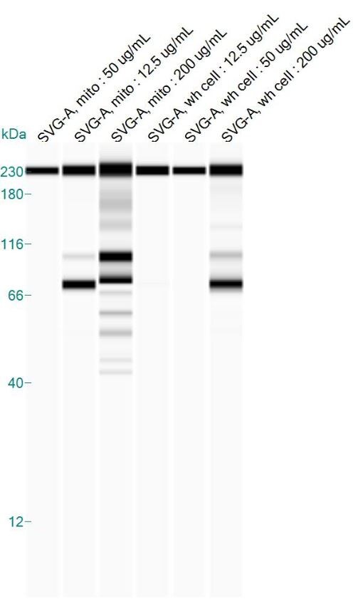
Application: Simple WesternSample Tested: SVG-A cells (different total protein concentrations of the mitochondria-enriched fraction versus whole-cell lysate)Species: HumanVerified Customer | Posted 08/22/2016PARK2 antibody on Simple Western using human SVG-A cells
There are no reviews that match your criteria.
Protocols
Find general support by application which include: protocols, troubleshooting, illustrated assays, videos and webinars.
- Appropriate Fixation of IHC/ICC Samples
- Cellular Response to Hypoxia Protocols
- ClariTSA™ Fluorophore Kits
- Detection & Visualization of Antibody Binding
- ELISA Sample Preparation & Collection Guide
- ELISA Troubleshooting Guide
- How to Run an R&D Systems DuoSet ELISA
- How to Run an R&D Systems Quantikine ELISA
- How to Run an R&D Systems Quantikine™ QuicKit™ ELISA
- ICC Cell Smear Protocol for Suspension Cells
- ICC Immunocytochemistry Protocol Videos
- ICC for Adherent Cells
- Immunocytochemistry (ICC) Protocol
- Immunocytochemistry Troubleshooting
- Immunofluorescence of Organoids Embedded in Cultrex Basement Membrane Extract
- Immunohistochemistry (IHC) and Immunocytochemistry (ICC) Protocols
- Preparing Samples for IHC/ICC Experiments
- Preventing Non-Specific Staining (Non-Specific Binding)
- Primary Antibody Selection & Optimization
- Protocol for VisUCyte™ HRP Polymer Detection Reagent
- Protocol for the Fluorescent ICC Staining of Cell Smears - Graphic
- Protocol for the Fluorescent ICC Staining of Cultured Cells on Coverslips - Graphic
- Protocol for the Preparation and Fluorescent ICC Staining of Cells on Coverslips
- Protocol for the Preparation and Fluorescent ICC Staining of Non-adherent Cells
- Protocol for the Preparation and Fluorescent ICC Staining of Stem Cells on Coverslips
- Protocol for the Preparation of a Cell Smear for Non-adherent Cell ICC - Graphic
- Quantikine HS ELISA Kit Assay Principle, Alkaline Phosphatase
- Quantikine HS ELISA Kit Principle, Streptavidin-HRP Polymer
- R&D Systems Quality Control Western Blot Protocol
- Sandwich ELISA (Colorimetric) – Biotin/Streptavidin Detection Protocol
- Sandwich ELISA (Colorimetric) – Direct Detection Protocol
- TUNEL and Active Caspase-3 Detection by IHC/ICC Protocol
- The Importance of IHC/ICC Controls
- Troubleshooting Guide: ELISA
- Troubleshooting Guide: Western Blot Figures
- Western Blot Conditions
- Western Blot Protocol
- Western Blot Protocol for Cell Lysates
- Western Blot Troubleshooting
- Western Blot Troubleshooting Guide
- View all Protocols, Troubleshooting, Illustrated assays and Webinars
Loading...