PAX8 Antibody (1F8-3A8)
Novus Biologicals | Catalog # NBP2-29903
![Western Blot: PAX8 Antibody (1F8-3A8) [NBP2-29903]](https://resources.rndsystems.com/images/products/PAX8-Antibody-1F8-3A8-Western-Blot-NBP2-29903-img0011.jpg "Western Blot: PAX8 Antibody (1F8-3A8) [NBP2-29903]")
Loading...
Key Product Details
Species Reactivity
Validated:
Human, Mouse, Monkey, Primate
Cited:
Mouse
Applications
Validated:
Immunohistochemistry, Immunohistochemistry-Paraffin, Western Blot, Immunocytochemistry/ Immunofluorescence
Cited:
Western Blot
Label
Unconjugated
Antibody Source
Monoclonal Mouse IgG1 Clone # 1F8-3A8
Loading...
Product Specifications
Immunogen
Purified internal fragment of human recombinant PAX8 expressed in E. coli.
Reactivity Notes
Mouse reactivity reported in a verified customer review.
Clonality
Monoclonal
Host
Mouse
Isotype
IgG1
Scientific Data Images for PAX8 Antibody (1F8-3A8)
Western Blot: PAX8 Antibody (1F8-3A8) [NBP2-29903]
Western Blot: PAX8 Antibody (1F8-3A8) [NBP2-29903] - Analysis of 20ug of the indicated whole cell lysates onto a 4-12% Novex Bis-Tris polyacrylamide gel. Data courtesy of the Innovators Program.![Immunocytochemistry/ Immunofluorescence: PAX8 Antibody (1F8-3A8) [NBP2-29903]](https://resources.rndsystems.com/images/products/PAX8-Antibody-1F8-3A8-Immunocytochemistry-Immunofluorescence-NBP2-29903-img0005.jpg "Immunocytochemistry/ Immunofluorescence: PAX8 Antibody (1F8-3A8) [NBP2-29903]")
Immunocytochemistry/ Immunofluorescence: PAX8 Antibody (1F8-3A8) [NBP2-29903]
Immunocytochemistry/Immunofluorescence: PAX8 Antibody (1F8-3A8) [NBP2-29903] - Analysis of PAX8 (green) in 293T (left panel) and COS7 cells (right panel). Formalin fixed cells were permeabilized with 0.1% Triton X-100 in TBS for 10 minutes at room temperature. Cells were blocked with 1% Blocker BSA for 15 minutes at room temperature. Cells were probed with a PAX8 monoclonal antibody at a dilution of 1:50 for at least 1 hour at room temperature, washed with PBS, and incubated with a DyLight 488-conjugated goat anti-mouse IgG secondary antibody at a dilution of 1:400 for 30 minutes at room temperature. F-Actin (red) was stained with DyLight-554 Phalloidin and nuclei (blue) were stained with Hoechst 33342 dye.![Immunohistochemistry: PAX8 Antibody (1F8-3A8) [NBP2-29903]](https://resources.rndsystems.com/images/products/PAX8-Antibody-1F8-3A8-Immunocytochemistry-NBP2-29903-img0012.jpg "Immunohistochemistry: PAX8 Antibody (1F8-3A8) [NBP2-29903]")
Immunohistochemistry: PAX8 Antibody (1F8-3A8) [NBP2-29903]
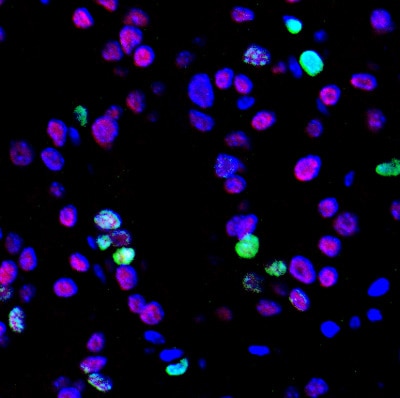
Immunohistochemistry: PAX8 Antibody (1F8-3A8) [NBP2-29903] - Staining in mouse thyroid tissue at pre-tumor stage (red). Dilution is 1:100. This image was submitted via customer Review.![Western Blot: PAX8 Antibody (1F8-3A8) [NBP2-29903]](https://resources.rndsystems.com/images/products/PAX8-Antibody-1F8-3A8-Western-Blot-NBP2-29903-img0009.jpg "Western Blot: PAX8 Antibody (1F8-3A8) [NBP2-29903]")
Western Blot: PAX8 Antibody (1F8-3A8) [NBP2-29903]
Western Blot: PAX8 Antibody (1F8-3A8) [NBP2-29903] - Analysis of 60ug of SKOV-3, COS7, and negative control HeLa whole cell lysates, and 10ul of PageRuler Plus Prestained Protein Ladder.![Western Blot: PAX8 Antibody (1F8-3A8) [NBP2-29903]](https://resources.rndsystems.com/images/products/PAX8-Antibody-1F8-3A8-Western-Blot-NBP2-29903-img0010.jpg "Western Blot: PAX8 Antibody (1F8-3A8) [NBP2-29903]")
Western Blot: PAX8 Antibody (1F8-3A8) [NBP2-29903]
Western Blot: PAX8 Antibody (1F8-3A8) [NBP2-29903] - Analysis of 25ug of HEK293T cell lysate overexpressing PAX8 (right lane) or empty vector control (left lane), and 10ul PageRuler Plus Prestained Protein Ladder.![Immunohistochemistry-Paraffin: PAX8 Antibody (1F8-3A8) [NBP2-29903]](https://resources.rndsystems.com/images/products/PAX8-Antibody-1F8-3A8-Immunohistochemistry-Paraffin-NBP2-29903-img0006.jpg "Immunohistochemistry-Paraffin: PAX8 Antibody (1F8-3A8) [NBP2-29903]")
Immunohistochemistry-Paraffin: PAX8 Antibody (1F8-3A8) [NBP2-29903]
Immunohistochemistry-Paraffin: PAX8 Antibody (1F8-3A8) [NBP2-29903] - Analysis showing staining in the nucleus of mouse kidney tissue (right) compared to a negative control without primary antibody (left).![Immunohistochemistry-Paraffin: PAX8 Antibody (1F8-3A8) [NBP2-29903]](https://resources.rndsystems.com/images/products/PAX8-Antibody-1F8-3A8-Immunohistochemistry-Paraffin-NBP2-29903-img0007.jpg "Immunohistochemistry-Paraffin: PAX8 Antibody (1F8-3A8) [NBP2-29903]")
Immunohistochemistry-Paraffin: PAX8 Antibody (1F8-3A8) [NBP2-29903]
Immunohistochemistry-Paraffin: PAX8 Antibody (1F8-3A8) [NBP2-29903] - Analysis of showing staining in the nucleus of human thyroid tissue (right) compared to a negative control without primary antibody (left).![Immunohistochemistry-Paraffin: PAX8 Antibody (1F8-3A8) [NBP2-29903]](https://resources.rndsystems.com/images/products/PAX8-Antibody-1F8-3A8-Immunohistochemistry-Paraffin-NBP2-29903-img0008.jpg "Immunohistochemistry-Paraffin: PAX8 Antibody (1F8-3A8) [NBP2-29903]")
Immunohistochemistry-Paraffin: PAX8 Antibody (1F8-3A8) [NBP2-29903]
Immunohistochemistry-Paraffin: PAX8 Antibody (1F8-3A8) [NBP2-29903] - Analysis showing staining in the nucleus of human kidney tissue (right) compared to a negative control without primary antibody (left).Applications for PAX8 Antibody (1F8-3A8)
Application
Recommended Usage
Immunocytochemistry/ Immunofluorescence
1:50 - 1:200
Immunohistochemistry
1:20 - 1:200
Immunohistochemistry-Paraffin
1:10 - 1:500
Western Blot
1:500 - 1:2000
Application Notes
WB: Detects a prominent approx. 48 kDa protein in human adenocarcinoma SKOV-3 and SV40 transformed African green monkey (Cercopithecus aethiops) kidney cells. In SKOV-3 cells, additional unknown low molecular bands are also detected. Specificity of the antibody was confirmed in HeLa (negative control) and 293T cells overexpressing full length PAX8. IHC reported in a review from a verified customer.
Reviewed Applications
Read 1 review rated 5 using NBP2-29903 in the following applications:
Formulation, Preparation, and Storage
Purification
Protein A purified
Formulation
PBS with 1 mg/ml BSA and 30% glycerol
Preservative
0.05% Sodium Azide
Concentration
1 mg/ml
Shipping
The product is shipped with polar packs. Upon receipt, store it immediately at the temperature recommended below.
Stability & Storage
Store at -20C. Avoid freeze-thaw cycles.
Background: PAX8
Alternate Names
paired box 8, paired box gene 8, paired box protein Pax-8, paired domain gene 8
Entrez Gene IDs
7849 (Human)
Gene Symbol
PAX8
UniProt
Additional PAX8 Products
Product Documents for PAX8 Antibody (1F8-3A8)
Certificate of Analysis
To download a Certificate of Analysis, please enter a lot or batch number in the search box below.
Product Specific Notices for PAX8 Antibody (1F8-3A8)
This product is for research use only and is not approved for use in humans or in clinical diagnosis. Primary Antibodies are guaranteed for 1 year from date of receipt.
Citations for PAX8 Antibody (1F8-3A8)
 Powered by Bioz
Customer Reviews for PAX8 Antibody (1F8-3A8) (1)
5 out of 5
1 Customer Rating
Have you used PAX8 Antibody (1F8-3A8)?
Submit a review and receive an Amazon gift card!
$25/€18/£15/$25CAN/¥2500 Yen for a review with an image
$10/€7/£6/$10CAN/¥1110 Yen for a review without an image
Submit a review
Customer Images
Showing
1
-
1 of
1 review
Showing All
Filter By:
-
Application: ImmunocytochemistrySample Tested: thyroid tumor tissueSpecies: MouseVerified Customer | Posted 05/30/2017Pax8 staining in mouse thyroid tissue at pre-tumor stage (red). Dilution is 1:100

There are no reviews that match your criteria.
Protocols
Find general support by application which include: protocols, troubleshooting, illustrated assays, videos and webinars.
- Antigen Retrieval Protocol (PIER)
- Antigen Retrieval for Frozen Sections Protocol
- Appropriate Fixation of IHC/ICC Samples
- Cellular Response to Hypoxia Protocols
- Chromogenic IHC Staining of Formalin-Fixed Paraffin-Embedded (FFPE) Tissue Protocol
- Chromogenic Immunohistochemistry Staining of Frozen Tissue
- ClariTSA™ Fluorophore Kits
- Detection & Visualization of Antibody Binding
- Fluorescent IHC Staining of Frozen Tissue Protocol
- Graphic Protocol for Heat-induced Epitope Retrieval
- Graphic Protocol for the Preparation and Fluorescent IHC Staining of Frozen Tissue Sections
- Graphic Protocol for the Preparation and Fluorescent IHC Staining of Paraffin-embedded Tissue Sections
- Graphic Protocol for the Preparation of Gelatin-coated Slides for Histological Tissue Sections
- ICC Cell Smear Protocol for Suspension Cells
- ICC Immunocytochemistry Protocol Videos
- ICC for Adherent Cells
- IHC Sample Preparation (Frozen sections vs Paraffin)
- Immunocytochemistry (ICC) Protocol
- Immunocytochemistry Troubleshooting
- Immunofluorescence of Organoids Embedded in Cultrex Basement Membrane Extract
- Immunofluorescent IHC Staining of Formalin-Fixed Paraffin-Embedded (FFPE) Tissue Protocol
- Immunohistochemistry (IHC) and Immunocytochemistry (ICC) Protocols
- Immunohistochemistry Frozen Troubleshooting
- Immunohistochemistry Paraffin Troubleshooting
- Preparing Samples for IHC/ICC Experiments
- Preventing Non-Specific Staining (Non-Specific Binding)
- Primary Antibody Selection & Optimization
- Protocol for Heat-Induced Epitope Retrieval (HIER)
- Protocol for Making a 4% Formaldehyde Solution in PBS
- Protocol for VisUCyte™ HRP Polymer Detection Reagent
- Protocol for the Fluorescent ICC Staining of Cell Smears - Graphic
- Protocol for the Fluorescent ICC Staining of Cultured Cells on Coverslips - Graphic
- Protocol for the Preparation & Fixation of Cells on Coverslips
- Protocol for the Preparation and Chromogenic IHC Staining of Frozen Tissue Sections
- Protocol for the Preparation and Chromogenic IHC Staining of Frozen Tissue Sections - Graphic
- Protocol for the Preparation and Chromogenic IHC Staining of Paraffin-embedded Tissue Sections
- Protocol for the Preparation and Chromogenic IHC Staining of Paraffin-embedded Tissue Sections - Graphic
- Protocol for the Preparation and Fluorescent ICC Staining of Cells on Coverslips
- Protocol for the Preparation and Fluorescent ICC Staining of Non-adherent Cells
- Protocol for the Preparation and Fluorescent ICC Staining of Stem Cells on Coverslips
- Protocol for the Preparation and Fluorescent IHC Staining of Frozen Tissue Sections
- Protocol for the Preparation and Fluorescent IHC Staining of Paraffin-embedded Tissue Sections
- Protocol for the Preparation of Gelatin-coated Slides for Histological Tissue Sections
- Protocol for the Preparation of a Cell Smear for Non-adherent Cell ICC - Graphic
- R&D Systems Quality Control Western Blot Protocol
- TUNEL and Active Caspase-3 Detection by IHC/ICC Protocol
- The Importance of IHC/ICC Controls
- Troubleshooting Guide: Immunohistochemistry
- Troubleshooting Guide: Western Blot Figures
- Western Blot Conditions
- Western Blot Protocol
- Western Blot Protocol for Cell Lysates
- Western Blot Troubleshooting
- Western Blot Troubleshooting Guide
- View all Protocols, Troubleshooting, Illustrated assays and Webinars
Loading...