PER2 Antibody - BSA Free
Novus Biologicals | Catalog # NB100-125
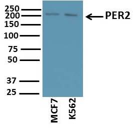
![Western Blot: PER2 AntibodyBSA Free [NB100-125]](https://resources.rndsystems.com/images/products/PER2-Antibody-Western-Blot-NB100-125-img0003.jpg "Western Blot: PER2 AntibodyBSA Free [NB100-125]")
Key Product Details
Species Reactivity
Validated:
Cited:
Applications
Validated:
Cited:
Label
Antibody Source
Format
Product Specifications
Immunogen
Reactivity Notes
Localization
Clonality
Host
Isotype
Scientific Data Images for PER2 Antibody - BSA Free
Western Blot: PER2 AntibodyBSA Free [NB100-125]
Western Blot: PER2 Antibody [NB100-125] - Western Blot Usage of NB100-125. Image submitted via verified customer review.![Immunocytochemistry/ Immunofluorescence: PER2 Antibody - BSA Free [NB100-125]](https://resources.rndsystems.com/images/products/PER2-Antibody-Immunocytochemistry-Immunofluorescence-NB100-125-img0005.jpg "Immunocytochemistry/ Immunofluorescence: PER2 Antibody - BSA Free [NB100-125]")
Immunocytochemistry/ Immunofluorescence: PER2 Antibody - BSA Free [NB100-125]
Immunocytochemistry/Immunofluorescence: PER2 Antibody [NB100-125] - U-87 cells were fixed for 10 minutes using 10% formalin and then permeabilized for 5 minutes using 1X PBS + 0.5% Triton-X100. The cells were incubated with anti-PER2 Antibody at 5 ug/ml overnight at 4C and detected with an anti-rabbit Dylight 488 (Green) at a 1:500 dilution. Nuclei were counterstained with DAPI (Blue). Cells were imaged using a 40X objective.![Western Blot: PER2 AntibodyBSA Free [NB100-125]](https://resources.rndsystems.com/images/products/PER2-Antibody-Western-Blot-NB100-125-img0002.jpg "Western Blot: PER2 AntibodyBSA Free [NB100-125]")
Western Blot: PER2 AntibodyBSA Free [NB100-125]
Western Blot: PER2 Antibody [NB100-125] - WB analysis of PER2 expression on ARPE-19 whole cell lysate.![Immunocytochemistry/ Immunofluorescence: PER2 Antibody - BSA Free [NB100-125]](https://resources.rndsystems.com/images/products/PER2-Antibody-Immunocytochemistry-Immunofluorescence-NB100-125-img0001.jpg "Immunocytochemistry/ Immunofluorescence: PER2 Antibody - BSA Free [NB100-125]")
Immunocytochemistry/ Immunofluorescence: PER2 Antibody - BSA Free [NB100-125]
Immunocytochemistry/Immunofluorescence: PER2 Antibody [NB100-125] - The PER2 antibody was tested in ARPE-19 cells at a 1:250 dilution against Dylight 488 (Green). Alpha-tubulin and nuclei were counterstained against Dylight 550 (Red) and DAPI (Blue), respectively.![Immunocytochemistry/ Immunofluorescence: PER2 Antibody - BSA Free [NB100-125]](https://resources.rndsystems.com/images/products/PER2-Antibody-Immunocytochemistry-Immunofluorescence-NB100-125-img0004.jpg "Immunocytochemistry/ Immunofluorescence: PER2 Antibody - BSA Free [NB100-125]")
Immunocytochemistry/ Immunofluorescence: PER2 Antibody - BSA Free [NB100-125]
Immunocytochemistry/Immunofluorescence: PER2 Antibody [NB100-125] - MCF7 cells were fixed for 10 minutes using 10% formalin and then permeabilized for 5 minutes using 1X PBS + 0.5% Triton-X100. The cells were incubated with anti-PER2 Antibody at 5 ug/ml overnight at 4C and detected with an anti-rabbit Dylight 488 (Green) at a 1:500 dilution. Nuclei were counterstained with DAPI (Blue). Cells were imaged using a 40X objective.
Western Blot: PER2 Antibody - BSA Free [NB100-125] -
Deletion of IL-1 beta signaling ameliorates expression of hepatic circadian transcription factors following DSS-PN.8–10 week old IL1KO mice were subjected to Chow feeding or DSS-PN. A. Liver tissue was harvested, and mRNA expression of transcription factors regulating hepatic CR analyzed by qRT-PCR. Expression was normalized against HPRT. Values are Mean+/- SEM, N = at least 3/condition. *p<0.05. **p<0.01, ***P<0.001, ****P<0.0001. B. Western analysis of circadian regulatory proteins. Expression was normalized using Gapdh expression for each blot. Values are Mean+/- SEM, N = 3/condition, a = significantly different from respective chow control, b = significantly different compared to WT DSS-PN. Image collected and cropped by CiteAb from the following open publication (https://pubmed.ncbi.nlm.nih.gov/37647292), licensed under a CC-BY license. Not internally tested by Novus Biologicals.
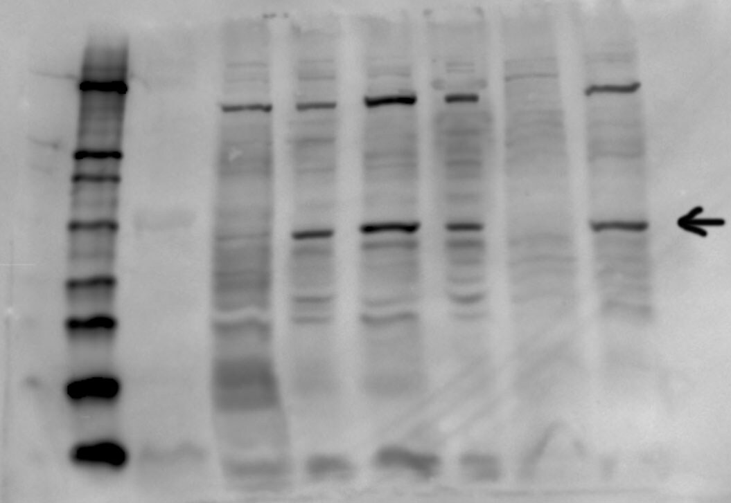
Western Blot: PER2 Antibody - BSA Free [NB100-125] -
Genetic inhibition of TNF alpha signaling normalizes expression of hepatic circadian transcription factors following DSS-PN.8–10 week old TNFRKO mice were subjected to Chow feeding or DSS-PN. A. Liver tissue was harvested, and mRNA expression of transcription factors regulating hepatic CR analyzed by qRT-PCR. Expression was normalized against HPRT. N = 4 per condition. Values are Mean+/- SEM. *p<0.05. **p<0.01, ***p<0.001. B. Western analysis of circadian regulatory proteins. Expression was normalized using Gapdh expression for each blot. Values are Mean+/- SEM, N = 3/condition, a = significantly different from respective chow control, b = significantly different compared to WT DSS-PN. Image collected and cropped by CiteAb from the following open publication (https://pubmed.ncbi.nlm.nih.gov/37647292), licensed under a CC-BY license. Not internally tested by Novus Biologicals.Applications for PER2 Antibody - BSA Free
Immunocytochemistry/ Immunofluorescence
Immunohistochemistry
Immunohistochemistry-Paraffin
Immunoprecipitation
Western Blot
Reviewed Applications
Read 3 reviews rated 3.7 using NB100-125 in the following applications:
Formulation, Preparation, and Storage
Purification
Formulation
Format
Preservative
Concentration
Shipping
Stability & Storage
Background: PER2
Alternate Names
Entrez Gene IDs
Gene Symbol
UniProt
Additional PER2 Products
Product Documents for PER2 Antibody - BSA Free
Certificate of Analysis
To download a Certificate of Analysis, please enter a lot or batch number in the search box below.
Product Specific Notices for PER2 Antibody - BSA Free
This product is for research use only and is not approved for use in humans or in clinical diagnosis. Primary Antibodies are guaranteed for 1 year from date of receipt.
Citations for PER2 Antibody - BSA Free
 Powered by Bioz
Customer Reviews for PER2 Antibody - BSA Free (3)
Have you used PER2 Antibody - BSA Free?
Submit a review and receive an Amazon gift card!
$25/€18/£15/$25CAN/¥2500 Yen for a review with an image
$10/€7/£6/$10CAN/¥1110 Yen for a review without an image
Submit a review
Customer Images

-
Application: Western BlotSample Tested: U-2 OS cellsSpecies: HumanVerified Customer | Posted 02/21/2019siRNA transfection optimisation: whole cell lysate of U-2 OS cells a) siRNA knockdown of PER1 (load 25 µg/well) b) wt U-2 OS cells with different protein amounts/well 1°AB over night, 4 °C 2°AB 1h, RT Nitrocellulose Membrane0unspecific bands with no obvious band of correct size

-
Application: Western BlotSample Tested: Human cancer cell lineSpecies: HumanVerified Customer | Posted 11/12/2017Sample: Whole cell lysates of cancer cell lines (10ug) Blocking: 5% nonfat dry milk reconstituted in TBS-T for 1 hour at room temperature Primary Antibody diluted (1:500) in 5% nonfat dry milk reconstituted in TBS-T overnight at 4deg C Secondary Antibody: Goat Anti-Rabbit IgG H&L (HRP) (ab97051) at 1/10000 dilution in TBS-T
-
Application: Western BlotSample Tested: Whole cell lysates of various cancer cell linesSpecies: HumanVerified Customer | Posted 12/10/2014PER2
There are no reviews that match your criteria.
Protocols
View specific protocols for PER2 Antibody - BSA Free (NB100-125):
Immunocytochemistry Protocol
Culture cells to appropriate density on suitable glass coverslips in 35 mm culture dishes or 6-well plates.
1. Remove culture medium and add 10% formalin to the dish. Fix at room temperature for 5-10 minutes.
2. Remove the formalin and add 0.5% Triton-X 100 in TBS to permeabilize the cells. Incubate for 5-10 minutes.
3. Remove the permeabilization buffer and add wash buffer (i.e. PBS or PBS with 0.1% Tween-20). Be sure to not let the specimen dry out. Gently wash three times for 10 minutes.
4. Alternatively, cells can be fixed with -20C methanol for 10 min at room temperature. Remove the methanol and rehydrate in PBS for 10 min before proceeding.
5. To block nonspecific antibody binding incubate in 10% normal goat serum for 1 hour at room temperature.
6. Add primary antibody at appropriate dilution and incubate at room temperature for 1 hour or at 4C overnight.
7. Remove primary antibody and replace with wash buffer. Gently wash three times for 10 minutes.
8. Add secondary antibody at the appropriate dilution. Incubate for 1 hour at room temperature.
9. Remove antibody and replace with wash buffer. Gently wash three times for 10 minutes.
10. Nuclei can be staining with 4',6' diamino phenylindole (DAPI) at 0.1 ug/ml, or coverslips can be directly mounted in media containing DAPI.
11. Cells can now be viewed with a fluorescence microscope.
*The above information is only intended as a guide. The researcher should determine what protocol best meets their needs. Please follow proper laboratory procedures for the disposal of formalin.
Western Blot Protocol
1. Perform SDS-PAGE on samples to be analyzed, loading 40 ug of total protein per lane.
2. Transfer proteins to membrane according to the instructions provided by the manufacturer of the membrane and transfer apparatus.
3. Stain according to standard Ponceau S procedure (or similar product) to assess transfer success, and mark molecular weight standards where appropriate.
4. Rinse the blot.
5. Block the membrane using standard blocking buffer for at least 1 hour.
6. Wash the membrane in wash buffer three times for 10 minutes each.
7. Dilute primary antibody in blocking buffer and incubate 1 hour at room temperature.
8. Wash the membrane in wash buffer three times for 10 minutes each.
9. Apply the diluted HRP conjugated secondary antibody in blocking buffer (as per manufacturers instructions) and incubate 1 hour at room temperature.
10. Wash the blot in wash buffer three times for 10 minutes each (this step can be repeated as required to reduce background).
11. Apply the detection reagent of choice in accordance with the manufacturers instructions.
Note: Tween-20 can be added to the blocking or antibody dilution buffer at a final concentration of 0.05-0.2%.
*The above information is only intended as a guide. The researcher should determine what protocol best meets their needs.
Find general support by application which include: protocols, troubleshooting, illustrated assays, videos and webinars.
- Antigen Retrieval Protocol (PIER)
- Antigen Retrieval for Frozen Sections Protocol
- Appropriate Fixation of IHC/ICC Samples
- Cellular Response to Hypoxia Protocols
- Chromogenic IHC Staining of Formalin-Fixed Paraffin-Embedded (FFPE) Tissue Protocol
- Chromogenic Immunohistochemistry Staining of Frozen Tissue
- ClariTSA™ Fluorophore Kits
- Detection & Visualization of Antibody Binding
- Fluorescent IHC Staining of Frozen Tissue Protocol
- Graphic Protocol for Heat-induced Epitope Retrieval
- Graphic Protocol for the Preparation and Fluorescent IHC Staining of Frozen Tissue Sections
- Graphic Protocol for the Preparation and Fluorescent IHC Staining of Paraffin-embedded Tissue Sections
- Graphic Protocol for the Preparation of Gelatin-coated Slides for Histological Tissue Sections
- ICC Cell Smear Protocol for Suspension Cells
- ICC Immunocytochemistry Protocol Videos
- ICC for Adherent Cells
- IHC Sample Preparation (Frozen sections vs Paraffin)
- Immunocytochemistry (ICC) Protocol
- Immunocytochemistry Troubleshooting
- Immunofluorescence of Organoids Embedded in Cultrex Basement Membrane Extract
- Immunofluorescent IHC Staining of Formalin-Fixed Paraffin-Embedded (FFPE) Tissue Protocol
- Immunohistochemistry (IHC) and Immunocytochemistry (ICC) Protocols
- Immunohistochemistry Frozen Troubleshooting
- Immunohistochemistry Paraffin Troubleshooting
- Immunoprecipitation Protocol
- Preparing Samples for IHC/ICC Experiments
- Preventing Non-Specific Staining (Non-Specific Binding)
- Primary Antibody Selection & Optimization
- Protocol for Heat-Induced Epitope Retrieval (HIER)
- Protocol for Making a 4% Formaldehyde Solution in PBS
- Protocol for VisUCyte™ HRP Polymer Detection Reagent
- Protocol for the Fluorescent ICC Staining of Cell Smears - Graphic
- Protocol for the Fluorescent ICC Staining of Cultured Cells on Coverslips - Graphic
- Protocol for the Preparation & Fixation of Cells on Coverslips
- Protocol for the Preparation and Chromogenic IHC Staining of Frozen Tissue Sections
- Protocol for the Preparation and Chromogenic IHC Staining of Frozen Tissue Sections - Graphic
- Protocol for the Preparation and Chromogenic IHC Staining of Paraffin-embedded Tissue Sections
- Protocol for the Preparation and Chromogenic IHC Staining of Paraffin-embedded Tissue Sections - Graphic
- Protocol for the Preparation and Fluorescent ICC Staining of Cells on Coverslips
- Protocol for the Preparation and Fluorescent ICC Staining of Non-adherent Cells
- Protocol for the Preparation and Fluorescent ICC Staining of Stem Cells on Coverslips
- Protocol for the Preparation and Fluorescent IHC Staining of Frozen Tissue Sections
- Protocol for the Preparation and Fluorescent IHC Staining of Paraffin-embedded Tissue Sections
- Protocol for the Preparation of Gelatin-coated Slides for Histological Tissue Sections
- Protocol for the Preparation of a Cell Smear for Non-adherent Cell ICC - Graphic
- R&D Systems Quality Control Western Blot Protocol
- TUNEL and Active Caspase-3 Detection by IHC/ICC Protocol
- The Importance of IHC/ICC Controls
- Troubleshooting Guide: Immunohistochemistry
- Troubleshooting Guide: Western Blot Figures
- Western Blot Conditions
- Western Blot Protocol
- Western Blot Protocol for Cell Lysates
- Western Blot Troubleshooting
- Western Blot Troubleshooting Guide
- View all Protocols, Troubleshooting, Illustrated assays and Webinars
FAQs for PER2 Antibody - BSA Free
-
Q: Would you please let me know if there is any updated data available regarding the testing of this antibody on mouse by WB?
A: We do not have an image showing the detection of NB100-125 in mouse tissues, however we will 100% guarantee this item to work as stated on the datasheet.