Pericentrin Antibody - BSA Free
Novus Biologicals | Catalog # NB100-61071
![Immunohistochemistry: Pericentrin Antibody [NB100-61071]](https://resources.rndsystems.com/images/products/Pericentrin-Antibody-Immunohistochemistry-NB100-61071-img0016.jpg "Immunohistochemistry: Pericentrin Antibody [NB100-61071]")
Key Product Details
Species Reactivity
Validated:
Cited:
Applications
Validated:
Cited:
Label
Antibody Source
Format
Product Specifications
Immunogen
Marker
Clonality
Host
Isotype
Theoretical MW
Disclaimer note: The observed molecular weight of the protein may vary from the listed predicted molecular weight due to post translational modifications, post translation cleavages, relative charges, and other experimental factors.
Scientific Data Images for Pericentrin Antibody - BSA Free
Immunohistochemistry: Pericentrin Antibody [NB100-61071]
Pericentrin-Antibody-Immunohistochemistry-NB100-61071-img0016.jpg![Western Blot: Pericentrin Antibody [NB100-61071]](https://resources.rndsystems.com/images/products/Pericentrin-Antibody-Western-Blot-NB100-61071-img0018.jpg "Western Blot: Pericentrin Antibody [NB100-61071]")
Western Blot: Pericentrin Antibody [NB100-61071]
Western Blot: Pericentrin Antibody [NB100-61071] - Whole cell lysate (10 ug) from HEK293T, HeLa, Hep-G2, and K-562 cells prepared using NETN lysis buffer. Antibody: Affinity purified rabbit anti-Pericentrin-Kendrinantibody used for WB at 0.04 ug/ml. Detection: Chemiluminescence with an exposure time of 30 second.![Immunocytochemistry/ Immunofluorescence: Pericentrin Antibody [NB100-61071]](https://resources.rndsystems.com/images/products/Pericentrin-Antibody-Immunocytochemistry-Immunofluorescence-NB100-61071-img0014.jpg "Immunocytochemistry/ Immunofluorescence: Pericentrin Antibody [NB100-61071]")
Immunocytochemistry/ Immunofluorescence: Pericentrin Antibody [NB100-61071]
Immunocytochemistry/Immunofluorescence: Pericentrin Antibody [NB100-61071] - Formaldehyde-fixed asynchronous HeLa cells. Antibody: Affinity purified rabbit anti-Pericentrin/Kendrin used at a dilution of 1:1,000 (1ug/ml). Detection: Red-fluorescent goat anti-rabbit IgG-heavy and light chain cross-adsorbed Antibody DyLight (R) 594 Conjugated used at a dilution of 1:100. Counterstain. DAPI (blue) and Phalloidin (green)![Immunoprecipitation: Pericentrin Antibody [NB100-61071]](https://resources.rndsystems.com/images/products/Pericentrin-Antibody-Immunoprecipitation-NB100-61071-img0015.jpg "Immunoprecipitation: Pericentrin Antibody [NB100-61071]")
Immunoprecipitation: Pericentrin Antibody [NB100-61071]
Immunoprecipitation: Pericentrin Antibody [NB100-61071] - Detection of human Pericentrin/Kendrin by western blot of immunoprecipitates. Samples: Whole cell lysate (0.5 or 1.0 mg per IP reaction; 20% of IP loaded) from HeLa cells prepared using NETN lysis buffer. Antibodies: Affinity purified rabbit anti-Pericentrin/Kendrin antibody NB100-61071 (lot 3) used for IP at 6 ug per reaction. Pericentrin/Kendrin was also immunoprecipitated by a previous lot of this antibody (lot 1). For blotting immunoprecipitated Pericentrin/Kendrin, NB100-61071 was used at 1 ug/ml. Detection: Chemiluminescence with an exposure time of 30 seconds.
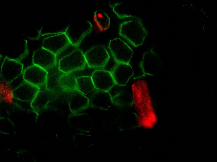
Immunocytochemistry/ Immunofluorescence: Pericentrin Antibody - BSA Free [NB100-61071] -
Immunofluorescent analysis of cilia in renal tissues. (A) Sections (4 μm) of renal parenchymal tissues and tumor tissues were stained with DAPI, acetylated-alpha -tubulin (Ac-tub) and pericentrin (PCNT) to mark cell nuclei and cilia. Presented images are maximal projections of confocal images of typical parenchymal tissue and a representative ccRCC. Scale bars 20 μm. (B) Normalized cilia frequencies of renal tumors, shown are paired quantifications of n = 20 samples. The plot compares the two cilia quantification methodologies described; data was obtained by immunofluorescent (IF) confocal image acquisition or scoring of immunohistochemical (IHC) stained sections. Statistics were determined by performing a paired t-test at a 95% confidence interval. Image collected and cropped by CiteAb from the following open publication (https://pubmed.ncbi.nlm.nih.gov/23369289), licensed under a CC-BY license. Not internally tested by Novus Biologicals.Applications for Pericentrin Antibody - BSA Free
Immunocytochemistry/ Immunofluorescence
Immunohistochemistry-Paraffin
Immunoprecipitation
Western Blot
Reviewed Applications
Read 1 review rated 1 using NB100-61071 in the following applications:
Formulation, Preparation, and Storage
Purification
Formulation
Format
Preservative
Concentration
Shipping
Stability & Storage
Background: Pericentrin
Alternate Names
Entrez Gene IDs
Gene Symbol
UniProt
Additional Pericentrin Products
Product Documents for Pericentrin Antibody - BSA Free
Certificate of Analysis
To download a Certificate of Analysis, please enter a lot or batch number in the search box below.
Product Specific Notices for Pericentrin Antibody - BSA Free
This product is for research use only and is not approved for use in humans or in clinical diagnosis. Primary Antibodies are guaranteed for 1 year from date of receipt.
Citations for Pericentrin Antibody - BSA Free
 Powered by Bioz
Customer Reviews for Pericentrin Antibody - BSA Free (1)
Have you used Pericentrin Antibody - BSA Free?
Submit a review and receive an Amazon gift card!
$25/€18/£15/$25CAN/¥2500 Yen for a review with an image
$10/€7/£6/$10CAN/¥1110 Yen for a review without an image
Submit a review
Customer Images
-
Application: ImmunocytochemistrySample Tested: Adult small intestineSpecies: MouseVerified Customer | Posted 08/16/2021Smear of 2% paraformaldehyde/lysine/periodate (PLP) fixed mouse small itestinal epithelial cells stained for ZO-1 (green) and pericentrin. No staining is detected.We tested the antibody on smears (cytology-like preps) of mouse small intestinal epithelial cells fixed with 2% paraformaldehyde/lysine/periodate (PLP; McLean and Nakane. J Histochem Cytochem, 1974) or-20°C methanol. After permeabilizing with 0.1% saponin (PLP-fixed only) and blocking with 5% donkey serum (in PBS), slides were incubated with antibodies at the dilutions indicated below and a rat monoclonal anti-ZO-1 (tight junction protein) antibody (clone R40.76) for 2 hours at room temperature. After washing, species-specific secondary antibodies (cross-adsorbed) produced in goat were applied.
 Bio-Techne ResponseThis review was submitted through the legacy Novus Innovators Program, reflecting a new species or application tested on a primary antibody.
There are no reviews that match your criteria.
Protocols
Find general support by application which include: protocols, troubleshooting, illustrated assays, videos and webinars.
- Antigen Retrieval Protocol (PIER)
- Antigen Retrieval for Frozen Sections Protocol
- Appropriate Fixation of IHC/ICC Samples
- Cellular Response to Hypoxia Protocols
- Chromogenic IHC Staining of Formalin-Fixed Paraffin-Embedded (FFPE) Tissue Protocol
- Chromogenic Immunohistochemistry Staining of Frozen Tissue
- ClariTSA™ Fluorophore Kits
- Detection & Visualization of Antibody Binding
- Fluorescent IHC Staining of Frozen Tissue Protocol
- Graphic Protocol for Heat-induced Epitope Retrieval
- Graphic Protocol for the Preparation and Fluorescent IHC Staining of Frozen Tissue Sections
- Graphic Protocol for the Preparation and Fluorescent IHC Staining of Paraffin-embedded Tissue Sections
- Graphic Protocol for the Preparation of Gelatin-coated Slides for Histological Tissue Sections
- ICC Cell Smear Protocol for Suspension Cells
- ICC Immunocytochemistry Protocol Videos
- ICC for Adherent Cells
- IHC Sample Preparation (Frozen sections vs Paraffin)
- Immunocytochemistry (ICC) Protocol
- Immunocytochemistry Troubleshooting
- Immunofluorescence of Organoids Embedded in Cultrex Basement Membrane Extract
- Immunofluorescent IHC Staining of Formalin-Fixed Paraffin-Embedded (FFPE) Tissue Protocol
- Immunohistochemistry (IHC) and Immunocytochemistry (ICC) Protocols
- Immunohistochemistry Frozen Troubleshooting
- Immunohistochemistry Paraffin Troubleshooting
- Immunoprecipitation Protocol
- Preparing Samples for IHC/ICC Experiments
- Preventing Non-Specific Staining (Non-Specific Binding)
- Primary Antibody Selection & Optimization
- Protocol for Heat-Induced Epitope Retrieval (HIER)
- Protocol for Making a 4% Formaldehyde Solution in PBS
- Protocol for VisUCyte™ HRP Polymer Detection Reagent
- Protocol for the Fluorescent ICC Staining of Cell Smears - Graphic
- Protocol for the Fluorescent ICC Staining of Cultured Cells on Coverslips - Graphic
- Protocol for the Preparation & Fixation of Cells on Coverslips
- Protocol for the Preparation and Chromogenic IHC Staining of Frozen Tissue Sections
- Protocol for the Preparation and Chromogenic IHC Staining of Frozen Tissue Sections - Graphic
- Protocol for the Preparation and Chromogenic IHC Staining of Paraffin-embedded Tissue Sections
- Protocol for the Preparation and Chromogenic IHC Staining of Paraffin-embedded Tissue Sections - Graphic
- Protocol for the Preparation and Fluorescent ICC Staining of Cells on Coverslips
- Protocol for the Preparation and Fluorescent ICC Staining of Non-adherent Cells
- Protocol for the Preparation and Fluorescent ICC Staining of Stem Cells on Coverslips
- Protocol for the Preparation and Fluorescent IHC Staining of Frozen Tissue Sections
- Protocol for the Preparation and Fluorescent IHC Staining of Paraffin-embedded Tissue Sections
- Protocol for the Preparation of Gelatin-coated Slides for Histological Tissue Sections
- Protocol for the Preparation of a Cell Smear for Non-adherent Cell ICC - Graphic
- R&D Systems Quality Control Western Blot Protocol
- TUNEL and Active Caspase-3 Detection by IHC/ICC Protocol
- The Importance of IHC/ICC Controls
- Troubleshooting Guide: Immunohistochemistry
- Troubleshooting Guide: Western Blot Figures
- Western Blot Conditions
- Western Blot Protocol
- Western Blot Protocol for Cell Lysates
- Western Blot Troubleshooting
- Western Blot Troubleshooting Guide
- View all Protocols, Troubleshooting, Illustrated assays and Webinars
FAQs for Pericentrin Antibody - BSA Free
-
Q: I ordered your antibody 3 weeks ago and I received it today. The product is Pericentrin Antibody (NB100-61071) (0.1 mg). I did not have experience for doing immunofluorescence with this antibody before. I note that you used acetone or NBF to fix cell in the manual of your product. But the description is too simple, so could you send me a more detailed protocol about doing immunofluorescence with pericentrin antibody?
A: Please see the following link for our ICC protocol. I hope this helps!