
Loading...
Key Product Details
Species Reactivity
Validated:
Human, Mouse
Cited:
Human, Mouse
Predicted:
Bovine (100%), Canine (100%), Rat (100%). Backed by our 100% Guarantee.
Applications
Validated:
Immunohistochemistry, Immunohistochemistry-Paraffin, Peptide ELISA, Flow Cytometry, Immunocytochemistry/ Immunofluorescence
Cited:
Immunohistochemistry-Paraffin, Western Blot, IF/IHC
Label
Unconjugated
Antibody Source
Polyclonal Goat IgG
Loading...
Product Specifications
Immunogen
Peptide with sequence C-DGLFDDSEDESDK, from the internal region of the protein sequence according to NP_037393.1; NP_001317680.1; NP_001317681.1; NP_001317682.1; NP_001341756.1.
Epitope
DGLFDDSEDESDK
Reactivity Notes
Mouse reactivity reported in scientific literature (PMID: 29020797).
Clonality
Polyclonal
Host
Goat
Isotype
IgG
Theoretical MW
91 kDa.
Disclaimer note: The observed molecular weight of the protein may vary from the listed predicted molecular weight due to post translational modifications, post translation cleavages, relative charges, and other experimental factors.
Disclaimer note: The observed molecular weight of the protein may vary from the listed predicted molecular weight due to post translational modifications, post translation cleavages, relative charges, and other experimental factors.
Scientific Data Images for PGC1 alpha Antibody
Flow Cytometry: PGC1 alpha Antibody [NB100-60955] -
Flow Cytometry: PGC1 alpha Antibody [NB100-60955] - Flow cytometric analysis of paraformaldehyde fixed HeLa cells (blue line), permeabilized with 0.5% Triton. Primary incubation 1hr (10ug/ml) followed by Alexa Fluor 488 secondary antibody (1ug/ml). IgG control: Unimmunized goat IgG (black line) followed by Alexa Fluor 488 secondary antibody.
Immunohistochemistry-Paraffin: PGC1 alpha Antibody [NB100-60955] -
Immunohistochemistry-Paraffin: PGC1 alpha Antibody [NB100-60955] - Negative Control showing staining of paraffin embedded Human Kidney, with no primary antibody.
Immunocytochemistry/Immunofluorescence: PGC1 alpha Antibody [NB100-60955] -
Immunocytochemistry/Immunofluorescence: PGC1 alpha Antibody [NB100-60955] - Immunofluorescence analysis of paraformaldehyde fixed U2OS cells, permeabilized with 0.15% Triton. Primary incubation 1hr (10ug/ml) followed by Alexa Fluor 488 secondary antibody (2ug/ml), showing nuclear and cytoplasmic staining. The nuclear stain is DAPI (blue). Negative control: Unimmunized goat IgG (10ug/ml) followed by Alexa Fluor 488 secondary antibody (2ug/ml).
Immunocytochemistry/Immunofluorescence: PGC1 alpha Antibody [NB100-60955] -
Immunocytochemistry/Immunofluorescence: PGC1 alpha Antibody [NB100-60955] - Immunofluorescence analysis of paraformaldehyde fixed HeLa cells, permeabilized with 0.15% Triton. Primary incubation 1hr (10ug/ml) followed by Alexa Fluor 488 secondary antibody (2ug/ml), showing nuclear and cytoplasmic staining. The nuclear stain is DAPI (blue). Negative control: Unimmunized goat IgG (10ug/ml) followed by Alexa Fluor 488 secondary antibody (2ug/ml).
Immunohistochemistry-Paraffin: PGC1 alpha Antibody [NB100-60955] -
Immunohistochemistry-Paraffin: PGC1 alpha Antibody [NB100-60955] - (6ug/ml) staining of paraffin embedded Human Kidney. Heat induced antigen retrieval with citrate buffer pH 6, HRP-staining.
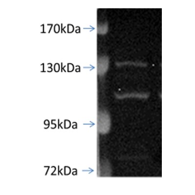
Western Blot: PGC1 alpha Antibody [NB100-60955] -
Changes in PGC-1 alpha mediated mitochondrial biogenesis in the process of cellular degeneration and recovery.A–C mRNA expression of PGC-1 alpha, AMPK-1 alpha, SIRT1 in neuronal PC12 cells measured by qPCR. Data are presented as mean +/- SEM. *P < 0.05, **P < 0.01, vs. control group; ##P < 0.01, ####P < 0.0001, vs. EtOH 3 h group. D–H Protein expression of PGC-1 alpha, AMPK-1 alpha, p-AMPK-1 alpha, and SIRT1 in neuronal PC12 cells detected by western blot. Data are presented as mean +/- SEM. *P < 0.05, **P < 0.01, ***P < 0.001, ****P < 0.0001, vs. control group; #P < 0.05, ##P < 0.01, ###P < 0.001, ####P < 0.0001, vs. EtOH 3 h group. EtOH ethanol. Image collected and cropped by CiteAb from the following open publication (https://www.nature.com/articles/s41420-024-01953-0), licensed under a CC-BY license. Not internally tested by Novus Biologicals.Applications for PGC1 alpha Antibody
Application
Recommended Usage
Flow Cytometry
10 ug/mL
Immunocytochemistry/ Immunofluorescence
10 ug/uL
Immunohistochemistry-Paraffin
6-8 ug/ml
Peptide ELISA
Detection limit 1:2000
Application Notes
Use in IHC-P reported in scientific literature (PMID: 23763823). Use in IHC reported in scientific literature (PMID: 22227854).
Reviewed Applications
Read 1 review rated 4 using NB100-60955 in the following applications:
Flow Cytometry Panel Builder
Bio-Techne Knows Flow Cytometry
Save time and reduce costly mistakes by quickly finding compatible reagents using the Panel Builder Tool.
Advanced Features
- Spectra Viewer - Custom analysis of spectra from multiple fluorochromes
- Spillover Popups - Visualize the spectra of individual fluorochromes
- Antigen Density Selector - Match fluorochrome brightness with antigen density

Formulation, Preparation, and Storage
Purification
Immunogen affinity purified
Formulation
Tris saline (20 mM Tris pH 7.3, 150 mM NaCl), 0.5% BSA
Preservative
0.02% Sodium Azide
Concentration
0.5 mg/ml
Shipping
The product is shipped with polar packs. Upon receipt, store it immediately at the temperature recommended below.
Stability & Storage
Store at -20C. Avoid freeze-thaw cycles.
Background: PGC1 alpha
PGC1 alpha is highly expressed in skeletal muscle, cardiac muscle, and brown adipose tissue, all tissues with high oxidative capacity (1, 2). PGC1 alpha is also induced by energy taxing physiological states, for example increased physical activity, reduced body temperature, and reduced food intake (3). Because of its role in pathways controlling cellular energy expenditure, PGC1 alpha dysfunction has been associated with pathophysiological conditions such as type 2 diabetes and the metabolic syndrome (3).
References
1.Liang, H., & Ward, W. F. (2006). PGC-1alpha: A key regulator of energy metabolism. American Journal of Physiology - Advances in Physiology Education. https://doi.org/10.1152/advan.00052.2006
2.Arany, Z., He, H., Lin, J., Hoyer, K., Handschin, C., Toka, O.,... Spiegelman, B. M. (2005). Transcriptional coactivator PGC-1alpha controls the energy state and contractile function of cardiac muscle. Cell Metabolism. https://doi.org/10.1016/j.cmet.2005.03.002
3.Canto, C., & Auwerx, J. (2009). PGC-1alpha, SIRT1 and AMPK, an energy sensing network that controls energy expenditure. Current Opinion in Lipidology. https://doi.org/10.1097/MOL.0b013e328328d0a4
Long Name
Peroxisome Proliferator-activated Receptor gamma, Coactivator 1 alpha
Alternate Names
LEM6, PPARGC1A
Gene Symbol
PPARGC1A
UniProt
Additional PGC1 alpha Products
Product Documents for PGC1 alpha Antibody
Certificate of Analysis
To download a Certificate of Analysis, please enter a lot or batch number in the search box below.
Product Specific Notices for PGC1 alpha Antibody
This product is for research use only and is not approved for use in humans or in clinical diagnosis. Primary Antibodies are guaranteed for 1 year from date of receipt.
Citations for PGC1 alpha Antibody
 Powered by Bioz
Customer Reviews for PGC1 alpha Antibody (1)
4 out of 5
1 Customer Rating
Have you used PGC1 alpha Antibody?
Submit a review and receive an Amazon gift card!
$25/€18/£15/$25CAN/¥2500 Yen for a review with an image
$10/€7/£6/$10CAN/¥1110 Yen for a review without an image
Submit a review
Customer Images
Showing
1
-
1 of
1 review
Showing All
Filter By:
-
Application: Western BlotSample Tested: Hela whole cell lysate, Sample Amount: 30ugSpecies: HumanVerified Customer | Posted 01/09/2011

There are no reviews that match your criteria.
Protocols
Find general support by application which include: protocols, troubleshooting, illustrated assays, videos and webinars.
- 7-Amino Actinomycin D (7-AAD) Cell Viability Flow Cytometry Protocol
- Antigen Retrieval Protocol (PIER)
- Antigen Retrieval for Frozen Sections Protocol
- Appropriate Fixation of IHC/ICC Samples
- Cellular Response to Hypoxia Protocols
- Chromogenic IHC Staining of Formalin-Fixed Paraffin-Embedded (FFPE) Tissue Protocol
- Chromogenic Immunohistochemistry Staining of Frozen Tissue
- ClariTSA™ Fluorophore Kits
- Detection & Visualization of Antibody Binding
- ELISA Sample Preparation & Collection Guide
- ELISA Troubleshooting Guide
- Extracellular Membrane Flow Cytometry Protocol
- Flow Cytometry Protocol for Cell Surface Markers
- Flow Cytometry Protocol for Staining Membrane Associated Proteins
- Flow Cytometry Staining Protocols
- Flow Cytometry Troubleshooting Guide
- Fluorescent IHC Staining of Frozen Tissue Protocol
- Graphic Protocol for Heat-induced Epitope Retrieval
- Graphic Protocol for the Preparation and Fluorescent IHC Staining of Frozen Tissue Sections
- Graphic Protocol for the Preparation and Fluorescent IHC Staining of Paraffin-embedded Tissue Sections
- Graphic Protocol for the Preparation of Gelatin-coated Slides for Histological Tissue Sections
- How to Run an R&D Systems DuoSet ELISA
- How to Run an R&D Systems Quantikine ELISA
- How to Run an R&D Systems Quantikine™ QuicKit™ ELISA
- ICC Cell Smear Protocol for Suspension Cells
- ICC Immunocytochemistry Protocol Videos
- ICC for Adherent Cells
- IHC Sample Preparation (Frozen sections vs Paraffin)
- Immunocytochemistry (ICC) Protocol
- Immunocytochemistry Troubleshooting
- Immunofluorescence of Organoids Embedded in Cultrex Basement Membrane Extract
- Immunofluorescent IHC Staining of Formalin-Fixed Paraffin-Embedded (FFPE) Tissue Protocol
- Immunohistochemistry (IHC) and Immunocytochemistry (ICC) Protocols
- Immunohistochemistry Frozen Troubleshooting
- Immunohistochemistry Paraffin Troubleshooting
- Intracellular Flow Cytometry Protocol Using Alcohol (Methanol)
- Intracellular Flow Cytometry Protocol Using Detergents
- Intracellular Nuclear Staining Flow Cytometry Protocol Using Detergents
- Intracellular Staining Flow Cytometry Protocol Using Alcohol Permeabilization
- Intracellular Staining Flow Cytometry Protocol Using Detergents to Permeabilize Cells
- Preparing Samples for IHC/ICC Experiments
- Preventing Non-Specific Staining (Non-Specific Binding)
- Primary Antibody Selection & Optimization
- Propidium Iodide Cell Viability Flow Cytometry Protocol
- Protocol for Heat-Induced Epitope Retrieval (HIER)
- Protocol for Liperfluo
- Protocol for Making a 4% Formaldehyde Solution in PBS
- Protocol for VisUCyte™ HRP Polymer Detection Reagent
- Protocol for the Characterization of Human Th22 Cells
- Protocol for the Characterization of Human Th9 Cells
- Protocol for the Fluorescent ICC Staining of Cell Smears - Graphic
- Protocol for the Fluorescent ICC Staining of Cultured Cells on Coverslips - Graphic
- Protocol for the Preparation & Fixation of Cells on Coverslips
- Protocol for the Preparation and Chromogenic IHC Staining of Frozen Tissue Sections
- Protocol for the Preparation and Chromogenic IHC Staining of Frozen Tissue Sections - Graphic
- Protocol for the Preparation and Chromogenic IHC Staining of Paraffin-embedded Tissue Sections
- Protocol for the Preparation and Chromogenic IHC Staining of Paraffin-embedded Tissue Sections - Graphic
- Protocol for the Preparation and Fluorescent ICC Staining of Cells on Coverslips
- Protocol for the Preparation and Fluorescent ICC Staining of Non-adherent Cells
- Protocol for the Preparation and Fluorescent ICC Staining of Stem Cells on Coverslips
- Protocol for the Preparation and Fluorescent IHC Staining of Frozen Tissue Sections
- Protocol for the Preparation and Fluorescent IHC Staining of Paraffin-embedded Tissue Sections
- Protocol for the Preparation of Gelatin-coated Slides for Histological Tissue Sections
- Protocol for the Preparation of a Cell Smear for Non-adherent Cell ICC - Graphic
- Protocol: Annexin V and PI Staining by Flow Cytometry
- Protocol: Annexin V and PI Staining for Apoptosis by Flow Cytometry
- Quantikine HS ELISA Kit Assay Principle, Alkaline Phosphatase
- Quantikine HS ELISA Kit Principle, Streptavidin-HRP Polymer
- Sandwich ELISA (Colorimetric) – Biotin/Streptavidin Detection Protocol
- Sandwich ELISA (Colorimetric) – Direct Detection Protocol
- TUNEL and Active Caspase-3 Detection by IHC/ICC Protocol
- The Importance of IHC/ICC Controls
- Troubleshooting Guide: ELISA
- Troubleshooting Guide: Fluorokine Flow Cytometry Kits
- Troubleshooting Guide: Immunohistochemistry
- View all Protocols, Troubleshooting, Illustrated assays and Webinars
FAQs for PGC1 alpha Antibody
Showing
1
-
3 of
3 FAQs
Showing All
-
Q: I wanted information on your primary antibody for PGC1 alpha NB100-60955, whether it detects the phosphorylated PGC1 alpha protein, or if it just detects the total PGC1 alpha protein.
A: NB100-60955 is not specific for a particular phosphorylated form of PGC1 alpha. This protein is only phosphorylated on two residues but has 13 known acetylation sites, which is why the protein is detected at around 100 kDa instead of the predicted 91 kDa. This antibody should detect the unphosphorylated and unacetylated form as well, if they are present in your sample.
-
Q: My question is whether this antibody (PGC1 alpha Antibody) detects the phosphorylated PGC1 alpha protein? Or if it just detects the total PGC1 alpha protein?
A: NB100-60955 is not specific for a particular phosphorylated form of PGC1 alpha. This protein is only phosphorylated on two residues but has 13 known acetylation sites, which is why the protein is detected at around 100 kDa instead of the predicted 91 kDa. This antibody should detect the unphosphorylated and unacetylated form as well, if they are present in your sample.
-
Q: Please differentiate to me between PPAR and PGC clearly. I am confused with the difference between these two
A:
Thank you very much for contacting Novus Biologicals technical support team and sharing your query on the differences between PGC-1 alpha and PPAR. These are two different proteins encoded by their respective genes and serves different functions. PGC-1 alpha (PGC1A or PPAR gamma coactivator 1-alpha) is a transcriptional co-activator for steroid receptors and nuclear receptors, and it regulates diverse aspects of cellular physiology. It up-regulates the transcriptional activity of PPAR-gamma /thyroid hormone receptor on the uncoupling protein promoter; regulates the key mitochondrial genes involved in adaptive thermogenesis; implicates in the metabolic reprogramming in response to nutrients availability through the coordination of the expression of a wide array of genes involved in the regulation of glucose and fatty acid metabolism. Among our PGC-1 alpha antibodies, NBP1-04676 is our best selling product with nice customer feedback and citations in at least 13 research publications. PPAR (PPAR alpha) on the other hand is a ligand-activated transcription factor which gets activated by the endogenous ligand 1-palmitoyl-2-oleoyl-sn-glycerol-3-phosphocholine, and oleylethanolamide (a naturally occurring lipid that regulates satiety), and acts as a key regulator of lipid metabolism. It also acts as a receptor for peroxisome proliferators such as hypolipidemic drugs and fatty acids. It regulates the peroxisomal beta-oxidation pathway of fatty acids, and also functions as transcription activator for the ACOX1 and P450 genes. We have a variety of PPAR alpha antibodies. I hope you will find this information helpful but please let me know if I can support you with anything else from my end. Thank you very much for choosing Novus Biologicals as your quality reagent supplier and we wish you the best with your research projects.
-
Q: I wanted information on your primary antibody for PGC1 alpha NB100-60955, whether it detects the phosphorylated PGC1 alpha protein, or if it just detects the total PGC1 alpha protein.
A: NB100-60955 is not specific for a particular phosphorylated form of PGC1 alpha. This protein is only phosphorylated on two residues but has 13 known acetylation sites, which is why the protein is detected at around 100 kDa instead of the predicted 91 kDa. This antibody should detect the unphosphorylated and unacetylated form as well, if they are present in your sample.
-
Q: My question is whether this antibody (PGC1 alpha Antibody) detects the phosphorylated PGC1 alpha protein? Or if it just detects the total PGC1 alpha protein?
A: NB100-60955 is not specific for a particular phosphorylated form of PGC1 alpha. This protein is only phosphorylated on two residues but has 13 known acetylation sites, which is why the protein is detected at around 100 kDa instead of the predicted 91 kDa. This antibody should detect the unphosphorylated and unacetylated form as well, if they are present in your sample.
-
Q: Please differentiate to me between PPAR and PGC clearly. I am confused with the difference between these two
A:
Thank you very much for contacting Novus Biologicals technical support team and sharing your query on the differences between PGC-1 alpha and PPAR. These are two different proteins encoded by their respective genes and serves different functions. PGC-1 alpha (PGC1A or PPAR gamma coactivator 1-alpha) is a transcriptional co-activator for steroid receptors and nuclear receptors, and it regulates diverse aspects of cellular physiology. It up-regulates the transcriptional activity of PPAR-gamma /thyroid hormone receptor on the uncoupling protein promoter; regulates the key mitochondrial genes involved in adaptive thermogenesis; implicates in the metabolic reprogramming in response to nutrients availability through the coordination of the expression of a wide array of genes involved in the regulation of glucose and fatty acid metabolism. Among our PGC-1 alpha antibodies, NBP1-04676 is our best selling product with nice customer feedback and citations in at least 13 research publications. PPAR (PPAR alpha) on the other hand is a ligand-activated transcription factor which gets activated by the endogenous ligand 1-palmitoyl-2-oleoyl-sn-glycerol-3-phosphocholine, and oleylethanolamide (a naturally occurring lipid that regulates satiety), and acts as a key regulator of lipid metabolism. It also acts as a receptor for peroxisome proliferators such as hypolipidemic drugs and fatty acids. It regulates the peroxisomal beta-oxidation pathway of fatty acids, and also functions as transcription activator for the ACOX1 and P450 genes. We have a variety of PPAR alpha antibodies. I hope you will find this information helpful but please let me know if I can support you with anything else from my end. Thank you very much for choosing Novus Biologicals as your quality reagent supplier and we wish you the best with your research projects.
Loading...
Associated Pathways
