PIEZO1 Antibody - BSA Free
Novus Biologicals | Catalog # NBP1-78537
![Immunocytochemistry/ Immunofluorescence: PIEZO1 Antibody - BSA Free [NBP1-78537]](https://resources.rndsystems.com/images/products/PIEZO1-Antibody-Immunocytochemistry-Immunofluorescence-NBP1-78537-img0009.jpg "Immunocytochemistry/ Immunofluorescence: PIEZO1 Antibody - BSA Free [NBP1-78537]")
Key Product Details
Species Reactivity
Validated:
Cited:
Applications
Validated:
Cited:
Label
Antibody Source
Format
Product Specifications
Immunogen
Localization
Clonality
Host
Isotype
Scientific Data Images for PIEZO1 Antibody - BSA Free
Immunocytochemistry/ Immunofluorescence: PIEZO1 Antibody - BSA Free [NBP1-78537]
PIEZO1-Antibody-Immunocytochemistry-Immunofluorescence-NBP1-78537-img0009.jpg![Immunocytochemistry/ Immunofluorescence: PIEZO1 Antibody - BSA Free [NBP1-78537]](https://resources.rndsystems.com/images/products/PIEZO1-Antibody-Immunocytochemistry-Immunofluorescence-NBP1-78537-img0008.jpg "Immunocytochemistry/ Immunofluorescence: PIEZO1 Antibody - BSA Free [NBP1-78537]")
Immunocytochemistry/ Immunofluorescence: PIEZO1 Antibody - BSA Free [NBP1-78537]
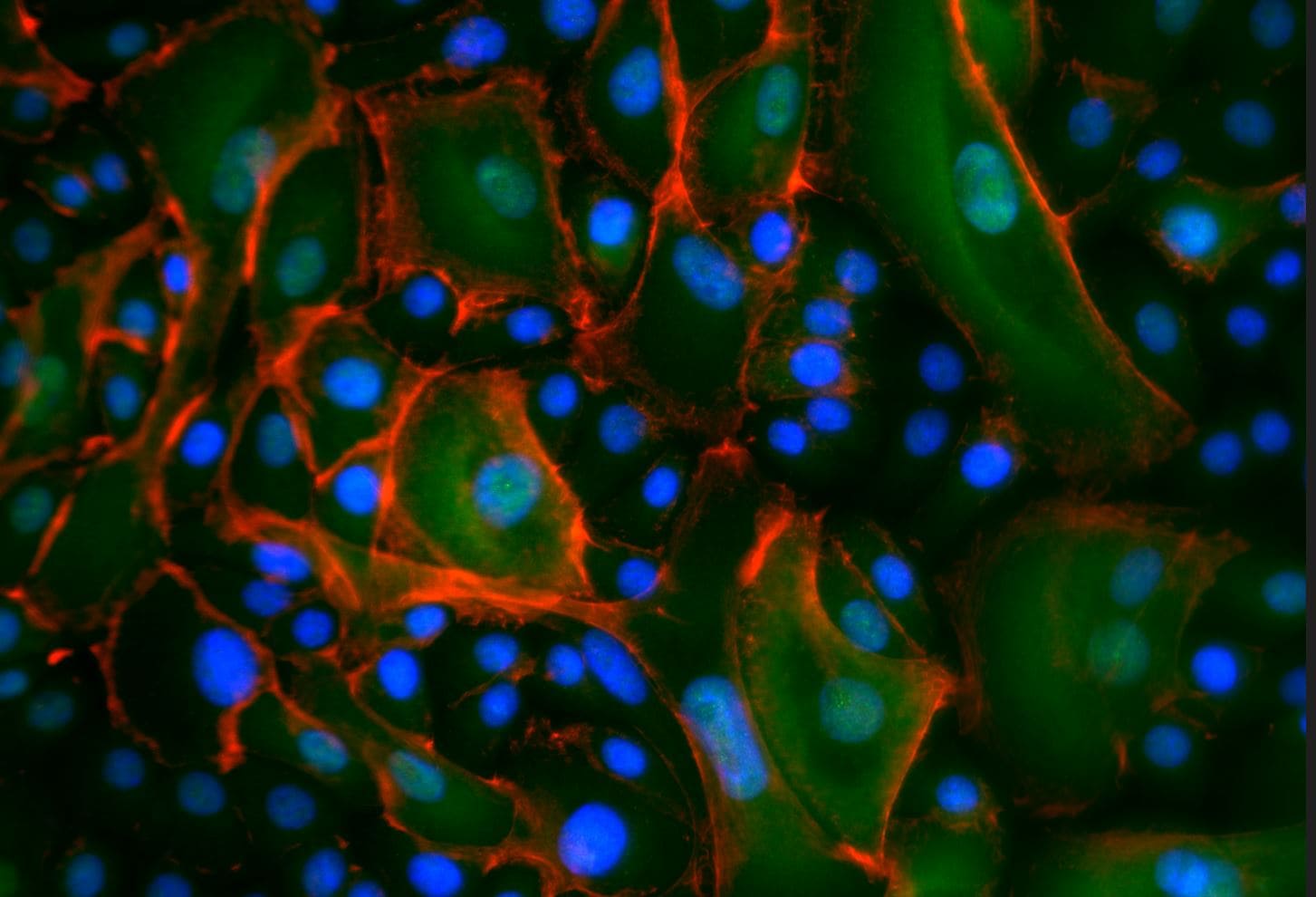
Immunocytochemistry/Immunofluorescence: PIEZO1 Antibody [NBP1-78537] - MCF7 cells were fixed in 4% paraformaldehyde for 10 minutes and permeabilized in 0.05% Triton X-100 in PBS for 5 minutes. The cells were incubated with anti- NBP1-78537 at 2 ug/ml overnight at 4C and detected with an anti-rabbit Dylight 488 (Green) at a 1:1000 dilution for 60 minutes. Nuclei were counterstained with DAPI (Blue). Cells were imaged using a 100X objective and digitally deconvolved.![Western Blot: PIEZO1 AntibodyBSA Free [NBP1-78537]](https://resources.rndsystems.com/images/products/PIEZO1-Antibody-Western-Blot-NBP1-78537-img0007.jpg "Western Blot: PIEZO1 AntibodyBSA Free [NBP1-78537]")
Western Blot: PIEZO1 AntibodyBSA Free [NBP1-78537]
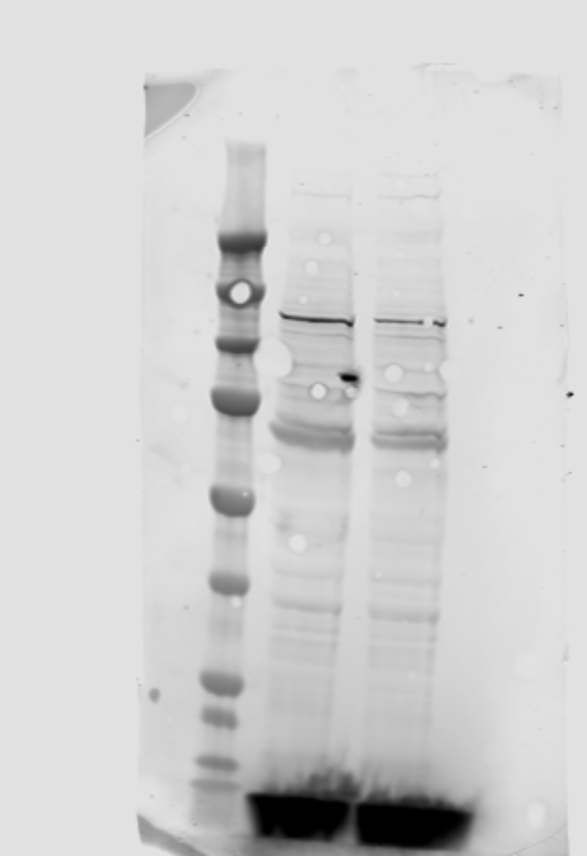
Western Blot: PIEZO1 Antibody [NBP1-78537] - Analysis of MCF7 cell lysate using PIEZ01 antibody (NBP1-78537) at 2 ug/mL.![Immunohistochemistry: PIEZO1 Antibody - BSA Free [NBP1-78537]](https://resources.rndsystems.com/images/products/PIEZO1-Antibody-Immunohistochemistry-NBP1-78537-img0006.jpg "Immunohistochemistry: PIEZO1 Antibody - BSA Free [NBP1-78537]")
Immunohistochemistry: PIEZO1 Antibody - BSA Free [NBP1-78537]
Immunohistochemistry: PIEZO1 Antibody [NBP1-78537] - Analysis of PIEZ01 in mouse epidermis using DAB with hematoxylin counterstain.
Immunohistochemistry: PIEZO1 Antibody [NBP1-78537] -
nbp1-78537_rabbit-polyclonal-piezo1-antibody-2552023153954.jpg
Immunocytochemistry/Immunofluorescence: PIEZO1 Antibody [NBP1-78537] -
Immunocytochemistry/Immunofluorescence: PIEZO1 Antibody [NBP1-78537] - Green: 2 hours at RT with CLTC antibody diluted 1:100 in PBS-Triton 0.2%X-100/BSA 1%. Red: Phalloidin Blue: DAPI. Image from verified customer review.
Simple Western: PIEZO1 Antibody - BSA Free [NBP1-78537] -
Simple Western: PIEZO1 Antibody - BSA Free [NBP1-78537] - Piezo1 expression profiling, cellular mechanotransduction, and biomechanical correlation analysis across brain regions of different donors. The relative abundance of (A) Piezo1, (B) YAP, (C) pYAP, and (D) beta-catenin in the regions of WM (magenta), GW junction (purple), and pons (green) for individual donors (D1-4). (E, top left) Spearman correlation between Piezo1 relative fluorescence units (RFU) and stiffness; GW Junction (orange) ns, WM (blue) r = -0.5341, p < 0.05, pons (green) ns. (E, top right) Correlation between Piezo1 regional RFU and spring term; GW junction r= 0.8791, and WM r = -0.5341, p < 0.05. (E, bottom left) Correlation between Piezo1 regional RFU and decay term (b); WM r = -0.6758, and pons r = 0.7692, p < 0.05. (E, bottom right) Correlation between Piezo1 regional RFU and equilibrium stress term; WM r = -0.8571, pons r = 0.7198, p < 0.05. (F) Comparison of protein expressed based on brain region for all donors, *p < 0.05 Image collected and cropped by CiteAb from the following publication (https://molecularbrain.biomedcentral.com/articles/10.1186/s13041-023-01… ), licensed under a CC-BY license. Not internally tested by Novus Biologicals.
Western Blot: PIEZO1 Antibody - BSA Free [NBP1-78537] -
Western Blot: PIEZO1 Antibody - BSA Free [NBP1-78537] - Expression & distribution of Piezo channels in mouse lenses. (A). RT-PCR-based confirmation of Piezo1 & Piezo2 expression in P1 & P30 mouse lenses. (B). qRT-PCR analysis revealed a relatively much higher level of Piezo1 expression in the lens (P30) compared to Piezo2. (C). Total lysates (800× g supernatants; 75 µg protein) derived from the P1, P14, & P16 mouse lenses analyzed using a Piezo1 polyclonal antibody exhibited immunopositive bands with an expected molecular mass of >250 kDa & >150 kDa. There was also a prominent immunopositive band at >75 kDa in the P1 & P14 lenses, the levels of which appeared to be decreased in the P16 lenses. (D). Piezo1 immunopositive bands of >250 & >75 kDa were present predominantly in the lens fiber samples (P21 & P27) compared to the lens epithelium (P21). (E,F). Immunofluorescence analysis of Piezo1 in the P1 mouse lens (the sagittal plane of the cryosection) revealed that the protein distributes predominantly to lens fibers relative to the epithelium (boxed area in panel (E) was magnified & shown in panel (F)). (G) Shows background immunofluorescence with secondary antibody alone. GAPDH: Loading control; Epi: Epithelium; Bars: Image magnification. Image collected & cropped by CiteAb from the following publication (https://pubmed.ncbi.nlm.nih.gov/35563101), licensed under a CC-BY license. Not internally tested by Novus Biologicals.
Immunohistochemistry: PIEZO1 Antibody - BSA Free [NBP1-78537] -
Immunohistochemistry: PIEZO1 Antibody - BSA Free [NBP1-78537] - Piezo1 expression & cell function of human nucleus pulposus cells under mechanical stress. A NP cells received various mechanical stress treatment. Cell viability was measured by MTT assay. B Gene expression of Piezo1 was measured by real-time PCR. C, D Western blot analysis of Piezo1’s protein level. Total beta -actin served as loading controls. E, F Immunofluorescence staining analysis of Piezo1 expression. Relative fluorescent levels of Piezo1 were measured by Image J software. G Concentrations of pro-inflammatory cytokines TNF-alpha, IL-1 beta, & IL-6 in supernatants were measured by ELISA. H Mitochondrial membrane potential was measured by JC-1 probe & flow cytometer. I OCR of cells were measured by Seahorse XFe96 Extracellular Flux Analyzer at basal conditions & with serial administration of oligomycin, FCCP & rotenone. J–L Gene & protein expressions of P53 & P16. Total beta -actin served as loading controls. *p < 0.05. All experiments were repeated at least three times Image collected & cropped by CiteAb from the following publication (https://pubmed.ncbi.nlm.nih.gov/35606793), licensed under a CC-BY license. Not internally tested by Novus Biologicals.
Western Blot: PIEZO1 Antibody - BSA Free [NBP1-78537] -
Western Blot: PIEZO1 Antibody - BSA Free [NBP1-78537] - The piezo1-tdT mouse confirms the expression & distribution of Piezo1 in lens fibers. (A). The Piezo1-tdT mouse model expressing a fusion protein of Piezo1 & tdTomato (Piezo1-tdT) was used to determine the distribution pattern of Piezo1 in the mouse lens. Similar to what was found in the wild-type lenses (Figure 3), Piezo1-tdT exhibiting the expected molecular mass of >250 kDa was detected only in P30 lens homogenates derived from the Piezo1-tdT mice, but not in the wild-type lens. The positive control (lung tissue lysate from the Piezo1-tdT mice) also showed a robust expression of Piezo1-tdT. Lanes 1 & 2 represent two different loads of the total protein (75 & 150 µg, respectively). (B) The Piezo1-tdT fusion protein was detected predominantly in fiber cell lysates compared to lens epithelial lysates. (C). Immunofluorescence analysis revealed the Piezo1-tdT fusion protein distributing to lens fibers with localization to both the short & long arms of the hexagonal lens fibers (Left & middle panels are with low & high magnification, respectively). The right panel shows a second antibody (Alexa Flour 488) background control fluorescence staining in the Piezo1-tdT mouse lens section. Bars: Image magnification. Image collected & cropped by CiteAb from the following publication (https://pubmed.ncbi.nlm.nih.gov/35563101), licensed under a CC-BY license. Not internally tested by Novus Biologicals.
Immunocytochemistry/ Immunofluorescence: PIEZO1 Antibody - BSA Free [NBP1-78537] -
Piezo1 is expressed in human leukemia K562 cells. (A) RT-PCR analysis revealed the presence of hPIEZO1 mRNA. Cropped gel with enhanced contrast is shown. (B) Immunofluorescent staining with specific antibodies detected Piezo1 proteins (Anti-Piezo1, red channel) in the cells. Cell nuclei were counterstained with DAPI (blue channel). Cells in white frame are shown in 2× zoom. No staining of the cells was observed after pre-incubation of the anti-Piezo antibody with the specific corresponding blocking peptide (Anti-Piezo+BP). (C) The single-channel activity of Piezo1 induced by Yoda1 (10 uM in the pipette solution) recorded in the representative cell-attached experiment at different membrane potentials. Here and elsewhere, the index shows a number of active channels (C—closed state (zero current), O—channel openings). Holding membrane potential is indicated near current traces. (D) The mean I-V relationship corresponds to a single-channel conductance of 19.2 +/- 1.2 pS (n = 9). Image collected and cropped by CiteAb from the following open publication (https://pubmed.ncbi.nlm.nih.gov/34360605), licensed under a CC-BY license. Not internally tested by Novus Biologicals.Applications for PIEZO1 Antibody - BSA Free
Immunocytochemistry/ Immunofluorescence
Immunohistochemistry
Immunohistochemistry-Paraffin
Simple Western
Western Blot
Reviewed Applications
Read 2 reviews rated 4 using NBP1-78537 in the following applications:
Formulation, Preparation, and Storage
Purification
Formulation
Format
Preservative
Concentration
Shipping
Stability & Storage
Background: PIEZO1
Long Name
Alternate Names
Entrez Gene IDs
Gene Symbol
UniProt
Additional PIEZO1 Products
Product Documents for PIEZO1 Antibody - BSA Free
Certificate of Analysis
To download a Certificate of Analysis, please enter a lot or batch number in the search box below.
Product Specific Notices for PIEZO1 Antibody - BSA Free
This product is for research use only and is not approved for use in humans or in clinical diagnosis. Primary Antibodies are guaranteed for 1 year from date of receipt.
Citations for PIEZO1 Antibody - BSA Free
 Powered by Bioz
Customer Reviews for PIEZO1 Antibody - BSA Free (2)
Have you used PIEZO1 Antibody - BSA Free?
Submit a review and receive an Amazon gift card!
$25/€18/£15/$25CAN/¥2500 Yen for a review with an image
$10/€7/£6/$10CAN/¥1110 Yen for a review without an image
Submit a review
Customer Images
-
Application: ImmunocytochemistrySample Tested: Cultured Human KeratinocytesSpecies: HumanVerified Customer | Posted 05/30/2023Green: 2 hours at RT with CLTC antibody diluted 1:100 in PBS-Triton 0.2%X-100/BSA 1%. Red: Phalloidin Blue: DAPI

-
Application: Western BlotSample Tested: Immortalized human podocytesSpecies: HumanVerified Customer | Posted 10/24/2022Western blot of PIEZO in human podocytes
There are no reviews that match your criteria.
Protocols
View specific protocols for PIEZO1 Antibody - BSA Free (NBP1-78537):
Immunocytochemistry Protocol
Culture cells to appropriate density in 35 mm culture dishes or 6-well plates.
1. Remove culture medium and add 10% formalin to the dish. Fix at room temperature for 30 minutes.
2. Remove the formalin and add ice cold methanol. Incubate for 5-10 minutes.
3. Remove methanol and add washing solution (i.e. PBS). Be sure to not let the specimen dry out. Wash three times for 10 minutes.
4. To block nonspecific antibody binding incubate in 10% normal goat serum from 1 hour to overnight at room temperature.
5. Add primary antibody at appropriate dilution and incubate at room temperature from 2 hours to overnight at room temperature.
6. Remove primary antibody and replace with washing solution. Wash three times for 10 minutes.
7. Add secondary antibody at appropriate dilution. Incubate for 1 hour at room temperature.
8. Remove antibody and replace with wash solution, then wash for 10 minutes. Add Hoechst 33258 to wash solution at 1:25,0000 and incubate for 10 minutes. Wash a third time for 10 minutes.
9. Cells can be viewed directly after washing. The plates can also be stored in PBS containing Azide covered in Parafilm (TM). Cells can also be cover-slipped using Fluoromount, with appropriate sealing.
*The above information is only intended as a guide. The researcher should determine what protocol best meets their needs. Please follow safe laboratory procedures.
Immunohistochemistry-Paraffin Embedded Sections
Antigen Unmasking:
Bring slides to a boil in 10 mM sodium citrate buffer (pH 6.0) then maintain at a sub-boiling temperature for 10 minutes. Cool slides on bench-top for 30 minutes.
Staining:
1. Wash sections in deionized water three times for 5 minutes each.
2. Wash sections in wash buffer for 5 minutes.
3. Block each section with 100-400 ul blocking solution for 1 hour at room temperature.
4. Remove blocking solution and add 100-400 ul diluted primary antibody. Incubate overnight at 4C.
5. Remove antibody solution and wash sections in wash buffer three times for 5 minutes each.
6. Add 100-400 ul biotinylated diluted secondary antibody. Incubate 30 minutes at room temperature.
7. Remove secondary antibody solution and wash sections three times with wash buffer for 5 minutes each.
8. Add 100-400 ul Streptavidin-HRP reagent to each section and incubate for 30 minutes at room temperature.
9. Wash sections three times in wash buffer for 5 minutes each.
10. Add 100-400 ul DAB substrate to each section and monitor staining closely.
11. As soon as the sections develop, immerse slides in deionized water.
12. Counterstain sections in hematoxylin.
13. Wash sections in deionized water two times for 5 minutes each.
14. Dehydrate sections.
15. Mount coverslips.
*The above information is only intended as a guide. The researcher should determine what protocol best meets their needs. Please follow safe laboratory procedures.
Western Blot Protocol
1. Perform SDS-PAGE on samples to be analyzed, loading 40 ug of total protein per lane.
2. Transfer proteins to membrane according to the instructions provided by the manufacturer of the membrane and transfer apparatus.
3. Stain according to standard Ponceau S procedure (or similar product) to assess transfer success, and mark molecular weight standards where appropriate.
4. Rinse the blot.
5. Block the membrane using standard blocking buffer for at least 1 hour.
6. Wash the membrane in wash buffer three times for 10 minutes each.
7. Dilute primary antibody in blocking buffer and incubate 1 hour at room temperature.
8. Wash the membrane in wash buffer three times for 10 minutes each.
9. Apply the diluted HRP conjugated secondary antibody in blocking buffer (as per manufacturers instructions) and incubate 1 hour at room temperature.
10. Wash the blot in wash buffer three times for 10 minutes each (this step can be repeated as required to reduce background).
11. Apply the detection reagent of choice in accordance with the manufacturers instructions.
*Note: Tween-20 can be added to the blocking or antibody dilution buffer at a final concentration of 0.05-0.2%.
*The above information is only intended as a guide. The researcher should determine what protocol best meets their needs. Please follow safe laboratory procedures.
Find general support by application which include: protocols, troubleshooting, illustrated assays, videos and webinars.
- Antigen Retrieval Protocol (PIER)
- Antigen Retrieval for Frozen Sections Protocol
- Appropriate Fixation of IHC/ICC Samples
- Cellular Response to Hypoxia Protocols
- Chromogenic IHC Staining of Formalin-Fixed Paraffin-Embedded (FFPE) Tissue Protocol
- Chromogenic Immunohistochemistry Staining of Frozen Tissue
- ClariTSA™ Fluorophore Kits
- Detection & Visualization of Antibody Binding
- Fluorescent IHC Staining of Frozen Tissue Protocol
- Graphic Protocol for Heat-induced Epitope Retrieval
- Graphic Protocol for the Preparation and Fluorescent IHC Staining of Frozen Tissue Sections
- Graphic Protocol for the Preparation and Fluorescent IHC Staining of Paraffin-embedded Tissue Sections
- Graphic Protocol for the Preparation of Gelatin-coated Slides for Histological Tissue Sections
- ICC Cell Smear Protocol for Suspension Cells
- ICC Immunocytochemistry Protocol Videos
- ICC for Adherent Cells
- IHC Sample Preparation (Frozen sections vs Paraffin)
- Immunocytochemistry (ICC) Protocol
- Immunocytochemistry Troubleshooting
- Immunofluorescence of Organoids Embedded in Cultrex Basement Membrane Extract
- Immunofluorescent IHC Staining of Formalin-Fixed Paraffin-Embedded (FFPE) Tissue Protocol
- Immunohistochemistry (IHC) and Immunocytochemistry (ICC) Protocols
- Immunohistochemistry Frozen Troubleshooting
- Immunohistochemistry Paraffin Troubleshooting
- Preparing Samples for IHC/ICC Experiments
- Preventing Non-Specific Staining (Non-Specific Binding)
- Primary Antibody Selection & Optimization
- Protocol for Heat-Induced Epitope Retrieval (HIER)
- Protocol for Making a 4% Formaldehyde Solution in PBS
- Protocol for VisUCyte™ HRP Polymer Detection Reagent
- Protocol for the Fluorescent ICC Staining of Cell Smears - Graphic
- Protocol for the Fluorescent ICC Staining of Cultured Cells on Coverslips - Graphic
- Protocol for the Preparation & Fixation of Cells on Coverslips
- Protocol for the Preparation and Chromogenic IHC Staining of Frozen Tissue Sections
- Protocol for the Preparation and Chromogenic IHC Staining of Frozen Tissue Sections - Graphic
- Protocol for the Preparation and Chromogenic IHC Staining of Paraffin-embedded Tissue Sections
- Protocol for the Preparation and Chromogenic IHC Staining of Paraffin-embedded Tissue Sections - Graphic
- Protocol for the Preparation and Fluorescent ICC Staining of Cells on Coverslips
- Protocol for the Preparation and Fluorescent ICC Staining of Non-adherent Cells
- Protocol for the Preparation and Fluorescent ICC Staining of Stem Cells on Coverslips
- Protocol for the Preparation and Fluorescent IHC Staining of Frozen Tissue Sections
- Protocol for the Preparation and Fluorescent IHC Staining of Paraffin-embedded Tissue Sections
- Protocol for the Preparation of Gelatin-coated Slides for Histological Tissue Sections
- Protocol for the Preparation of a Cell Smear for Non-adherent Cell ICC - Graphic
- R&D Systems Quality Control Western Blot Protocol
- TUNEL and Active Caspase-3 Detection by IHC/ICC Protocol
- The Importance of IHC/ICC Controls
- Troubleshooting Guide: Immunohistochemistry
- Troubleshooting Guide: Western Blot Figures
- Western Blot Conditions
- Western Blot Protocol
- Western Blot Protocol for Cell Lysates
- Western Blot Troubleshooting
- Western Blot Troubleshooting Guide
- View all Protocols, Troubleshooting, Illustrated assays and Webinars
FAQs for PIEZO1 Antibody - BSA Free
-
Q: What is the expected molecular weight of the detected band in WB (mouse protein) in NBP1-78537? Has there been detection at lower molecular weight?
A:
This human protein has at least one isoform with the major one has 2,521 a.a. with the calculated size of 286,790 Da. If considering the post-translational modifications (phosphorylated a.a. and glycosylations), the apparent size could be considerable larger (https://www.uniprot.org/uniprot/Q92508#sequences). Above website also shows some potential isoforms with the much smaller sizes (in 300 a.a or less). Whereas the counterpart in mouse has the major isoform of 2,547 a.a with 292,002 Da, and 4 potential isoforms (ranging from 1000 a.a. - 2500+ a.a.; https://www.uniprot.org/uniprot/E2JF22#sequences) [however it doesn't mean that there is no other smaller ones like human; just that probably no scientist has looked for them, because the proteins in the high mammals are very conserved]. 1) In terms of detecting the smaller PIEZO1 species, the WB image (fig# 1) done by our manufacturing lab on the MCF7 cell lysate shows that many smaller bands below the expected PIEZO1 band (above 251 kDa). 2) I think they could not assign any of the smaller bands to the protein target was because a, for expressing the large proteins in vivo and in vitro, they intend to be pre-mature terminated of the chain synthesis. Therefore the shorter chain(s) can vary in size(s) in the different cases. 3) I checked online and found that https://www.frontiersin.org/articles/10.3389/fnagi.2018.00332/full shows rat PIEZO1 has many smaller bands (Fig 1); seems that it was a wide-spread observation.
4) All rabbit polyclonal antibodies, more or less, have some backgrounds/non-specific bands, because of the nature of the host. Only performing the steps to block the membranes well and incubate the Abs at room temp for 1 hr (or less) [and use less Ab as possible] to reduce the backgrounds/non-specific bands.