PIEZO2 Antibody - BSA Free
Novus Biologicals | Catalog # NBP1-78624

Key Product Details
Species Reactivity
Validated:
Cited:
Applications
Validated:
Cited:
Label
Antibody Source
Format
Product Specifications
Immunogen
Reactivity Notes
Localization
Clonality
Host
Isotype
Scientific Data Images for PIEZO2 Antibody - BSA Free
Immunofluorescent Staining of PIEZO2 in Mouse Neurons
PIEZO2-Antibody-Immunocytochemistry-Immunofluorescence-NBP1-78624-img0008.jpg
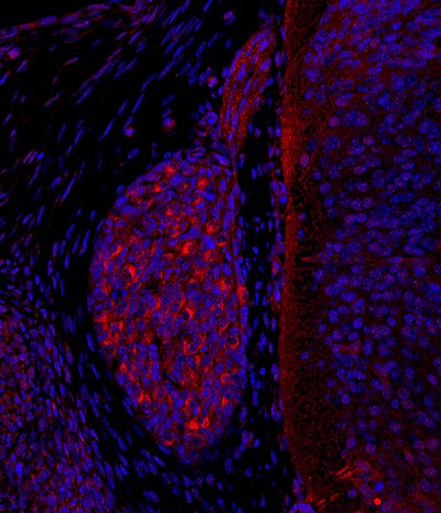
Immunohistochemical Analysis of PIEZO2 in Paraffin Embedded Mouse Dorsal Root Ganglion
Analysis of PIEZO2 in dorsal root ganglion of embryonic day (E) 13.5 mouse. PIEZO2 in red, DAPI in blue. Antigen retrieval with 1x DAKO antigen retrieval solution, by heating in a microwave oven, for total time of 6 minutes 10 seconds. Washing buffer 1 x PBS with 0.1% Triton X-100. Blocking in 5% donkey serum (Jackson lab) in washing buffer. Primary antibody diluted 1:1000, incubation 18 hours in room temperature. Secondary antibody donkey anti-rabbit Cy3, 1:400, 3 hours in room temperature. Image from verified customer review.
Immunocytochemistry/Immunofluorescence Staining of PIEZO2 in A431 Cells
Immunocytochemistry/Immunofluorescence: PIEZO2 Antibody [NBP1-78624] - PIEZO2 antibody was tested at 1:50 in A431 cells with FITC (green).Nuclei and alpha-tubulin were counterstained with DAPI (blue) and DyLight 550 (red).
Immunohistochemical Analysis of PIEZO2 in Mouse Epidermis
Analysis of PIEZ02 in mouse epidermis using DAB with hematoxylin counterstain.
Immunohistochemical Staining of PIEZO2 in Paraffin Embedded Human Breast Tissue
Analysis of a FFPE tissue section of human breast using 1:200 dilution of PIEZO2 antibody. The staining was developed using HRP labeled anti-rabbit secondary antibody and DAB reagent, and nuclei of cells were counter-stained with hematoxylin.
Immunocytochemistry/ Immunofluorescence: PIEZO2 Antibody - BSA Free [NBP1-78624] -
Immunocytochemistry/ Immunofluorescence: PIEZO2 Antibody - BSA Free [NBP1-78624] - Suppression of MA in MTN neurons from adult C57BL/6J mice by silencing Piezo2.(a) Schematic representation of the unilateral adeno-associated viral (AAV) injection site targeting MTN neurons. (b) Fluorescence images showing the expression of GFP-tagged MTN neurons in brain slices following AAV delivery. (b’) Higher magnification of the boxed area in (b). (c) The AAV-Piezo2-sh-1 construct. (d) Brainstem coronal sections of C57BL/6J mice infected with the AVV-shRNA against Piezo2 (middle) & the scrambled AVV (lower), showing MTN neurons immunolabeled for Piezo2 (left, red) or GFP (middle, green), & the merged image of both, with the nuclei stained with DAPI (blue). Arrowheads indicate representative MTN neurons. Scale bar 50 μm. (e) Traces of MA currents evoked by mechanical indentation in MTN neurons infected with AAV-Piezo2-sh-1 or with a scrambled AVV (f). (g) Trace from a MTN neuron infected with AAV-Piezo2-sh-1 that responded with RA mechanical activated currents ( tau = 6.2 ms). The inset shows a higher magnification of the current trace corresponding to a 7 μm displacement. The black upper trace shows the 250 ms mechanical stimulus steps in 1 μm increments applied every 10 s, the breaks in the trace corresponding to equivalent breaks in the recording. (h) Histogram summarizing the mean amplitude of the RA currents in MTN neurons from WT mice (n = 69) & those infected with AAV-Piezo2-sh-1 (n = 20) or AAV-shScr (n = 8). The data are expressed as the means ± s.e.m.: *p < 0.05 (Student’s t-test). Image collected & cropped by CiteAb from the following publication (https://www.nature.com/articles/srep25923), licensed under a CC-BY license. Not internally tested by Novus Biologicals.Applications for PIEZO2 Antibody - BSA Free
Immunocytochemistry/ Immunofluorescence
Immunohistochemistry
Immunohistochemistry-Paraffin
Simple Western
Western Blot
Reviewed Applications
Read 1 review rated 4 using NBP1-78624 in the following applications:
Formulation, Preparation, and Storage
Purification
Formulation
Format
Preservative
Concentration
Shipping
Stability & Storage
Background: PIEZO2
Long Name
Alternate Names
Entrez Gene IDs
Gene Symbol
UniProt
Additional PIEZO2 Products
Product Documents for PIEZO2 Antibody - BSA Free
Certificate of Analysis
To download a Certificate of Analysis, please enter a lot or batch number in the search box below.
Product Specific Notices for PIEZO2 Antibody - BSA Free
This product is for research use only and is not approved for use in humans or in clinical diagnosis. Primary Antibodies are guaranteed for 1 year from date of receipt.
Citations for PIEZO2 Antibody - BSA Free
 Powered by Bioz
Customer Reviews for PIEZO2 Antibody - BSA Free (1)
Have you used PIEZO2 Antibody - BSA Free?
Submit a review and receive an Amazon gift card!
$25/€18/£15/$25CAN/¥2500 Yen for a review with an image
$10/€7/£6/$10CAN/¥1110 Yen for a review without an image
Submit a review
Customer Images
-
Application: Immunohistochemistry-ParaffinSample Tested: Paraffin section of mouse embryoSpecies: MouseVerified Customer | Posted 03/30/2022Dorsal root ganglion of embryonic day (E) 13.5 mouse. Piezo2 in red, DAPI in blue.Antigen retrieval with 1x DAKO antigen retrieval solution, by heating in a microwave oven, for total time of 6 minutes 10 seconds. (0.01M citric acid, pH 6.0, can also be used.) Washing buffer 1 x PBS with 0.1% Triton X-100. Blocking in 5% donkey serum (Jackson lab) in washing buffer. Primary antibody diluted 1:1000, incubation 18 hours in room temperature. Secondary antibody donkey anti-rabbit Cy3 (Jackson lab), 1:400, 3 hours in room temperature.

There are no reviews that match your criteria.
Protocols
View specific protocols for PIEZO2 Antibody - BSA Free (NBP1-78624):
Immunocytochemistry Protocol
Culture cells to appropriate density in 35 mm culture dishes or 6-well plates.
1. Remove culture medium and add 10% formalin to the dish. Fix at room temperature for 30 minutes.
2. Remove the formalin and add ice cold methanol. Incubate for 5-10 minutes.
3. Remove methanol and add washing solution (i.e. PBS). Be sure to not let the specimen dry out. Wash three times for 10 minutes.
4. To block nonspecific antibody binding incubate in 10% normal goat serum from 1 hour to overnight at room temperature.
5. Add primary antibody at appropriate dilution and incubate at room temperature from 2 hours to overnight at room temperature.
6. Remove primary antibody and replace with washing solution. Wash three times for 10 minutes.
7. Add secondary antibody at appropriate dilution. Incubate for 1 hour at room temperature.
8. Remove antibody and replace with wash solution, then wash for 10 minutes. Add Hoechst 33258 to wash solution at 1:25,0000 and incubate for 10 minutes. Wash a third time for 10 minutes.
9. Cells can be viewed directly after washing. The plates can also be stored in PBS containing Azide covered in Parafilm (TM). Cells can also be cover-slipped using Fluoromount, with appropriate sealing.
*The above information is only intended as a guide. The researcher should determine what protocol best meets their needs. Please follow safe laboratory procedures.
Immunohistochemistry-Paraffin Embedded Sections
Antigen Unmasking:
Bring slides to a boil in 10 mM sodium citrate buffer (pH 6.0) then maintain at a sub-boiling temperature for 10 minutes. Cool slides on bench-top for 30 minutes.
Staining:
1. Wash sections in deionized water three times for 5 minutes each.
2. Wash sections in wash buffer for 5 minutes.
3. Block each section with 100-400 ul blocking solution for 1 hour at room temperature.
4. Remove blocking solution and add 100-400 ul diluted primary antibody. Incubate overnight at 4C.
5. Remove antibody solution and wash sections in wash buffer three times for 5 minutes each.
6. Add 100-400 ul biotinylated diluted secondary antibody. Incubate 30 minutes at room temperature.
7. Remove secondary antibody solution and wash sections three times with wash buffer for 5 minutes each.
8. Add 100-400 ul Streptavidin-HRP reagent to each section and incubate for 30 minutes at room temperature.
9. Wash sections three times in wash buffer for 5 minutes each.
10. Add 100-400 ul DAB substrate to each section and monitor staining closely.
11. As soon as the sections develop, immerse slides in deionized water.
12. Counterstain sections in hematoxylin.
13. Wash sections in deionized water two times for 5 minutes each.
14. Dehydrate sections.
15. Mount coverslips.
*The above information is only intended as a guide. The researcher should determine what protocol best meets their needs. Please follow safe laboratory procedures.
Western Blot Protocol
1. Perform SDS-PAGE on samples to be analyzed, loading 40 ug of total protein per lane.
2. Transfer proteins to membrane according to the instructions provided by the manufacturer of the membrane and transfer apparatus.
3. Stain according to standard Ponceau S procedure (or similar product) to assess transfer success, and mark molecular weight standards where appropriate.
4. Rinse the blot.
5. Block the membrane using standard blocking buffer for at least 1 hour.
6. Wash the membrane in wash buffer three times for 10 minutes each.
7. Dilute primary antibody in blocking buffer and incubate 1 hour at room temperature.
8. Wash the membrane in wash buffer three times for 10 minutes each.
9. Apply the diluted HRP conjugated secondary antibody in blocking buffer (as per manufacturers instructions) and incubate 1 hour at room temperature.
10. Wash the blot in wash buffer three times for 10 minutes each (this step can be repeated as required to reduce background).
11. Apply the detection reagent of choice in accordance with the manufacturers instructions.
*Note: Tween-20 can be added to the blocking or antibody dilution buffer at a final concentration of 0.05-0.2%.
*The above information is only intended as a guide. The researcher should determine what protocol best meets their needs. Please follow safe laboratory procedures.
Find general support by application which include: protocols, troubleshooting, illustrated assays, videos and webinars.
- Antigen Retrieval Protocol (PIER)
- Antigen Retrieval for Frozen Sections Protocol
- Appropriate Fixation of IHC/ICC Samples
- Cellular Response to Hypoxia Protocols
- Chromogenic IHC Staining of Formalin-Fixed Paraffin-Embedded (FFPE) Tissue Protocol
- Chromogenic Immunohistochemistry Staining of Frozen Tissue
- ClariTSA™ Fluorophore Kits
- Detection & Visualization of Antibody Binding
- Fluorescent IHC Staining of Frozen Tissue Protocol
- Graphic Protocol for Heat-induced Epitope Retrieval
- Graphic Protocol for the Preparation and Fluorescent IHC Staining of Frozen Tissue Sections
- Graphic Protocol for the Preparation and Fluorescent IHC Staining of Paraffin-embedded Tissue Sections
- Graphic Protocol for the Preparation of Gelatin-coated Slides for Histological Tissue Sections
- ICC Cell Smear Protocol for Suspension Cells
- ICC Immunocytochemistry Protocol Videos
- ICC for Adherent Cells
- IHC Sample Preparation (Frozen sections vs Paraffin)
- Immunocytochemistry (ICC) Protocol
- Immunocytochemistry Troubleshooting
- Immunofluorescence of Organoids Embedded in Cultrex Basement Membrane Extract
- Immunofluorescent IHC Staining of Formalin-Fixed Paraffin-Embedded (FFPE) Tissue Protocol
- Immunohistochemistry (IHC) and Immunocytochemistry (ICC) Protocols
- Immunohistochemistry Frozen Troubleshooting
- Immunohistochemistry Paraffin Troubleshooting
- Preparing Samples for IHC/ICC Experiments
- Preventing Non-Specific Staining (Non-Specific Binding)
- Primary Antibody Selection & Optimization
- Protocol for Heat-Induced Epitope Retrieval (HIER)
- Protocol for Making a 4% Formaldehyde Solution in PBS
- Protocol for VisUCyte™ HRP Polymer Detection Reagent
- Protocol for the Fluorescent ICC Staining of Cell Smears - Graphic
- Protocol for the Fluorescent ICC Staining of Cultured Cells on Coverslips - Graphic
- Protocol for the Preparation & Fixation of Cells on Coverslips
- Protocol for the Preparation and Chromogenic IHC Staining of Frozen Tissue Sections
- Protocol for the Preparation and Chromogenic IHC Staining of Frozen Tissue Sections - Graphic
- Protocol for the Preparation and Chromogenic IHC Staining of Paraffin-embedded Tissue Sections
- Protocol for the Preparation and Chromogenic IHC Staining of Paraffin-embedded Tissue Sections - Graphic
- Protocol for the Preparation and Fluorescent ICC Staining of Cells on Coverslips
- Protocol for the Preparation and Fluorescent ICC Staining of Non-adherent Cells
- Protocol for the Preparation and Fluorescent ICC Staining of Stem Cells on Coverslips
- Protocol for the Preparation and Fluorescent IHC Staining of Frozen Tissue Sections
- Protocol for the Preparation and Fluorescent IHC Staining of Paraffin-embedded Tissue Sections
- Protocol for the Preparation of Gelatin-coated Slides for Histological Tissue Sections
- Protocol for the Preparation of a Cell Smear for Non-adherent Cell ICC - Graphic
- R&D Systems Quality Control Western Blot Protocol
- TUNEL and Active Caspase-3 Detection by IHC/ICC Protocol
- The Importance of IHC/ICC Controls
- Troubleshooting Guide: Immunohistochemistry
- Troubleshooting Guide: Western Blot Figures
- Western Blot Conditions
- Western Blot Protocol
- Western Blot Protocol for Cell Lysates
- Western Blot Troubleshooting
- Western Blot Troubleshooting Guide
- View all Protocols, Troubleshooting, Illustrated assays and Webinars
FAQs for PIEZO2 Antibody - BSA Free
-
Q: I am interested in purchasing the PIEZO 1 and PIEZO 2 antibodies and their blocking peptides from Novus. I have read your data sheets that in WBs the bands seen are approximately 286 kDa and 318 kDa, respectively. I would greatly appreciate if you could show me these WBs on human cells For PIEZO1 and 2 and the effects of the blocking peptides (or point me to publications using your Abs).
A: We offer a PIEZO1 blocking peptide specific to product NBP1-78446. We unfortunately do not offer a PIEZO2 blocking peptide. Our western blot data for NBP1-78446, NBP1-78624, and NBP1-78538 was performed by an outside collaborator and they have not given permission to us to release their images. That is why we do not have them available on our website. We will fully guarantee all of our products for any listed application or species. We unfortunately do not have any publications for these products either.
-
Q: In my project we need Anti Piezo2 and we find it in your catalogs. PIEZO2 Antibody. I want to know the prediction size of your antibody. The size of the protein is more than 200 kd. but in this photo there there are some sharp band are there the actual bands?
A: The predicted molecular weights for this protein are based on the multiple transcripts associated with this protein. Please see Ensembl for detailed information. Those expected molecular weights are the following: 318.1, 80.8, 73.8, 62.7 kDa. We see this kind of staining across many of our antibodies for PIEZO2. It would appear that the transcript present is dependent on the tissue type. Based on protein arrays, we do believe that this antibody is specific.
-
Q: I am interested in purchasing the PIEZO 1 and PIEZO 2 antibodies and their blocking peptides from Novus. I have read your data sheets that in WBs the bands seen are approximately 286 kDa and 318 kDa, respectively. I would greatly appreciate if you could show me these WBs on human cells For PIEZO1 and 2 and the effects of the blocking peptides (or point me to publications using your Abs).
A: We offer a PIEZO1 blocking peptide specific to product NBP1-78446. We unfortunately do not offer a PIEZO2 blocking peptide. Our western blot data for NBP1-78446, NBP1-78624, and NBP1-78538 was performed by an outside collaborator and they have not given permission to us to release their images. That is why we do not have them available on our website. We will fully guarantee all of our products for any listed application or species. We unfortunately do not have any publications for these products either.
-
Q: In my project we need Anti Piezo2 and we find it in your catalogs. PIEZO2 Antibody. I want to know the prediction size of your antibody. The size of the protein is more than 200 kd. but in this photo there there are some sharp band are there the actual bands?
A: The predicted molecular weights for this protein are based on the multiple transcripts associated with this protein. Please see Ensembl for detailed information. Those expected molecular weights are the following: 318.1, 80.8, 73.8, 62.7 kDa. We see this kind of staining across many of our antibodies for PIEZO2. It would appear that the transcript present is dependent on the tissue type. Based on protein arrays, we do believe that this antibody is specific.