PMCA4 Antibody (2G8) - Azide and BSA Free
Novus Biologicals | Catalog # H00000493-M07
![Western Blot: PMCA4 Antibody (2G8) [H00000493-M07]](https://resources.rndsystems.com/images/products/PMCA4-Antibody-2G8-Western-Blot-H00000493-M07-img0005.jpg "Western Blot: PMCA4 Antibody (2G8) [H00000493-M07]")
Loading...
Key Product Details
Species Reactivity
Validated:
Human
Cited:
Human
Applications
Validated:
Western Blot, ELISA, Sandwich ELISA, Immunocytochemistry/ Immunofluorescence
Cited:
Western Blot
Label
Unconjugated
Antibody Source
Monoclonal Mouse IgG1 kappa Clone # 2G8
Format
Azide and BSA Free
Loading...
Product Specifications
Immunogen
ATP2B4 (NP_001675, 1 a.a. ~ 92 a.a) partial recombinant protein with GST tag. MW of the GST tag alone is 26 KDa. MTNPSDRVLPANSMAESREGDFGCTVMELRKLMELRSRDALTQINVHYGGVQNLCSRLKTSPVEGLSGNPADLEKRRQVFGHNVIPPKKPKT
Specificity
This product is specific for Human ATP2B4 monoclonal antibody (M07), clone 2G8 [Gene ID: 493].
Clonality
Monoclonal
Host
Mouse
Isotype
IgG1 kappa
Description
Quality control test: Antibody Reactive Against Recombinant Protein.
Scientific Data Images for PMCA4 Antibody (2G8) - Azide and BSA Free
Western Blot: PMCA4 Antibody (2G8) [H00000493-M07]
PMCA4-Antibody-2G8-Western-Blot-H00000493-M07-img0005.jpg![Immunocytochemistry/ Immunofluorescence: PMCA4 Antibody (2G8) [H00000493-M07]](https://resources.rndsystems.com/images/products/PMCA4-Antibody-2G8-Immunocytochemistry-Immunofluorescence-H00000493-M07-img0006.jpg "Immunocytochemistry/ Immunofluorescence: PMCA4 Antibody (2G8) [H00000493-M07]")
Immunocytochemistry/ Immunofluorescence: PMCA4 Antibody (2G8) [H00000493-M07]
PMCA4-Antibody-2G8-Immunocytochemistry-Immunofluorescence-H00000493-M07-img0006.jpg![Western Blot: PMCA4 Antibody (2G8) [H00000493-M07]](https://resources.rndsystems.com/images/products/PMCA4-Antibody-2G8-Western-Blot-H00000493-M07-img0002.jpg "Western Blot: PMCA4 Antibody (2G8) [H00000493-M07]")
Western Blot: PMCA4 Antibody (2G8) [H00000493-M07]
PMCA4-Antibody-2G8-Western-Blot-H00000493-M07-img0002.jpg![Western Blot: PMCA4 Antibody (2G8) [H00000493-M07]](https://resources.rndsystems.com/images/products/PMCA4-Antibody-2G8-Western-Blot-H00000493-M07-img0003.jpg "Western Blot: PMCA4 Antibody (2G8) [H00000493-M07]")
Western Blot: PMCA4 Antibody (2G8) [H00000493-M07]
PMCA4-Antibody-2G8-Western-Blot-H00000493-M07-img0003.jpg![Western Blot: PMCA4 Antibody (2G8) [H00000493-M07]](https://resources.rndsystems.com/images/products/PMCA4-Antibody-2G8-Western-Blot-H00000493-M07-img0004.jpg "Western Blot: PMCA4 Antibody (2G8) [H00000493-M07]")
Western Blot: PMCA4 Antibody (2G8) [H00000493-M07]
PMCA4-Antibody-2G8-Western-Blot-H00000493-M07-img0004.jpg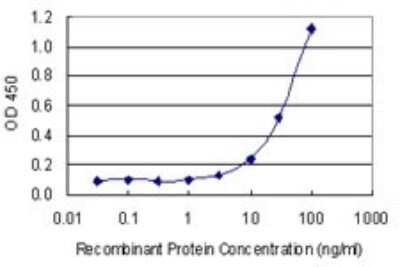
Sandwich ELISA: PMCA4 Antibody (2G8) [H00000493-M07] - Detection limit for recombinant GST tagged ATP2B4 is 1 ng/ml as a capture antibody.
 [H00000493-M07] -")
Western Blot: PMCA4 Antibody (2G8) [H00000493-M07] -
Western Blot: PMCA4 Antibody (2G8) [H00000493-M07] - Identification of plasma membrane calcium ATPase isoform PMCA4b as a renalase binding protein.A, HK-2 cells incubated with either labeled RP-Scr220 or RP-220, biotin-labeled proteins purified using streptavidin column, separated by SDS-PAGE & visualized by western blot using streptavidin-HRP; * = regions evaluated by mass spectrometry in samples labeled with either RP-Scr220 or RP-220; # = RP-220 band containing the plasma membrane calcium ATPase isoform PMCA4b. B, Endogenous expression of PMCA4b in HK-2 cells, western immunoblot using isoform specific monoclonal; CCL-119: human leukemic cell line; thyroid tumor = human thyroid tumor cell line (ATCC, CRL-1803) 10 μg protein loaded in each lane. C, co-immunolocalization of PMCA4b & renalase in HK-2 cells, images acquired using a Zeiss laser scanning confocal microscope, scale bar = 9 μm; arrow = plasma membrane. D, Co-Immunoprecipitation of PMCA4b & renalase from HK-2 cell lysates; renalase-Ab-beads = renalase antibody coated beads; PMCA4b-Ab-beads = PMCA4b antibody coated beads. Image collected & cropped by CiteAb from the following publication (https://pubmed.ncbi.nlm.nih.gov/25906147), licensed under a CC0-1.0 license. Not internally tested by Novus Biologicals. - Azide and BSA Free [H00000493-M07] -")
Immunocytochemistry/ Immunofluorescence: PMCA4 Antibody (2G8) - Azide and BSA Free [H00000493-M07] -
Identification of plasma membrane calcium ATPase isoform PMCA4b as a renalase binding protein.A, HK-2 cells incubated with either labeled RP-Scr220 or RP-220, biotin-labeled proteins purified using streptavidin column, separated by SDS-PAGE and visualized by western blot using streptavidin-HRP; * = regions evaluated by mass spectrometry in samples labeled with either RP-Scr220 or RP-220; # = RP-220 band containing the plasma membrane calcium ATPase isoform PMCA4b. B, Endogenous expression of PMCA4b in HK-2 cells, western immunoblot using isoform specific monoclonal; CCL-119: human leukemic cell line; thyroid tumor = human thyroid tumor cell line (ATCC, CRL-1803) 10 μg protein loaded in each lane. C, co-immunolocalization of PMCA4b and renalase in HK-2 cells, images acquired using a Zeiss laser scanning confocal microscope, scale bar = 9 μm; arrow = plasma membrane. D, Co-Immunoprecipitation of PMCA4b and renalase from HK-2 cell lysates; renalase-Ab-beads = renalase antibody coated beads; PMCA4b-Ab-beads = PMCA4b antibody coated beads. Image collected and cropped by CiteAb from the following open publication (https://pubmed.ncbi.nlm.nih.gov/25906147), licensed under a CC0-1.0 license. Not internally tested by Novus Biologicals.Applications for PMCA4 Antibody (2G8) - Azide and BSA Free
Application
Recommended Usage
ELISA
Optimal dilutions of this antibody should be experimentally determined.
Immunocytochemistry/ Immunofluorescence
Optimal dilutions of this antibody should be experimentally determined.
Sandwich ELISA
Optimal dilutions of this antibody should be experimentally determined.
Western Blot
Optimal dilutions of this antibody should be experimentally determined.
Formulation, Preparation, and Storage
Purification
IgG purified
Formulation
In 1x PBS, pH 7.4
Format
Azide and BSA Free
Preservative
No Preservative
Concentration
Concentrations vary lot to lot. See vial label for concentration. If unlisted please contact technical services.
Shipping
The product is shipped with polar packs. Upon receipt, store it immediately at the temperature recommended below.
Stability & Storage
Aliquot and store at -20C or -80C. Avoid freeze-thaw cycles.
Background: PMCA4
Alternate Names
ATP2B2plasma membrane calcium ATPase, ATPase, Ca++ transporting, plasma membrane 4, DKFZp686G08106, DKFZp686M088, EC 3.6.3, EC 3.6.3.8, Matrix-remodeling-associated protein 1, matrix-remodelling associated 1, MXRA1, Plasma membrane calcium ATPase isoform 4, plasma membrane calcium pump, Plasma membrane calcium pump isoform 4, plasma membrane calcium-transporting ATPase 4, PMCA4b, PMCA4PMCA4x, sarcolemmal calcium pump
Gene Symbol
ATP2B4
UniProt
Additional PMCA4 Products
Product Documents for PMCA4 Antibody (2G8) - Azide and BSA Free
Certificate of Analysis
To download a Certificate of Analysis, please enter a lot or batch number in the search box below.
Product Specific Notices for PMCA4 Antibody (2G8) - Azide and BSA Free
This product is produced by and distributed for Abnova, a company based in Taiwan.
This product is for research use only and is not approved for use in humans or in clinical diagnosis. Primary Antibodies are guaranteed for 1 year from date of receipt.
Citations for PMCA4 Antibody (2G8) - Azide and BSA Free
 Powered by Bioz
Customer Reviews for PMCA4 Antibody (2G8) - Azide and BSA Free
There are currently no reviews for this product. Be the first to review PMCA4 Antibody (2G8) - Azide and BSA Free and earn rewards!
Have you used PMCA4 Antibody (2G8) - Azide and BSA Free?
Submit a review and receive an Amazon gift card!
$25/€18/£15/$25CAN/¥2500 Yen for a review with an image
$10/€7/£6/$10CAN/¥1110 Yen for a review without an image
Submit a review
Protocols
Find general support by application which include: protocols, troubleshooting, illustrated assays, videos and webinars.
- Appropriate Fixation of IHC/ICC Samples
- Cellular Response to Hypoxia Protocols
- ClariTSA™ Fluorophore Kits
- Detection & Visualization of Antibody Binding
- ELISA Sample Preparation & Collection Guide
- ELISA Troubleshooting Guide
- How to Run an R&D Systems DuoSet ELISA
- How to Run an R&D Systems Quantikine ELISA
- How to Run an R&D Systems Quantikine™ QuicKit™ ELISA
- ICC Cell Smear Protocol for Suspension Cells
- ICC Immunocytochemistry Protocol Videos
- ICC for Adherent Cells
- Immunocytochemistry (ICC) Protocol
- Immunocytochemistry Troubleshooting
- Immunofluorescence of Organoids Embedded in Cultrex Basement Membrane Extract
- Immunohistochemistry (IHC) and Immunocytochemistry (ICC) Protocols
- Preparing Samples for IHC/ICC Experiments
- Preventing Non-Specific Staining (Non-Specific Binding)
- Primary Antibody Selection & Optimization
- Protocol for VisUCyte™ HRP Polymer Detection Reagent
- Protocol for the Fluorescent ICC Staining of Cell Smears - Graphic
- Protocol for the Fluorescent ICC Staining of Cultured Cells on Coverslips - Graphic
- Protocol for the Preparation and Fluorescent ICC Staining of Cells on Coverslips
- Protocol for the Preparation and Fluorescent ICC Staining of Non-adherent Cells
- Protocol for the Preparation and Fluorescent ICC Staining of Stem Cells on Coverslips
- Protocol for the Preparation of a Cell Smear for Non-adherent Cell ICC - Graphic
- Quantikine HS ELISA Kit Assay Principle, Alkaline Phosphatase
- Quantikine HS ELISA Kit Principle, Streptavidin-HRP Polymer
- R&D Systems Quality Control Western Blot Protocol
- Sandwich ELISA (Colorimetric) – Biotin/Streptavidin Detection Protocol
- Sandwich ELISA (Colorimetric) – Direct Detection Protocol
- TUNEL and Active Caspase-3 Detection by IHC/ICC Protocol
- The Importance of IHC/ICC Controls
- Troubleshooting Guide: ELISA
- Troubleshooting Guide: Western Blot Figures
- Western Blot Conditions
- Western Blot Protocol
- Western Blot Protocol for Cell Lysates
- Western Blot Troubleshooting
- Western Blot Troubleshooting Guide
- View all Protocols, Troubleshooting, Illustrated assays and Webinars
Loading...