PPAR alpha/NR1C1 Antibody - BSA Free
Novus Biologicals | Catalog # NBP1-31834
Key Product Details
Validated by
Species Reactivity
Validated:
Predicted:
Applications
Label
Antibody Source
Format
Product Specifications
Immunogen
Reactivity Notes
Localization
Clonality
Host
Isotype
Theoretical MW
Disclaimer note: The observed molecular weight of the protein may vary from the listed predicted molecular weight due to post translational modifications, post translation cleavages, relative charges, and other experimental factors.
Scientific Data Images for PPAR alpha/NR1C1 Antibody - BSA Free

Immunohistochemistry-Paraffin: PPAR alpha/NR1C1 Antibody [NBP1-31834] -
Immunohistochemistry-Paraffin: PPAR alpha/NR1C1 Antibody [NBP1-31834] - PPAR alpha/NR1C1 antibody detects PPAR alpha/NR1C1 protein at nucleus by immunohistochemical analysis.Sample: Paraffin-embedded rat brain.
PPAR alpha/NR1C1 stained by PPAR alpha/NR1C1 antibody (NBP1-31834) diluted at 1:165.
Antigen Retrieval: Citrate buffer, pH 6.0, 15 min

Immunohistochemistry-Paraffin: PPAR alpha/NR1C1 Antibody [NBP1-31834] -
Immunohistochemistry-Paraffin: PPAR alpha/NR1C1 Antibody [NBP1-31834] - PPAR alpha/NR1C1 antibody detects PPAR alpha/NR1C1 protein at nucleus by immunohistochemical analysis.Sample: Paraffin-embedded mouse intestine.
PPAR alpha/NR1C1 stained by PPAR alpha/NR1C1 antibody (NBP1-31834) diluted at 1:165.
Antigen Retrieval: Citrate buffer, pH 6.0, 15 min

Western Blot: PPAR alpha/NR1C1 Antibody [NBP1-31834] -
Non-transfected (-) and transfected (+) unboiled HepG2 whole cell extracts (30 ug) were separated by 10% SDS-PAGE, and the membrane was blotted with PPAR alpha/NR1C1 antibody (NBP1-31834) diluted at 1:1000. The HRP-conjugated anti-rabbit IgG antibody was used to detect the primary antibody.
Western Blot: PPAR alpha/NR1C1 Antibody [NBP1-31834] -
Various tissue extracts (50 ug) were separated by 10% SDS-PAGE, and the membrane was blotted with PPAR alpha/NR1C1 antibody (NBP1-31834) diluted at 1:2000. The HRP-conjugated anti-rabbit IgG antibody was used to detect the primary antibody.
Western Blot: PPAR alpha/NR1C1 Antibody [NBP1-31834] -
Mouse tissue extract (50 ug) was separated by 10% SDS-PAGE, and the membrane was blotted with PPAR alpha/NR1C1 antibody (NBP1-31834) diluted at 1:1000. The HRP-conjugated anti-rabbit IgG antibody was used to detect the primary antibody.
Western Blot: PPAR alpha/NR1C1 Antibody [NBP1-31834] -
Whole cell extract (30 ug) was separated by 10% SDS-PAGE, and the membrane was blotted with PPAR alpha/NR1C1 antibody (NBP1-31834) diluted at 1:500. The HRP-conjugated anti-rabbit IgG antibody was used to detect the primary antibody.Applications for PPAR alpha/NR1C1 Antibody - BSA Free
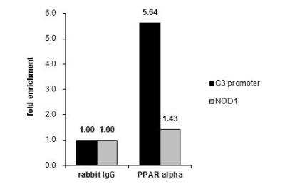
Chromatin Immunoprecipitation (ChIP)
Immunohistochemistry-Paraffin
Western Blot
Formulation, Preparation, and Storage
Purification
Formulation
Format
Preservative
Concentration
Shipping
Stability & Storage
Background: PPAR alpha/NR1C1
Long Name
Alternate Names
Gene Symbol
Additional PPAR alpha/NR1C1 Products
Product Documents for PPAR alpha/NR1C1 Antibody - BSA Free
Certificate of Analysis
To download a Certificate of Analysis, please enter a lot or batch number in the search box below.
Product Specific Notices for PPAR alpha/NR1C1 Antibody - BSA Free
This product is for research use only and is not approved for use in humans or in clinical diagnosis. Primary Antibodies are guaranteed for 1 year from date of receipt.
⚠ WARNING: This product can expose you to chemicals including mercury, which is known to the State of California to cause reproductive toxicity with developmental effects. For more information go to www.P65Warnings.ca.gov.Related Research Areas
Customer Reviews for PPAR alpha/NR1C1 Antibody - BSA Free
There are currently no reviews for this product. Be the first to review PPAR alpha/NR1C1 Antibody - BSA Free and earn rewards!
Have you used PPAR alpha/NR1C1 Antibody - BSA Free?
Submit a review and receive an Amazon gift card!
$25/€18/£15/$25CAN/¥2500 Yen for a review with an image
$10/€7/£6/$10CAN/¥1110 Yen for a review without an image
Submit a review
Protocols
Find general support by application which include: protocols, troubleshooting, illustrated assays, videos and webinars.
- Antigen Retrieval Protocol (PIER)
- Antigen Retrieval for Frozen Sections Protocol
- Appropriate Fixation of IHC/ICC Samples
- Cellular Response to Hypoxia Protocols
- ChIP Protocol Video
- Chromatin Immunoprecipitation (ChIP) Protocol
- Chromatin Immunoprecipitation Protocol
- Chromogenic IHC Staining of Formalin-Fixed Paraffin-Embedded (FFPE) Tissue Protocol
- Chromogenic Immunohistochemistry Staining of Frozen Tissue
- ClariTSA™ Fluorophore Kits
- Detection & Visualization of Antibody Binding
- Fluorescent IHC Staining of Frozen Tissue Protocol
- Graphic Protocol for Heat-induced Epitope Retrieval
- Graphic Protocol for the Preparation and Fluorescent IHC Staining of Frozen Tissue Sections
- Graphic Protocol for the Preparation and Fluorescent IHC Staining of Paraffin-embedded Tissue Sections
- Graphic Protocol for the Preparation of Gelatin-coated Slides for Histological Tissue Sections
- IHC Sample Preparation (Frozen sections vs Paraffin)
- Immunofluorescent IHC Staining of Formalin-Fixed Paraffin-Embedded (FFPE) Tissue Protocol
- Immunohistochemistry (IHC) and Immunocytochemistry (ICC) Protocols
- Immunohistochemistry Frozen Troubleshooting
- Immunohistochemistry Paraffin Troubleshooting
- Preparing Samples for IHC/ICC Experiments
- Preventing Non-Specific Staining (Non-Specific Binding)
- Primary Antibody Selection & Optimization
- Protocol for Heat-Induced Epitope Retrieval (HIER)
- Protocol for Making a 4% Formaldehyde Solution in PBS
- Protocol for VisUCyte™ HRP Polymer Detection Reagent
- Protocol for the Preparation & Fixation of Cells on Coverslips
- Protocol for the Preparation and Chromogenic IHC Staining of Frozen Tissue Sections
- Protocol for the Preparation and Chromogenic IHC Staining of Frozen Tissue Sections - Graphic
- Protocol for the Preparation and Chromogenic IHC Staining of Paraffin-embedded Tissue Sections
- Protocol for the Preparation and Chromogenic IHC Staining of Paraffin-embedded Tissue Sections - Graphic
- Protocol for the Preparation and Fluorescent IHC Staining of Frozen Tissue Sections
- Protocol for the Preparation and Fluorescent IHC Staining of Paraffin-embedded Tissue Sections
- Protocol for the Preparation of Gelatin-coated Slides for Histological Tissue Sections
- R&D Systems Quality Control Western Blot Protocol
- TUNEL and Active Caspase-3 Detection by IHC/ICC Protocol
- The Importance of IHC/ICC Controls
- Troubleshooting Guide: Immunohistochemistry
- Troubleshooting Guide: Western Blot Figures
- Western Blot Conditions
- Western Blot Protocol
- Western Blot Protocol for Cell Lysates
- Western Blot Troubleshooting
- Western Blot Troubleshooting Guide
- View all Protocols, Troubleshooting, Illustrated assays and Webinars
FAQs for PPAR alpha/NR1C1 Antibody - BSA Free
-
Q: Please differentiate to me between PPAR and PGC clearly. I am confused with the difference between these two
A:
Thank you very much for contacting Novus Biologicals technical support team and sharing your query on the differences between PGC-1 alpha and PPAR. These are two different proteins encoded by their respective genes and serves different functions. PGC-1 alpha (PGC1A or PPAR gamma coactivator 1-alpha) is a transcriptional co-activator for steroid receptors and nuclear receptors, and it regulates diverse aspects of cellular physiology. It up-regulates the transcriptional activity of PPAR-gamma /thyroid hormone receptor on the uncoupling protein promoter; regulates the key mitochondrial genes involved in adaptive thermogenesis; implicates in the metabolic reprogramming in response to nutrients availability through the coordination of the expression of a wide array of genes involved in the regulation of glucose and fatty acid metabolism. Among our PGC-1 alpha antibodies, NBP1-04676 is our best selling product with nice customer feedback and citations in at least 13 research publications. PPAR (PPAR alpha) on the other hand is a ligand-activated transcription factor which gets activated by the endogenous ligand 1-palmitoyl-2-oleoyl-sn-glycerol-3-phosphocholine, and oleylethanolamide (a naturally occurring lipid that regulates satiety), and acts as a key regulator of lipid metabolism. It also acts as a receptor for peroxisome proliferators such as hypolipidemic drugs and fatty acids. It regulates the peroxisomal beta-oxidation pathway of fatty acids, and also functions as transcription activator for the ACOX1 and P450 genes. We have a variety of PPAR alpha antibodies. I hope you will find this information helpful but please let me know if I can support you with anything else from my end. Thank you very much for choosing Novus Biologicals as your quality reagent supplier and we wish you the best with your research projects.