PPAR gamma/NR1C3 Antibody - BSA Free
Novus Biologicals | Catalog # NBP2-22106
![Western Blot: PPAR gamma/NR1C3 AntibodyBSA Free [NBP2-22106]](https://resources.rndsystems.com/images/products/PPAR-gamma-NR1C3-Antibody-Western-Blot-NBP2-22106-img0003.jpg "Western Blot: PPAR gamma/NR1C3 AntibodyBSA Free [NBP2-22106]")
Key Product Details
Species Reactivity
Validated:
Human, Mouse, Rat
Cited:
Human, Mouse, Rat
Applications
Validated:
Immunohistochemistry, Immunohistochemistry-Paraffin, Western Blot, Immunoprecipitation
Cited:
Western Blot, IF/IHC
Label
Unconjugated
Antibody Source
Polyclonal Rabbit IgG
Format
BSA Free
Loading...
Product Specifications
Immunogen
A synthetic peptide made to an internal portion of the human PPAR gamma protein (between residues 20-120) [UniProt P37231]
Reactivity Notes
Rat reactivity reported in scientific literature (PMID: 27862692).
Clonality
Polyclonal
Host
Rabbit
Isotype
IgG
Theoretical MW
55 kDa.
Disclaimer note: The observed molecular weight of the protein may vary from the listed predicted molecular weight due to post translational modifications, post translation cleavages, relative charges, and other experimental factors.
Disclaimer note: The observed molecular weight of the protein may vary from the listed predicted molecular weight due to post translational modifications, post translation cleavages, relative charges, and other experimental factors.
Scientific Data Images for PPAR gamma/NR1C3 Antibody - BSA Free
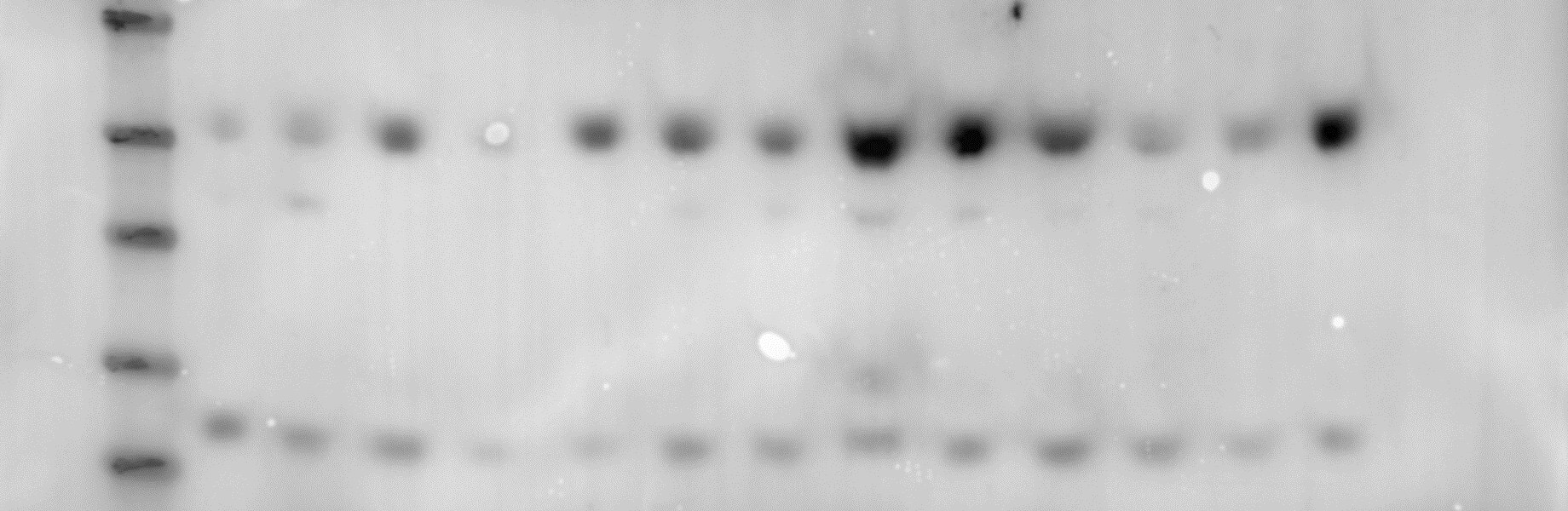
Western Blot: PPAR gamma/NR1C3 AntibodyBSA Free [NBP2-22106]
Western Blot: PPAR gamma/NR1C3 Antibody [NBP2-22106] - Total protein from 3T3-L1 mouse embryonic fibroblast adipose-like cell line, separated by 4-12% SDS-PAGE, transferred to nitrocellulose membrane and blocked in 5% non-fat milk for 1h at room temperature. The membrane was probed with anti-PPAR gamma 1:800 in non-fat milk. Lane 2-7 increasing protein concentration, undifferentiated adipocytes. Lane 8-13 increasing protein concentration, differentiated adipocytes. This image was submitted via customer review.![Immunohistochemistry-Paraffin: PPAR gamma/NR1C3 Antibody - BSA Free [NBP2-22106]](https://resources.rndsystems.com/images/products/PPAR-gamma-Antibody-Immunohistochemistry-Paraffin-NBP2-22106-img0001.jpg "Immunohistochemistry-Paraffin: PPAR gamma/NR1C3 Antibody - BSA Free [NBP2-22106]")
Immunohistochemistry-Paraffin: PPAR gamma/NR1C3 Antibody - BSA Free [NBP2-22106]
Immunohistochemistry-Paraffin: PPAR gamma Antibody [NBP2-22106] - IHC analysis of PPAR gamma in mouse liver.![Western Blot: PPAR gamma/NR1C3 AntibodyBSA Free [NBP2-22106]](https://resources.rndsystems.com/images/products/PPAR-gamma-Antibody-Western-Blot-NBP2-22106-img0002.jpg "Western Blot: PPAR gamma/NR1C3 AntibodyBSA Free [NBP2-22106]")
Western Blot: PPAR gamma/NR1C3 AntibodyBSA Free [NBP2-22106]
Western Blot: PPAR gamma Antibody [NBP2-22106] - Western blot analysis of PPAR gamma in 1. human adipose and 2. human adrenal lysate.Applications for PPAR gamma/NR1C3 Antibody - BSA Free
Application
Recommended Usage
Immunohistochemistry
1:100
Immunohistochemistry-Paraffin
1:100
Immunoprecipitation
reported in scientific literature (PMID 27862692)
Western Blot
2 ug/ml
Application Notes
In Western blot, a band is detected at ~55 kDa. Prior to immunostaining paraffin tissues, antigen retrieval with sodium citrate buffer (pH 6.0) is recommended. The observed molecular weight of the protein may vary from the listed predicted molecular weight due to post translational modifications, post translation cleavages, relative charges, and other experimental factors.
Reviewed Applications
Read 2 reviews rated 4 using NBP2-22106 in the following applications:
Formulation, Preparation, and Storage
Purification
Immunogen affinity purified
Formulation
PBS
Format
BSA Free
Preservative
0.05% Sodium Azide
Concentration
1.0 mg/ml
Shipping
The product is shipped with polar packs. Upon receipt, store it immediately at the temperature recommended below.
Stability & Storage
Store at 4C short term. Aliquot and store at -20C long term. Avoid freeze-thaw cycles.
Background: PPAR gamma/NR1C3
Long Name
Peroxisome Proliferator-activated Receptor gamma
Alternate Names
NR1C3, PPARG
Entrez Gene IDs
5468 (Human)
Gene Symbol
PPARG
UniProt
Additional PPAR gamma/NR1C3 Products
Product Documents for PPAR gamma/NR1C3 Antibody - BSA Free
Certificate of Analysis
To download a Certificate of Analysis, please enter a lot or batch number in the search box below.
Product Specific Notices for PPAR gamma/NR1C3 Antibody - BSA Free
This product is for research use only and is not approved for use in humans or in clinical diagnosis. Primary Antibodies are guaranteed for 1 year from date of receipt.
Citations for PPAR gamma/NR1C3 Antibody - BSA Free
 Powered by Bioz
Customer Reviews for PPAR gamma/NR1C3 Antibody - BSA Free (2)
4 out of 5
2 Customer Ratings
Have you used PPAR gamma/NR1C3 Antibody - BSA Free?
Submit a review and receive an Amazon gift card!
$25/€18/£15/$25CAN/¥2500 Yen for a review with an image
$10/€7/£6/$10CAN/¥1110 Yen for a review without an image
Submit a review
Customer Images
Showing
1
-
2 of
2 reviews
Showing All
Filter By:
-
Application: Western BlotSample Tested: Adipose tissueSpecies: MouseVerified Customer | Posted 10/25/2019Mouse small intestine was homogenised and protein content was quantified by a BCA assay. Twenty micrograms of protein were resolved on a 4-12% Bis-Tris gel and transferred to nitrocellulose membranes. Membranes were probed with primary antibody PPAR gamma diluted 1:1000 in 5% BSA 2, before incubation with Anti-rabbit secondary horseradish peroxidase-conjugated antibody 1:5000. Blots were visualised with Immobilon Western Chemiluminescence HRP Substrate and imaged with Syngene chemiluminescence imaging system.

-
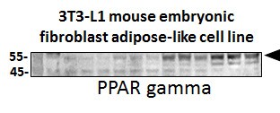
Application: Western BlotSample Tested: 3T3-L1 mouse embryonic fibroblast adipose-like cell lineSpecies: MouseVerified Customer | Posted 03/27/2018Western Blot: PPAR gamma/NR1C3 Antibody [NBP2-422106SS] - Total protein from 3T3-L1 mouse embryonic fibroblast adipose-like cell line, separated on a 4-12% gel by SDS-PAGE, transferred to nitrocellulose membrane and blocked in 5% non-fat milk for 1h at room temperature. The membrane was probed with anti-PPAR gamma 1:800 in non-fat milk.
There are no reviews that match your criteria.
Protocols
View specific protocols for PPAR gamma/NR1C3 Antibody - BSA Free (NBP2-22106):
PPAR gamma/NR1C3 Antibody:
Immunohistochemistry-Paraffin Embedded Sections
Antigen Unmasking:
Bring slides to a boil in 10 mM sodium citrate buffer (pH 6.0) then maintain at a sub-boiling temperature for 10 minutes. Cool slides on bench-top for 30 minutes.
Staining:
1. Wash sections in deionized water three times for 5 minutes each.
2. Wash sections in wash buffer for 5 minutes.
3. Block each section with 100-400 ul blocking solution for 1 hour at room temperature.
4. Remove blocking solution and add 100-400 ul diluted primary antibody. Incubate overnight at 4 C.
5. Remove antibody solution and wash sections in wash buffer three times for 5 minutes each.
6. Add 100-400 ul biotinylated diluted secondary antibody. Incubate 30 minutes at room temperature.
7. Remove secondary antibody solution and wash sections three times with wash buffer for 5 minutes each.
8. Add 100-400 ul Streptavidin-HRP reagent to each section and incubate for 30 minutes at room temperature.
9. Wash sections three times in wash buffer for 5 minutes each.
10. Add 100-400 ul DAB substrate to each section and monitor staining closely.
11. As soon as the sections develop, immerse slides in deionized water.
12. Counterstain sections in hematoxylin.
13. Wash sections in deionized water two times for 5 minutes each.
14. Dehydrate sections.
15. Mount coverslips.
PPAR gamma/NR1C3 Antibody:
Western Blot Protocol
1. Perform SDS-PAGE on samples to be analyzed, loading 25 ug of total protein per lane.
2. Transfer proteins to membrane according to the instructions provided by the manufacturer of the membrane and transfer apparatus.
3. Stain according to standard Ponceau S procedure (or similar product) to assess transfer success, and mark molecular weight standards where appropriate.
4. Rinse the blot.
5. Block the membrane using standard blocking buffer for at least 1 hour.
6. Wash the membrane in wash buffer three times for 10 minutes each.
7. Dilute anti-PPAR gamma primary antibody in blocking buffer and incubate 1 hour at room temperature.
8. Wash the membrane in wash buffer three times for 10 minutes each.
9. Apply the diluted HRP conjugated secondary antibody in blocking buffer (as per manufacturers instructions) and incubate 1 hour at room temperature.
10. Wash the blot in wash buffer three times for 10 minutes each (this step can be repeated as required to reduce background).
11. Apply the detection reagent of choice in accordance with the manufacturers instructions.
Note: Tween-20 can be added to the blocking or antibody dilution buffer at a final concentration of 0.05-0.2%.
Find general support by application which include: protocols, troubleshooting, illustrated assays, videos and webinars.
- Antigen Retrieval Protocol (PIER)
- Antigen Retrieval for Frozen Sections Protocol
- Appropriate Fixation of IHC/ICC Samples
- Cellular Response to Hypoxia Protocols
- Chromogenic IHC Staining of Formalin-Fixed Paraffin-Embedded (FFPE) Tissue Protocol
- Chromogenic Immunohistochemistry Staining of Frozen Tissue
- ClariTSA™ Fluorophore Kits
- Detection & Visualization of Antibody Binding
- Fluorescent IHC Staining of Frozen Tissue Protocol
- Graphic Protocol for Heat-induced Epitope Retrieval
- Graphic Protocol for the Preparation and Fluorescent IHC Staining of Frozen Tissue Sections
- Graphic Protocol for the Preparation and Fluorescent IHC Staining of Paraffin-embedded Tissue Sections
- Graphic Protocol for the Preparation of Gelatin-coated Slides for Histological Tissue Sections
- IHC Sample Preparation (Frozen sections vs Paraffin)
- Immunofluorescent IHC Staining of Formalin-Fixed Paraffin-Embedded (FFPE) Tissue Protocol
- Immunohistochemistry (IHC) and Immunocytochemistry (ICC) Protocols
- Immunohistochemistry Frozen Troubleshooting
- Immunohistochemistry Paraffin Troubleshooting
- Immunoprecipitation Protocol
- Preparing Samples for IHC/ICC Experiments
- Preventing Non-Specific Staining (Non-Specific Binding)
- Primary Antibody Selection & Optimization
- Protocol for Heat-Induced Epitope Retrieval (HIER)
- Protocol for Making a 4% Formaldehyde Solution in PBS
- Protocol for VisUCyte™ HRP Polymer Detection Reagent
- Protocol for the Preparation & Fixation of Cells on Coverslips
- Protocol for the Preparation and Chromogenic IHC Staining of Frozen Tissue Sections
- Protocol for the Preparation and Chromogenic IHC Staining of Frozen Tissue Sections - Graphic
- Protocol for the Preparation and Chromogenic IHC Staining of Paraffin-embedded Tissue Sections
- Protocol for the Preparation and Chromogenic IHC Staining of Paraffin-embedded Tissue Sections - Graphic
- Protocol for the Preparation and Fluorescent IHC Staining of Frozen Tissue Sections
- Protocol for the Preparation and Fluorescent IHC Staining of Paraffin-embedded Tissue Sections
- Protocol for the Preparation of Gelatin-coated Slides for Histological Tissue Sections
- R&D Systems Quality Control Western Blot Protocol
- TUNEL and Active Caspase-3 Detection by IHC/ICC Protocol
- The Importance of IHC/ICC Controls
- Troubleshooting Guide: Immunohistochemistry
- Troubleshooting Guide: Western Blot Figures
- Western Blot Conditions
- Western Blot Protocol
- Western Blot Protocol for Cell Lysates
- Western Blot Troubleshooting
- Western Blot Troubleshooting Guide
- View all Protocols, Troubleshooting, Illustrated assays and Webinars
Loading...
Associated Pathways

