PTGFR Antibody - BSA Free
Novus Biologicals | Catalog # NLS1049
![Immunocytochemistry/ Immunofluorescence: PTGFR Antibody - BSA Free [NLS1049]](https://resources.rndsystems.com/images/products/PTGFR-Antibody-Immunocytochemistry-Immunofluorescence-NLS1049-img0007.jpg "Immunocytochemistry/ Immunofluorescence: PTGFR Antibody - BSA Free [NLS1049]")
Loading...
Key Product Details
Species Reactivity
Human, Porcine, Equine, Monkey, Primate
Applications
Immunohistochemistry, Immunohistochemistry-Paraffin, Immunocytochemistry, Immunocytochemistry/ Immunofluorescence
Label
Unconjugated
Antibody Source
Polyclonal Rabbit IgG
Format
BSA Free
Loading...
Product Specifications
Immunogen
Synthetic 16 amino acid peptide from 2nd extracellular domain of human PTGFR.
Epitope
2nd extracellular domain
Reactivity Notes
Predicted cross-reactivity based on sequence identity: Mouse (88%), Rat (88%), Canine (88%), Sheep (81%), Bovine (81%), Hamster (81%), Rabbit (81%).
Specificity
Human PTGFR. BLAST analysis of the peptide immunogen showed no homology with other human proteins.
Clonality
Polyclonal
Host
Rabbit
Isotype
IgG
Description
Product can be stored undiluted at 4C for up to 1 month.
Scientific Data Images for PTGFR Antibody - BSA Free
Immunocytochemistry/ Immunofluorescence: PTGFR Antibody - BSA Free [NLS1049]
PTGFR-Antibody-Immunocytochemistry-Immunofluorescence-NLS1049-img0007.jpg![Immunohistochemistry-Paraffin: PTGFR Antibody - BSA Free [NLS1049]](https://resources.rndsystems.com/images/products/PTGFR-Antibody-Immunohistochemistry-Paraffin-NLS1049-img0005.jpg "Immunohistochemistry-Paraffin: PTGFR Antibody - BSA Free [NLS1049]")
Immunohistochemistry-Paraffin: PTGFR Antibody - BSA Free [NLS1049]
Immunohistochemistry-Paraffin: PTGFR Antibody [NLS1049] - Analysis of anti-FP / PTGFR antibody with human skin, melanoma.![Immunohistochemistry-Paraffin: PTGFR Antibody - BSA Free [NLS1049]](https://resources.rndsystems.com/images/products/PTGFR-Antibody-Immunohistochemistry-Paraffin-NLS1049-img0001.jpg "Immunohistochemistry-Paraffin: PTGFR Antibody - BSA Free [NLS1049]")
Immunohistochemistry-Paraffin: PTGFR Antibody - BSA Free [NLS1049]
Immunohistochemistry-Paraffin: PTGFR Antibody [NLS1049] - Analysis of anti-FP / PTGFR antibody with transfection control.![Immunohistochemistry-Paraffin: PTGFR Antibody - BSA Free [NLS1049]](https://resources.rndsystems.com/images/products/PTGFR-Antibody-Immunohistochemistry-Paraffin-NLS1049-img0002.jpg "Immunohistochemistry-Paraffin: PTGFR Antibody - BSA Free [NLS1049]")
Immunohistochemistry-Paraffin: PTGFR Antibody - BSA Free [NLS1049]
Immunohistochemistry-Paraffin: PTGFR Antibody [NLS1049] - Analysis of anti-FP / PTGFR antibody with human ovary.![Immunohistochemistry-Paraffin: PTGFR Antibody - BSA Free [NLS1049]](https://resources.rndsystems.com/images/products/PTGFR-Antibody-Immunohistochemistry-Paraffin-NLS1049-img0003.jpg "Immunohistochemistry-Paraffin: PTGFR Antibody - BSA Free [NLS1049]")
Immunohistochemistry-Paraffin: PTGFR Antibody - BSA Free [NLS1049]
Immunohistochemistry-Paraffin: PTGFR Antibody [NLS1049] - Analysis of anti-FP / PTGFR antibody with transfected cells expressing Prostaglandin F2 alpha Receptor at 4 ug/ ml.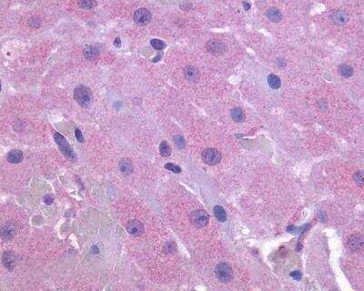
PTGFR Antibody [NLS1049] - Anti-PTGFR antibody IHC of human ovary. Immunohistochemistry of formalin-fixed, paraffin-embedded tissue after heat-induced antigen retrieval.

Western Blot: PTGFR Antibody - BSA Free [NLS1049] -
Presence of FPr in porcine corpora lutea (pCLs) at the early-luteal phase (EL-p), the mid-luteal phase (ML-p) and during pregnancy (P-p). A) Relative FPr mRNA expression. The results are presented as Delta Ct (HPRT Ct - FPr Ct). B) FPr protein content. The results are presented as AU (arbitrary units). Data represent mean ± SD. Different letters indicate statistically significant differences (P < 0.05). C) Representative Western blotting of FPr.
Western Blot: PTGFR Antibody - BSA Free [NLS1049] -
Presence of FPr in porcine corpora lutea-derived microvascular endothelial cells (pCL-MVECs) and porcine Aortic Endothelial Cells (pAECs). A) Relative FPr mRNA expression. The results are presented as Delta Ct (HPRT Ct - FPr Ct). B) FPr protein content. The results are presented as AU (arbitrary units). Data represent mean ± SD. Different letters indicate statistically significant differences (P < 0.05). C) Representative Western blotting of FPr.
Western Blot: PTGFR Antibody - BSA Free [NLS1049] -
Representative FPr immunofluorescent staining on porcine corpus luteum-microvascular endothelial cells (pCL-MVECs) isolated from the early luteal phase (EL-p) (A), the mid-luteal phase (ML-p) (B) and during pregnancy (P-p) (C) (×400). D) porcine Aortic Endothelial Cell (pAEC) culture (x400). Representative immunostaining of the FPr on non-fixed EL-p pCL-MVECs is shown (E).
Western Blot: PTGFR Antibody - BSA Free [NLS1049] -
Western Blot: PTGFR Antibody - BSA Free [NLS1049] - Presence of FPr in porcine corpora lutea (pCLs) at the early-luteal phase (EL-p), the mid-luteal phase (ML-p) & during pregnancy (P-p). A) Relative FPr mRNA expression. The results are presented as Delta Ct (HPRT Ct - FPr Ct). B) FPr protein content. The results are presented as AU (arbitrary units). Data represent mean ± SD. Different letters indicate statistically significant differences (P < 0.05). C) Representative Western blotting of FPr. Image collected & cropped by CiteAb from the following publication (https://rbej.biomedcentral.com/articles/10.1186/1477-7827-5-31), licensed under a CC-BY license. Not internally tested by Novus Biologicals.
Western Blot: PTGFR Antibody - BSA Free [NLS1049] -
Western Blot: PTGFR Antibody - BSA Free [NLS1049] - Representative FPr immunofluorescent staining on porcine corpus luteum-microvascular endothelial cells (pCL-MVECs) isolated from the early luteal phase (EL-p) (A), the mid-luteal phase (ML-p) (B) & during pregnancy (P-p) (C) (×400). D) porcine Aortic Endothelial Cell (pAEC) culture (x400). Representative immunostaining of the FPr on non-fixed EL-p pCL-MVECs is shown (E). Image collected & cropped by CiteAb from the following publication (https://rbej.biomedcentral.com/articles/10.1186/1477-7827-5-31), licensed under a CC-BY license. Not internally tested by Novus Biologicals.
Immunocytochemistry/ Immunofluorescence: PTGFR Antibody - BSA Free [NLS1049] -
Immunocytochemistry/ Immunofluorescence: PTGFR Antibody - BSA Free [NLS1049] - Representative FPr immunofluorescent staining on porcine corpus luteum-microvascular endothelial cells (pCL-MVECs) isolated from the early luteal phase (EL-p) (A), the mid-luteal phase (ML-p) (B) & during pregnancy (P-p) (C) (×400). D) porcine Aortic Endothelial Cell (pAEC) culture (x400). Representative immunostaining of the FPr on non-fixed EL-p pCL-MVECs is shown (E). Image collected & cropped by CiteAb from the following publication (https://rbej.biomedcentral.com/articles/10.1186/1477-7827-5-31), licensed under a CC-BY license. Not internally tested by Novus Biologicals.
Western Blot: PTGFR Antibody - BSA Free [NLS1049] -
Western Blot: PTGFR Antibody - BSA Free [NLS1049] - Presence of FPr in porcine corpora lutea-derived microvascular endothelial cells (pCL-MVECs) & porcine Aortic Endothelial Cells (pAECs). A) Relative FPr mRNA expression. The results are presented as Delta Ct (HPRT Ct - FPr Ct). B) FPr protein content. The results are presented as AU (arbitrary units). Data represent mean ± SD. Different letters indicate statistically significant differences (P < 0.05). C) Representative Western blotting of FPr. Image collected & cropped by CiteAb from the following publication (https://rbej.biomedcentral.com/articles/10.1186/1477-7827-5-31), licensed under a CC-BY license. Not internally tested by Novus Biologicals.Applications for PTGFR Antibody - BSA Free
Application
Recommended Usage
Immunocytochemistry
1:10 - 1:500
Immunohistochemistry-Paraffin
4-8 ug/ml
Application Notes
.
Formulation, Preparation, and Storage
Purification
Immunogen affinity purified
Formulation
PBS
Format
BSA Free
Preservative
0.1% Sodium Azide
Concentration
1.0 mg/ml
Shipping
The product is shipped with polar packs. Upon receipt, store it immediately at the temperature recommended below.
Stability & Storage
Keep as concentrated solution. Aliquot and store at -20C or below. Avoid multiple freeze-thaw cycles.
Background: PTGFR
Long Name
Prostaglandin F Receptor
Alternate Names
FP, PGF2-alpha Receptor
Entrez Gene IDs
5737 (Human)
Gene Symbol
PTGFR
Additional PTGFR Products
Product Documents for PTGFR Antibody - BSA Free
Certificate of Analysis
To download a Certificate of Analysis, please enter a lot or batch number in the search box below.
Product Specific Notices for PTGFR Antibody - BSA Free
This product is for research use only and is not approved for use in humans or in clinical diagnosis. Primary Antibodies are guaranteed for 1 year from date of receipt.
Related Research Areas
Citations for PTGFR Antibody - BSA Free
 Powered by Bioz
Customer Reviews for PTGFR Antibody - BSA Free
There are currently no reviews for this product. Be the first to review PTGFR Antibody - BSA Free and earn rewards!
Have you used PTGFR Antibody - BSA Free?
Submit a review and receive an Amazon gift card!
$25/€18/£15/$25CAN/¥2500 Yen for a review with an image
$10/€7/£6/$10CAN/¥1110 Yen for a review without an image
Submit a review
Protocols
View specific protocols for PTGFR Antibody - BSA Free (NLS1049):
Immunohistochemistry Protocol for Prostaglandin F2 alpha Receptor Antibody (NLS1049):
Immunohistochemistry
1. Prepare tissue with formalin fixation and by embedding it in paraffin wax.
2. Make 4-um sections and place on pre-cleaned and charged microscope slides.
3. Heat in a tissue-drying oven for 45 minutes at 60 degrees Celcius.
4. Deparaffinize the tissues by wash drying the slides in 3 changes of xylene approximately 5 minutes each @ RT.
5. Rehydrate the tissues by washing the slides in 3 changes of 100% alcohol approximately 3 minutes each @ RT.
6. Wash the slides in 2 changes of 95% alcohol approximately 3 minutes each @ RT.
7. Wash the slides in 1 change of 80% alcohol approximately 3 minutes @ RT.
8. Rinse the slides in gentle running distilled water approximately 5 minutes @ RT.
9. Perform antigen retrieval by steaming the slides in 0.01M sodium citrate buffer (pH 6.0) @ 99-100 degrees Celcius for 20 minutes.
10. Remove the slides from the heat and let stand in buffer @ RT for 20 minutes.
11. Rinse the slides in 1X TBS-T for 1 minute @ RT.
**Do not allow the tissues to dry at any time during the staining procedure**
12. Begin the immunostaining by applying a universal protein block approximately 20 minutes @ RT.
13. Drain protein block from the slides and apply the diluted primary antibody approximately 45 minutes @ RT.
14. Rinse the slide in 1X TBS-T approximately 1 minute @ RT.
15. Apply a biotinylated anti-rabbit IgG (H+L) secondary approximately 30 minutes @ RT.
16. Rinse the slide in 1X TBS-T approximately 1 minute at RT.
17. Apply an alkaline phosphatase steptavidin approximately 30 minutes at RT.
18. Rinse the slide in 1X TBS-T approximately 1 minute at RT.
19. Apply an alkaline phosphatase chromagen substrate approximately 30 minutes at RT.
20. Rinse the slide in distilled water approximately 1 minute @ RT.
**This method should only be used if the chromagen substrate is alcohol insoluble (ie: Vector Red, DAB)**
21. Dehydrate the tissue by washing the slides in 2 changes of 80% alcohol approximately 1 minute each @ RT.
22. Wash the slides in 2 changes of 95% alcohol approximately 1 minute each @ RT.
23. Wash the slides in 3 changes of 100% alcohol approximately 1 minute each @ RT.
24. Wash the slides in 3 changes of xyleneapproximately 1 minute each @ RT.
25. Apply cover slip.
Find general support by application which include: protocols, troubleshooting, illustrated assays, videos and webinars.
- Antigen Retrieval Protocol (PIER)
- Antigen Retrieval for Frozen Sections Protocol
- Appropriate Fixation of IHC/ICC Samples
- Cellular Response to Hypoxia Protocols
- Chromogenic IHC Staining of Formalin-Fixed Paraffin-Embedded (FFPE) Tissue Protocol
- Chromogenic Immunohistochemistry Staining of Frozen Tissue
- ClariTSA™ Fluorophore Kits
- Detection & Visualization of Antibody Binding
- Fluorescent IHC Staining of Frozen Tissue Protocol
- Graphic Protocol for Heat-induced Epitope Retrieval
- Graphic Protocol for the Preparation and Fluorescent IHC Staining of Frozen Tissue Sections
- Graphic Protocol for the Preparation and Fluorescent IHC Staining of Paraffin-embedded Tissue Sections
- Graphic Protocol for the Preparation of Gelatin-coated Slides for Histological Tissue Sections
- ICC Cell Smear Protocol for Suspension Cells
- ICC Immunocytochemistry Protocol Videos
- ICC for Adherent Cells
- IHC Sample Preparation (Frozen sections vs Paraffin)
- Immunocytochemistry (ICC) Protocol
- Immunocytochemistry Troubleshooting
- Immunofluorescence of Organoids Embedded in Cultrex Basement Membrane Extract
- Immunofluorescent IHC Staining of Formalin-Fixed Paraffin-Embedded (FFPE) Tissue Protocol
- Immunohistochemistry (IHC) and Immunocytochemistry (ICC) Protocols
- Immunohistochemistry Frozen Troubleshooting
- Immunohistochemistry Paraffin Troubleshooting
- Preparing Samples for IHC/ICC Experiments
- Preventing Non-Specific Staining (Non-Specific Binding)
- Primary Antibody Selection & Optimization
- Protocol for Heat-Induced Epitope Retrieval (HIER)
- Protocol for Making a 4% Formaldehyde Solution in PBS
- Protocol for VisUCyte™ HRP Polymer Detection Reagent
- Protocol for the Fluorescent ICC Staining of Cell Smears - Graphic
- Protocol for the Fluorescent ICC Staining of Cultured Cells on Coverslips - Graphic
- Protocol for the Preparation & Fixation of Cells on Coverslips
- Protocol for the Preparation and Chromogenic IHC Staining of Frozen Tissue Sections
- Protocol for the Preparation and Chromogenic IHC Staining of Frozen Tissue Sections - Graphic
- Protocol for the Preparation and Chromogenic IHC Staining of Paraffin-embedded Tissue Sections
- Protocol for the Preparation and Chromogenic IHC Staining of Paraffin-embedded Tissue Sections - Graphic
- Protocol for the Preparation and Fluorescent ICC Staining of Cells on Coverslips
- Protocol for the Preparation and Fluorescent ICC Staining of Non-adherent Cells
- Protocol for the Preparation and Fluorescent ICC Staining of Stem Cells on Coverslips
- Protocol for the Preparation and Fluorescent IHC Staining of Frozen Tissue Sections
- Protocol for the Preparation and Fluorescent IHC Staining of Paraffin-embedded Tissue Sections
- Protocol for the Preparation of Gelatin-coated Slides for Histological Tissue Sections
- Protocol for the Preparation of a Cell Smear for Non-adherent Cell ICC - Graphic
- TUNEL and Active Caspase-3 Detection by IHC/ICC Protocol
- The Importance of IHC/ICC Controls
- Troubleshooting Guide: Immunohistochemistry
- View all Protocols, Troubleshooting, Illustrated assays and Webinars
Loading...