PTPN13/PTPL1 Antibody - BSA Free
Novus Biologicals | Catalog # NB100-56139
![Western Blot: PTPN13/PTPL1 Antibody [NB100-56139]](https://resources.rndsystems.com/images/products/PTPN13-PTPL1-Antibody-Western-Blot-NB100-56139-img0015.jpg "Western Blot: PTPN13/PTPL1 Antibody [NB100-56139]")
Loading...
Key Product Details
Species Reactivity
Validated:
Human
Cited:
Human
Applications
Validated:
Immunohistochemistry, Immunohistochemistry-Paraffin, Immunohistochemistry-Frozen, Western Blot, Immunocytochemistry/ Immunofluorescence, Immunoprecipitation
Cited:
Western Blot, IF/IHC
Label
Unconjugated
Antibody Source
Polyclonal Rabbit IgG
Format
BSA Free
Loading...
Product Specifications
Immunogen
A recombinant protein corresponding to amino acids 1279 to 1883 of human FAP-1 protein was used as immunogen; GenBank no. NP_542414.1.
Clonality
Polyclonal
Host
Rabbit
Isotype
IgG
Scientific Data Images for PTPN13/PTPL1 Antibody - BSA Free
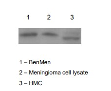
Western Blot: PTPN13/PTPL1 Antibody [NB100-56139]
Western Blot: PTPN13/PTPL1 Antibody - Unpurified [NB100-56139] - PTPN13/PTPL1 Antibody [NB100-56139] - Analysis of PTPN13/PTPL1 using this antibody. In ovarian carcinoma cell lines PTPN13/PTPL1 expression was detected in SK-OV-3 and OVCAR-3, but not in BG-1 or HEY. Human Jurkat T and 293 kidney cell lines were used as negative and positive controls, respectively.![Immunohistochemistry-Paraffin: PTPN13/PTPL1 Antibody [NB100-56139]](https://resources.rndsystems.com/images/products/PTPN13-PTPL1-Antibody-Immunohistochemistry-Paraffin-NB100-56139-img0013.jpg "Immunohistochemistry-Paraffin: PTPN13/PTPL1 Antibody [NB100-56139]")
Immunohistochemistry-Paraffin: PTPN13/PTPL1 Antibody [NB100-56139]
Immunohistochemistry-Paraffin: PTPN13/PTPL1 Antibody - Unpurified [NB100-56139] - PTPN13/PTPL1 Antibody [NB100-56139] - Analysis of PTPN13/PTPL1 in cell lines using this antibody at 1:2000. In ovarian carcinoma cell lines PTPN13/PTPL1, expression was detected in SK-OV-3 and OVCAR-3, but not in BG-1 or HEY. Human 293 kidney and Jurkat T cell lines were used as positive and negative controls, respectively.![Immunohistochemistry-Paraffin: PTPN13/PTPL1 Antibody [NB100-56139]](https://resources.rndsystems.com/images/products/PTPN13-PTPL1-Antibody-Immunohistochemistry-Paraffin-NB100-56139-img0011.jpg "Immunohistochemistry-Paraffin: PTPN13/PTPL1 Antibody [NB100-56139]")
Immunohistochemistry-Paraffin: PTPN13/PTPL1 Antibody [NB100-56139]
Immunohistochemistry-Paraffin: PTPN13/PTPL1 Antibody - Unpurified [NB100-56139] - PTPN13/PTPL1 Antibody [NB100-56139] - Ovarian carcinoma cores from a tissue microarray at 1:2000. A-D, samples are from four different patients. A1-D1 are high magnification images from A-D, respectively. Hematoxylin-eosin counterstain.Applications for PTPN13/PTPL1 Antibody - BSA Free
Application
Recommended Usage
Immunocytochemistry/ Immunofluorescence
1:500-1:3000
Immunohistochemistry
1:10-1:500
Immunohistochemistry-Frozen
1:10-1:500
Immunohistochemistry-Paraffin
1:1000-1:5000
Immunoprecipitation
1:50-1:200
Western Blot
1:1000-1:2000
Application Notes
FAP-1 typically migrates at ~270 kDa on western blots. Thus for SDS-PAGE and western blot, the researcher should use techniques optimized for the transfer of proteins with high molecular weights. The actual molecular weight of FAP-1 observed depends on the isoform expression.
Reviewed Applications
Read 1 review rated 4 using NB100-56139 in the following applications:
Formulation, Preparation, and Storage
Purification
Unpurified
Formulation
Whole antisera
Format
BSA Free
Preservative
0.05% Sodium Azide
Concentration
This product is unpurified. The exact concentration of antibody is not quantifiable.
Shipping
The product is shipped with polar packs. Upon receipt, store it immediately at the temperature recommended below.
Stability & Storage
Store at -20C. Avoid freeze-thaw cycles.
Background: PTPN13/PTPL1
Long Name
Protein Tyrosine Phosphatase, Non-receptor Type 13
Alternate Names
EAP-1, FAP-1, PNP1, PTP-BAS, PTP-BL, PTP1E, PTPL1, PTPLE
Entrez Gene IDs
5783 (Human)
Gene Symbol
PTPN13
UniProt
Additional PTPN13/PTPL1 Products
Product Documents for PTPN13/PTPL1 Antibody - BSA Free
Certificate of Analysis
To download a Certificate of Analysis, please enter a lot or batch number in the search box below.
Product Specific Notices for PTPN13/PTPL1 Antibody - BSA Free
This product is for research use only and is not approved for use in humans or in clinical diagnosis. Primary Antibodies are guaranteed for 1 year from date of receipt.
Related Research Areas
Citations for PTPN13/PTPL1 Antibody - BSA Free
 Powered by Bioz
Customer Reviews for PTPN13/PTPL1 Antibody - BSA Free (1)
4 out of 5
1 Customer Rating
Have you used PTPN13/PTPL1 Antibody - BSA Free?
Submit a review and receive an Amazon gift card!
$25/€18/£15/$25CAN/¥2500 Yen for a review with an image
$10/€7/£6/$10CAN/¥1110 Yen for a review without an image
Submit a review
Customer Images
Showing
1
-
1 of
1 review
Showing All
Filter By:
-
Application: Western BlotSample Tested: 293T cell lysateSpecies: HumanVerified Customer | Posted 09/30/2019The protein content of all samples were quantified prior to loading, and standardized at concentrations of 40ug.

There are no reviews that match your criteria.
Protocols
Find general support by application which include: protocols, troubleshooting, illustrated assays, videos and webinars.
- Antigen Retrieval Protocol (PIER)
- Antigen Retrieval for Frozen Sections Protocol
- Appropriate Fixation of IHC/ICC Samples
- Cellular Response to Hypoxia Protocols
- Chromogenic IHC Staining of Formalin-Fixed Paraffin-Embedded (FFPE) Tissue Protocol
- Chromogenic Immunohistochemistry Staining of Frozen Tissue
- ClariTSA™ Fluorophore Kits
- Detection & Visualization of Antibody Binding
- Fluorescent IHC Staining of Frozen Tissue Protocol
- Graphic Protocol for Heat-induced Epitope Retrieval
- Graphic Protocol for the Preparation and Fluorescent IHC Staining of Frozen Tissue Sections
- Graphic Protocol for the Preparation and Fluorescent IHC Staining of Paraffin-embedded Tissue Sections
- Graphic Protocol for the Preparation of Gelatin-coated Slides for Histological Tissue Sections
- ICC Cell Smear Protocol for Suspension Cells
- ICC Immunocytochemistry Protocol Videos
- ICC for Adherent Cells
- IHC Sample Preparation (Frozen sections vs Paraffin)
- Immunocytochemistry (ICC) Protocol
- Immunocytochemistry Troubleshooting
- Immunofluorescence of Organoids Embedded in Cultrex Basement Membrane Extract
- Immunofluorescent IHC Staining of Formalin-Fixed Paraffin-Embedded (FFPE) Tissue Protocol
- Immunohistochemistry (IHC) and Immunocytochemistry (ICC) Protocols
- Immunohistochemistry Frozen Troubleshooting
- Immunohistochemistry Paraffin Troubleshooting
- Immunoprecipitation Protocol
- Preparing Samples for IHC/ICC Experiments
- Preventing Non-Specific Staining (Non-Specific Binding)
- Primary Antibody Selection & Optimization
- Protocol for Heat-Induced Epitope Retrieval (HIER)
- Protocol for Making a 4% Formaldehyde Solution in PBS
- Protocol for VisUCyte™ HRP Polymer Detection Reagent
- Protocol for the Fluorescent ICC Staining of Cell Smears - Graphic
- Protocol for the Fluorescent ICC Staining of Cultured Cells on Coverslips - Graphic
- Protocol for the Preparation & Fixation of Cells on Coverslips
- Protocol for the Preparation and Chromogenic IHC Staining of Frozen Tissue Sections
- Protocol for the Preparation and Chromogenic IHC Staining of Frozen Tissue Sections - Graphic
- Protocol for the Preparation and Chromogenic IHC Staining of Paraffin-embedded Tissue Sections
- Protocol for the Preparation and Chromogenic IHC Staining of Paraffin-embedded Tissue Sections - Graphic
- Protocol for the Preparation and Fluorescent ICC Staining of Cells on Coverslips
- Protocol for the Preparation and Fluorescent ICC Staining of Non-adherent Cells
- Protocol for the Preparation and Fluorescent ICC Staining of Stem Cells on Coverslips
- Protocol for the Preparation and Fluorescent IHC Staining of Frozen Tissue Sections
- Protocol for the Preparation and Fluorescent IHC Staining of Paraffin-embedded Tissue Sections
- Protocol for the Preparation of Gelatin-coated Slides for Histological Tissue Sections
- Protocol for the Preparation of a Cell Smear for Non-adherent Cell ICC - Graphic
- R&D Systems Quality Control Western Blot Protocol
- TUNEL and Active Caspase-3 Detection by IHC/ICC Protocol
- The Importance of IHC/ICC Controls
- Troubleshooting Guide: Immunohistochemistry
- Troubleshooting Guide: Western Blot Figures
- Western Blot Conditions
- Western Blot Protocol
- Western Blot Protocol for Cell Lysates
- Western Blot Troubleshooting
- Western Blot Troubleshooting Guide
- View all Protocols, Troubleshooting, Illustrated assays and Webinars
Loading...