Pyruvate Dehydrogenase E1-alpha subunit [p Ser293] Antibody - BSA Free
Novus Biologicals | Catalog # NB110-93479
Key Product Details
Validated by
Species Reactivity
Validated:
Cited:
Applications
Validated:
Cited:
Label
Antibody Source
Format
Product Specifications
Immunogen
Modification
Localization
Specificity
Clonality
Host
Isotype
Theoretical MW
Disclaimer note: The observed molecular weight of the protein may vary from the listed predicted molecular weight due to post translational modifications, post translation cleavages, relative charges, and other experimental factors.
Scientific Data Images
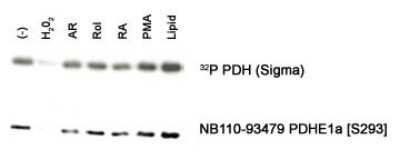
![Western Blot: Pyruvate Dehydrogenase E1-alpha subunit [p Ser293] AntibodyBSA Free [NB110-93479]](https://resources.rndsystems.com/images/products/Pyruvate-Dehydrogenase-E1-alpha-subunit-[p-Ser293]-Antibody-Western-Blot-NB110-93479-img0008.jpg "Western Blot: Pyruvate Dehydrogenase E1-alpha subunit [p Ser293] AntibodyBSA Free [NB110-93479]")
Western Blot: Pyruvate Dehydrogenase E1-alpha subunit [p Ser293] AntibodyBSA Free [NB110-93479]
Pyruvate-Dehydrogenase-E1-alpha-subunit-[p-Ser293]-Antibody-Western-Blot-NB110-93479-img0008.jpg![Pyruvate Dehydrogenase E1-alpha subunit [p Ser293] Antibody - BSA Free](https://resources.rndsystems.com/images/products/nb110-93479_rabbit-polyclonal-pyruvate-dehydrogenase-e1-alpha-subunit-p-ser293-antibody-310202416212313.jpg "Western Blot: Pyruvate Dehydrogenase E1-alpha subunit [p Ser293] Antibody - BSA Free [NB110-93479] -")
Western Blot: Pyruvate Dehydrogenase E1-alpha subunit [p Ser293] Antibody - BSA Free [NB110-93479] -
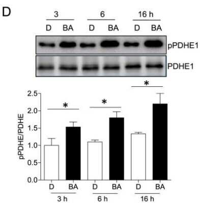
Western Blot: Pyruvate Dehydrogenase E1-alpha subunit [p Ser293] Antibody - BSA Free [NB110-93479] - PDK4 expression during syncytialization of human placental trophoblasts. (A) Changes of the RPKM values for PDK family members in trophoblasts before (red column, 3 hours) & after (blue column, 48 hours) syncytialization. (B) Changes in PDK4 mRNA (black column, n = 4) & protein (white column, n = 4) abundance during syncytialization. (C) Changes in the phosphorylation of PDHE1 alpha during syncytialization n = 4. (D) Representative images showing intense staining of PDK4 (red) in the cytotrophoblast layer & weak staining of PDK4 in the syncytial layer of human chorionic villi at early gestation. The syncytial & cytotrophoblast layers were labeled with beta -hCG (green) & SPINT1 (green) respectively. Nuclei were counterstained with DAPI (blue). n = 3; *P < 0.05; **P < 0.01; ***P < 0.001 against 3 hours; n.s., not significant. Image collected & cropped by CiteAb from the following publication (https://www.nature.com/articles/s41598-017-09163-8), licensed under a CC-BY license. Not internally tested by Novus Biologicals.Applications
Flow Cytometry
Immunocytochemistry/ Immunofluorescence
Immunoprecipitation
Knockout Validated
Western Blot
Reviewed Applications
Read 5 reviews rated 4.8 using NB110-93479 in the following applications:
Flow Cytometry Panel Builder
Bio-Techne Knows Flow Cytometry
Save time and reduce costly mistakes by quickly finding compatible reagents using the Panel Builder Tool.
Advanced Features
- Spectra Viewer - Custom analysis of spectra from multiple fluorochromes
- Spillover Popups - Visualize the spectra of individual fluorochromes
- Antigen Density Selector - Match fluorochrome brightness with antigen density

Formulation, Preparation, and Storage
Purification
Formulation
Format
Preservative
Concentration
Shipping
Stability & Storage
Background: Pyruvate Dehydrogenase E1-alpha subunit
Alternate Names
Gene Symbol
Additional Pyruvate Dehydrogenase E1-alpha subunit Products
Product Documents
Certificate of Analysis
To download a Certificate of Analysis, please enter a lot or batch number in the search box below.
Product Specific Notices
This product is for research use only and is not approved for use in humans or in clinical diagnosis. Primary Antibodies are guaranteed for 1 year from date of receipt.
Citations for Pyruvate Dehydrogenase E1-alpha subunit [p Ser293] Antibody - BSA Free
 Powered by Bioz
Customer Reviews (5)
Have you used Pyruvate Dehydrogenase E1-alpha subunit [p Ser293] Antibody - BSA Free?
Submit a review and receive an Amazon gift card!
$25/€18/£15/$25CAN/¥2500 Yen for a review with an image
$10/€7/£6/$10CAN/¥1110 Yen for a review without an image
Submit a review
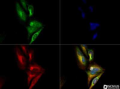
Customer Images
![Pyruvate Dehydrogenase E1-alpha subunit [p Ser293] Antibody - BSA Free NB110-93479](https://resources.rndsystems.com/images/reviews/review_nb110-93479_42236_0_0.png)
![Pyruvate Dehydrogenase E1-alpha subunit [p Ser293] Antibody - BSA Free NB110-93479](https://resources.rndsystems.com/images/reviews/review_nb110-93479_41841_0_0_0_0_0.jpg)
![Pyruvate Dehydrogenase E1-alpha subunit [p Ser293] Antibody - BSA Free NB110-93479](https://resources.rndsystems.com/images/reviews/Western-Blot_Pyruvate_Dehydrogenase_E1-alpha_subunit_NB110-93479_24181.jpg)
![Pyruvate Dehydrogenase E1-alpha subunit [p Ser293] Antibody - BSA Free NB110-93479](https://resources.rndsystems.com/images/reviews/review_image_314831_5846_1389721268.jpg)
![Pyruvate Dehydrogenase E1-alpha subunit [p Ser293] Antibody - BSA Free NB110-93479](https://resources.rndsystems.com/images/reviews/review_image_9127_411_1281624005.jpeg)
-
Application: Western BlotSample Tested: placenta cellsSpecies: HumanVerified Customer | Posted 07/10/2018
![Pyruvate Dehydrogenase E1-alpha subunit [p Ser293] Antibody - BSA Free NB110-93479](data:image/png;base64,R0lGODlhAQABAAD/ACwAAAAAAQABAAACADs=)
-
Application: Western BlotSample Tested: NTera-2 human testicular embryonic carcinoma cell line and Differentiated Neuronal NTERA2 cellsSpecies: HumanVerified Customer | Posted 06/12/2018The site was similar in PDHA2 and we IP PDHA2 and its mutants at the site and probed with the antibody
-
Application: Western BlotSample Tested: primary mouse macrophage lysatesSpecies: MouseVerified Customer | Posted 05/12/2016PDHE1a Ser 293 antibody- mouse macrophages
-
Application: Western BlotSample Tested: rat Sertoli cellsSpecies: RatVerified Customer | Posted 01/14/2014
-
Application: Western BlotSample Tested: MCF10A whole cell lysate, Sample Amount: 30ugSpecies: HumanVerified Customer | Posted 08/12/2010
There are no reviews that match your criteria.
Protocols
Find general support by application which include: protocols, troubleshooting, illustrated assays, videos and webinars.
- 7-Amino Actinomycin D (7-AAD) Cell Viability Flow Cytometry Protocol
- Appropriate Fixation of IHC/ICC Samples
- Cellular Response to Hypoxia Protocols
- ClariTSA™ Fluorophore Kits
- Detection & Visualization of Antibody Binding
- Extracellular Membrane Flow Cytometry Protocol
- Flow Cytometry Protocol for Cell Surface Markers
- Flow Cytometry Protocol for Staining Membrane Associated Proteins
- Flow Cytometry Staining Protocols
- Flow Cytometry Troubleshooting Guide
- ICC Cell Smear Protocol for Suspension Cells
- ICC Immunocytochemistry Protocol Videos
- ICC for Adherent Cells
- Immunocytochemistry (ICC) Protocol
- Immunocytochemistry Troubleshooting
- Immunofluorescence of Organoids Embedded in Cultrex Basement Membrane Extract
- Immunohistochemistry (IHC) and Immunocytochemistry (ICC) Protocols
- Immunoprecipitation Protocol
- Intracellular Flow Cytometry Protocol Using Alcohol (Methanol)
- Intracellular Flow Cytometry Protocol Using Detergents
- Intracellular Nuclear Staining Flow Cytometry Protocol Using Detergents
- Intracellular Staining Flow Cytometry Protocol Using Alcohol Permeabilization
- Intracellular Staining Flow Cytometry Protocol Using Detergents to Permeabilize Cells
- Preparing Samples for IHC/ICC Experiments
- Preventing Non-Specific Staining (Non-Specific Binding)
- Primary Antibody Selection & Optimization
- Propidium Iodide Cell Viability Flow Cytometry Protocol
- Protocol for Liperfluo
- Protocol for VisUCyte™ HRP Polymer Detection Reagent
- Protocol for the Characterization of Human Th22 Cells
- Protocol for the Characterization of Human Th9 Cells
- Protocol for the Fluorescent ICC Staining of Cell Smears - Graphic
- Protocol for the Fluorescent ICC Staining of Cultured Cells on Coverslips - Graphic
- Protocol for the Preparation and Fluorescent ICC Staining of Cells on Coverslips
- Protocol for the Preparation and Fluorescent ICC Staining of Non-adherent Cells
- Protocol for the Preparation and Fluorescent ICC Staining of Stem Cells on Coverslips
- Protocol for the Preparation of a Cell Smear for Non-adherent Cell ICC - Graphic
- Protocol: Annexin V and PI Staining by Flow Cytometry
- Protocol: Annexin V and PI Staining for Apoptosis by Flow Cytometry
- R&D Systems Quality Control Western Blot Protocol
- TUNEL and Active Caspase-3 Detection by IHC/ICC Protocol
- The Importance of IHC/ICC Controls
- Troubleshooting Guide: Fluorokine Flow Cytometry Kits
- Troubleshooting Guide: Western Blot Figures
- Western Blot Conditions
- Western Blot Protocol
- Western Blot Protocol for Cell Lysates
- Western Blot Troubleshooting
- Western Blot Troubleshooting Guide
- View all Protocols, Troubleshooting, Illustrated assays and Webinars