Best Seller
RBFOX3/NeuN Antibody (1B7) - BSA Free
Novus Biologicals | Catalog # NBP1-92693

Key Product Details
Species Reactivity
Validated:
Human, Mouse, Rat
Cited:
Human, Mouse, Rat
Applications
Validated:
Immunohistochemistry, Immunohistochemistry-Paraffin, Immunohistochemistry-Frozen, Western Blot, Flow Cytometry, Flow (Intracellular), Immunocytochemistry/ Immunofluorescence, CyTOF-ready
Cited:
Immunohistochemistry, Immunohistochemistry-Paraffin, Immunohistochemistry-Frozen, Immunohistochemistry Free-Floating, Western Blot, Flow Cytometry, Immunofluorescence, Immunocytochemistry/ Immunofluorescence, Chromatin Immunoprecipitation (ChIP), IF/IHC
Label
Unconjugated
Antibody Source
Monoclonal Mouse IgG2B Clone # 1B7
Format
BSA Free
Loading...
Product Specifications
Immunogen
N-terminal 99 amino acids of human FOX3 as expressed in and purified from E. coli
Localization
Nucleus. Cytoplasm.
Marker
Neuronal Marker
Clonality
Monoclonal
Host
Mouse
Isotype
IgG2B
Theoretical MW
33.8 kDa.
Disclaimer note: The observed molecular weight of the protein may vary from the listed predicted molecular weight due to post translational modifications, post translation cleavages, relative charges, and other experimental factors.
Disclaimer note: The observed molecular weight of the protein may vary from the listed predicted molecular weight due to post translational modifications, post translation cleavages, relative charges, and other experimental factors.
Scientific Data Images for RBFOX3/NeuN Antibody (1B7) - BSA Free
Immunohistochemical Staining of RBFOX3/NeuN in Paraffin Embedded Rat Brain Stem
Formalin fixed rat brain stem neurons stained with to NeuN in green and counter stained with, our chicken polyclonal antibody to microtubule associated protein 2 in red. The nuclei of cells are revealed with DAPI in blue. The antibody reveals strong nuclear and distal cytoplasmic staining for Fox3/NeuN and the complete absence of staining of other cell types. The MAP2 antibody binds to dendrites and overlaps with Fox3 staining in perikarya. This Fox3/NeuN antibody is therefore an excellent marker of neuronal cells.
Immunohistochemical Staining of RBFOX3/NeuN in Mouse Spinal Cords
RBFOX3-NeuN-Antibody-1B7-Immunohistochemistry-NBP1-92693-img0023.jpg
Western Blot Detection of RBFOX3/NeuN Mouse and Rat Brain Tissue Lysates
Analysis of whole brain tissue lysates using mouse mAb to FOX3/NeuN NBP1-92693, dilution 1:1,000 in green: [1] protein standard (red), [2] adult rat brain, [3] embryonic E20 rat brain, [4] adult mouse brain. Note the strong twin bands corresponding to the two alternate transcripts of FOX3/NeuN protein with apparent SDS-PAGE molecular weights of 46 and 48kDa. As with other FOX3/NeuN antibodies, an additional band at ~70kDa is revealed in some lysates.
Flow Cytometry of SH-SY5Y Cells Stained with Allophycocyanin Conjugated RBFOX3/NeuN Antibody
Using the Allophycocyanin direct conjugate An intracellular stain was performed on SH-SY5Y cells with RBFOXP3/NeuN (1B7) antibody NBP1-92693APC (blue) and a matched isotype control NB600-986APC (orange). Cells were fixed with 4% PFA and then permeablized with 0.1% saponin. Cells were incubated in an antibody dilution of 1 ug/mL for 30 minutes at room temperature. Both antibodies were conjugated to Allophycocyanin.
Flow Cytometry of SH-SY5Y Cells Stained with Alexa Fluor 647 Conjugated RBFOX3/NeuN Antibody
An intracellular stain was performed on SH-SY5Y cells with RBFOXP3/NeuN (1B7) antibody NBP1-92693AF647 (blue) and a matched isotype control (orange). Cells were fixed with 4% PFA and then permeablized with 0.1% saponin. Cells were incubated in an antibody dilution of 2.5 ug/mL for 30 minutes at room temperature. Both antibodies were conjugated to Alexa Fluor 647.
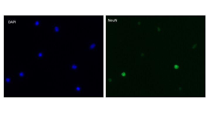
Immunocytochemistry/Immunofluorescence Imaging of RBFOX3/NeuN in Dissociated Spinal Cord Nuclei
Dissociated spinal cord nuclei immunostained with anti-NeuN antibody and Alexa 488 secondary. DAPI (Blue) to stain nuclei. This image was submitted by customer review.
Flow Cytometry of U87-MG Cells Stained with Alexa Fluor 700 Conjugated RBFOX3/NeuN Antibody
An intracellular stain was performed on U87-MG cells with RBFOXP3/NeuN (1B7) antibody NBP1-92693AF700 (blue) and a matched isotype control (orange). Cells were fixed with 4% PFA and then permeablized with 0.1% saponin. Cells were incubated in an antibody dilution of 10 ug/mL for 30 minutes at room temperature. Both antibodies were conjugated to Alexa Fluor 700.
Immunocytochemistry/Immunofluorescence Staining of RBFOX3/NeuN in Rat Brain Neural Cultures
Rat brain neural cultures stained with NBP1-92693 (red), a chicken polyclonal antibody to GFAP (NBP1-05198, green) and DAPI (blue). NBP1-92693 reveals strong nuclear and distal cytoplasmic staining for RBFOX/Neun and the complete absence of staining of astrocytes, which are staining with the GFAP antibody, and other kinds of non-neuronal cells. This RBFOX/Neun (1B7) antibody is therefore an excellent marker of neuronal cells.
Immunohistochemical Analysis of RBFOX3/NeuN in Frozen Mouse Spinal Cord
IHC-Fr analysis of a 4% PFA fixed mouse spinal cord tissue section using RBFOX3/NeuN antibody (clone 1B7) at 1:500 dilution with overnight 4C incubation in a dilution buffer which contained BSA 1% and 0.2% TritonX100 in PBS. The signal was detected using Alexa Fluor 594 conjugated goat anti-mouse IgG (H+L) secondary antibody. The antibody generated a specific cytoplasmic-nuclear signal and the staining was more intense in the nuclei of the neurons. This image was submitted as a review via a verified end user of this product.
Flow Cytometry of SH-SY5Y Cells Stained with Phycoerythrin Conjugated RBFOX3/NeuN Antibody
Analysis of PE conjugate of NBP1-92693. An intracellular stain was performed on SH-SY5Y cells with RBFOX3 (1B7) antibody NBP1-92693PE (blue) and a matched isotype control NB600-986PE (orange). Cells were fixed with 4% PFA and then permeablized with 0.1% saponin. Cells were incubated in an antibody dilution of 1 ug/mL for 30 minutes at room temperature. Both antibodies were conjugated to Phycoerythrin.
Flow Cytometry of SH-SY5Y Cells Stained with PerCP Conjugated RBFOX3/NeuN Antibody
Using the PerCP direct conjugate An intracellular stain was performed on SH-SY5Y cells with RBFOXP3/NeuN (1B7) antibody NBP1-92693PCP (blue) and a matched isotype control NB600-986PCP (orange). Cells were fixed with 4% PFA and then permeablized with 0.1% saponin. Cells were incubated in an antibody dilution of 10 ug/mL for 30 minutes at room temperature. Both antibodies were conjugated to Peridinin-Chlorophyll-protein.
Flow Cytometry of SH-SY5Y Cells Stained with Alexa Fluor 700 Conjugated RBFOX3/NeuN Antibody
An intracellular stain was performed on SH-SY5Y cells with RBFOXP3/NeuN (1B7) antibody NBP1-92693AF700 (blue) and a matched isotype control (orange). Cells were fixed with 4% PFA and then permeablized with 0.1% saponin. Cells were incubated in an antibody dilution of 10 ug/mL for 30 minutes at room temperature. Both antibodies were conjugated to Alexa Fluor 700. Image from the Alexa Fluor 700 version of this antibody.
Flow Cytometry of A549 Cells Stained with FITC Conjugated RBFOX3/NeuN Antibody
An intracellular stain was performed on A549 cells with RBFOXP3/NeuN (1B7) antibody NBP1-92693F (blue) and a matched isotype control (orange). Cells were fixed with 4% PFA and then permeablized with 0.1% saponin. Cells were incubated in an antibody dilution of 10 ug/mL for 30 minutes at room temperature. Both antibodies were conjugated to FITC. [NBP1-92693] -")
Immunocytochemistry/ Immunofluorescence: RBFOX3/NeuN Antibody (1B7) [NBP1-92693] -
Immunocytochemistry/ Immunofluorescence: RBFOX3/NeuN Antibody (1B7) [NBP1-92693] - NF kappa B localizes to both neuronal & glial nuclei in superior colliculus. Representative confocal images of NF kappa B in the superior colliculus (SC) of vehicle- & HE3286-treated rats with all nuclei indicated (DAPI). Sections were also labeled for astrocytes (A, GFAP), microglia (B, Iba1), or neurons using antibodies against either NeuN (C) or phosphorylated neurofilament heavy (pNFH; D). Representative nuclei from each cell class demonstrated NF kappa B localization (arrows). Insets (solid white, B) show region contained within dashed box with red channel removed to better visualize nuclear localization of NF kappa B. Scale: 10 μm. Image collected & cropped by CiteAb from the following publication (http://journal.frontiersin.org/article/10.3389/fnins.2017.00045/full), licensed under a CC-BY license. Not internally tested by Novus Biologicals.Applications for RBFOX3/NeuN Antibody (1B7) - BSA Free
Application
Recommended Usage
Flow Cytometry
1:10
Immunocytochemistry/ Immunofluorescence
1:500-1:1000
Immunohistochemistry
1:400
Immunohistochemistry-Frozen
Reported in scientific literature (PMID: 30654114/ 23776455)
Western Blot
1:5000-1:10000
Application Notes
This RBFOX3/Neun (1B7) antibody is useful for Immunocytochemistry/Immunofluorescence, Immunohistochemistry, and Western blot, where bands can be seen at 46 and 48 kDa. Flow Cytometry was reported in verified customer review using Alexa Fluor 700 conjugated form of this antibody, NBP1-92693AF700. This antibody is CyTOF ready.
Reviewed Applications
Read 2 reviews rated 4.5 using NBP1-92693 in the following applications:
Flow Cytometry Panel Builder
Bio-Techne Knows Flow Cytometry
Save time and reduce costly mistakes by quickly finding compatible reagents using the Panel Builder Tool.
Advanced Features
- Spectra Viewer - Custom analysis of spectra from multiple fluorochromes
- Spillover Popups - Visualize the spectra of individual fluorochromes
- Antigen Density Selector - Match fluorochrome brightness with antigen density

Formulation, Preparation, and Storage
Purification
Immunogen affinity purified
Formulation
50% PBS, 50% glycerol
Format
BSA Free
Preservative
5mM Sodium Azide
Concentration
1 mg/ml
Shipping
The product is shipped with polar packs. Upon receipt, store it immediately at the temperature recommended below.
Stability & Storage
Store at 4C short term. Aliquot and store at -20C long term. Avoid freeze-thaw cycles.
Background: RBFOX3/NeuN
Rbfox proteins participate in regulation of alternative splicing between family members as well as in autoregulation. While the breadth of brain and muscle specific targets for alternative splicing is well established for Rbfox proteins, those specific to Rbfox3/NeuN along with its alternative splicing mechanism are less understood. Rbfox3 has been shown to regulate neuronal differentiation by alternative splicing of Numb pre-mRNA and has a role in adult neurogenesis. Dysfunctional Rbfox3/NeuN has been associated with various neurological disorders such as neurodevelopmental delay, autism spectrum disorder, Benign rolandic epilepsy (BRE), and cognitive impairments (1,2).
References
1. Duan W, Zhang YP, Hou Z, Huang C, Zhu H, Zhang CQ, Yin Q. (2016) Novel Insights into NeuN: from Neuronal Marker to Splicing Regulator. Mol Neurobiol. 53(3):1637-1647. PMID: 25680637.
2. Su CH, D D, Tarn WY. (2018) Alternative Splicing in Neurogenesis and Brain Development. Front Mol Biosci. 5:12. PMID: 29484299
Long Name
RNA binding fox-1 homolog 3
Alternate Names
FOX-3, FOX3, HRNBP3, NEUN, NeuN flow cytometry
Entrez Gene IDs
146713 (Human)
Gene Symbol
RBFOX3
UniProt
Additional RBFOX3/NeuN Products
Product Documents for RBFOX3/NeuN Antibody (1B7) - BSA Free
Certificate of Analysis
To download a Certificate of Analysis, please enter a lot or batch number in the search box below.
Product Specific Notices for RBFOX3/NeuN Antibody (1B7) - BSA Free
This product is for research use only and is not approved for use in humans or in clinical diagnosis. Primary Antibodies are guaranteed for 1 year from date of receipt.
Citations for RBFOX3/NeuN Antibody (1B7) - BSA Free
 Powered by Bioz
Customer Reviews for RBFOX3/NeuN Antibody (1B7) - BSA Free (2)
4.5 out of 5
2 Customer Ratings
Have you used RBFOX3/NeuN Antibody (1B7) - BSA Free?
Submit a review and receive an Amazon gift card!
$25/€18/£15/$25CAN/¥2500 Yen for a review with an image
$10/€7/£6/$10CAN/¥1110 Yen for a review without an image
Submit a review
Customer Images
Showing
1
-
2 of
2 reviews
Showing All
Filter By:
-
Application: Immunostaining of dissociated spinal nucleiSample Tested: Dissociated spinal cord nucleiSpecies: MouseVerified Customer | Posted 01/26/2018Dissociated spinal cord nuclei immunostained with anti-NeuN antibody and Alexa 488 secondary. DAPI (Blue) to stain nuclei.

-
Application: Immunohistochemistry-FrozenSample Tested: mouse spinal cordSpecies: MouseVerified Customer | Posted 05/11/2016
There are no reviews that match your criteria.
Protocols
Find general support by application which include: protocols, troubleshooting, illustrated assays, videos and webinars.
- 7-Amino Actinomycin D (7-AAD) Cell Viability Flow Cytometry Protocol
- Antigen Retrieval Protocol (PIER)
- Antigen Retrieval for Frozen Sections Protocol
- Appropriate Fixation of IHC/ICC Samples
- Cellular Response to Hypoxia Protocols
- Chromogenic IHC Staining of Formalin-Fixed Paraffin-Embedded (FFPE) Tissue Protocol
- Chromogenic Immunohistochemistry Staining of Frozen Tissue
- ClariTSA™ Fluorophore Kits
- Detection & Visualization of Antibody Binding
- Extracellular Membrane Flow Cytometry Protocol
- Flow Cytometry Protocol for Cell Surface Markers
- Flow Cytometry Protocol for Staining Membrane Associated Proteins
- Flow Cytometry Staining Protocols
- Flow Cytometry Troubleshooting Guide
- Fluorescent IHC Staining of Frozen Tissue Protocol
- Graphic Protocol for Heat-induced Epitope Retrieval
- Graphic Protocol for the Preparation and Fluorescent IHC Staining of Frozen Tissue Sections
- Graphic Protocol for the Preparation and Fluorescent IHC Staining of Paraffin-embedded Tissue Sections
- Graphic Protocol for the Preparation of Gelatin-coated Slides for Histological Tissue Sections
- ICC Cell Smear Protocol for Suspension Cells
- ICC Immunocytochemistry Protocol Videos
- ICC for Adherent Cells
- IHC Sample Preparation (Frozen sections vs Paraffin)
- Immunocytochemistry (ICC) Protocol
- Immunocytochemistry Troubleshooting
- Immunofluorescence of Organoids Embedded in Cultrex Basement Membrane Extract
- Immunofluorescent IHC Staining of Formalin-Fixed Paraffin-Embedded (FFPE) Tissue Protocol
- Immunohistochemistry (IHC) and Immunocytochemistry (ICC) Protocols
- Immunohistochemistry Frozen Troubleshooting
- Immunohistochemistry Paraffin Troubleshooting
- Intracellular Flow Cytometry Protocol Using Alcohol (Methanol)
- Intracellular Flow Cytometry Protocol Using Detergents
- Intracellular Nuclear Staining Flow Cytometry Protocol Using Detergents
- Intracellular Staining Flow Cytometry Protocol Using Alcohol Permeabilization
- Intracellular Staining Flow Cytometry Protocol Using Detergents to Permeabilize Cells
- Preparing Samples for IHC/ICC Experiments
- Preventing Non-Specific Staining (Non-Specific Binding)
- Primary Antibody Selection & Optimization
- Propidium Iodide Cell Viability Flow Cytometry Protocol
- Protocol for Heat-Induced Epitope Retrieval (HIER)
- Protocol for Liperfluo
- Protocol for Making a 4% Formaldehyde Solution in PBS
- Protocol for VisUCyte™ HRP Polymer Detection Reagent
- Protocol for the Characterization of Human Th22 Cells
- Protocol for the Characterization of Human Th9 Cells
- Protocol for the Fluorescent ICC Staining of Cell Smears - Graphic
- Protocol for the Fluorescent ICC Staining of Cultured Cells on Coverslips - Graphic
- Protocol for the Preparation & Fixation of Cells on Coverslips
- Protocol for the Preparation and Chromogenic IHC Staining of Frozen Tissue Sections
- Protocol for the Preparation and Chromogenic IHC Staining of Frozen Tissue Sections - Graphic
- Protocol for the Preparation and Chromogenic IHC Staining of Paraffin-embedded Tissue Sections
- Protocol for the Preparation and Chromogenic IHC Staining of Paraffin-embedded Tissue Sections - Graphic
- Protocol for the Preparation and Fluorescent ICC Staining of Cells on Coverslips
- Protocol for the Preparation and Fluorescent ICC Staining of Non-adherent Cells
- Protocol for the Preparation and Fluorescent ICC Staining of Stem Cells on Coverslips
- Protocol for the Preparation and Fluorescent IHC Staining of Frozen Tissue Sections
- Protocol for the Preparation and Fluorescent IHC Staining of Paraffin-embedded Tissue Sections
- Protocol for the Preparation of Gelatin-coated Slides for Histological Tissue Sections
- Protocol for the Preparation of a Cell Smear for Non-adherent Cell ICC - Graphic
- Protocol: Annexin V and PI Staining by Flow Cytometry
- Protocol: Annexin V and PI Staining for Apoptosis by Flow Cytometry
- R&D Systems Quality Control Western Blot Protocol
- TUNEL and Active Caspase-3 Detection by IHC/ICC Protocol
- The Importance of IHC/ICC Controls
- Troubleshooting Guide: Fluorokine Flow Cytometry Kits
- Troubleshooting Guide: Immunohistochemistry
- Troubleshooting Guide: Western Blot Figures
- Western Blot Conditions
- Western Blot Protocol
- Western Blot Protocol for Cell Lysates
- Western Blot Troubleshooting
- Western Blot Troubleshooting Guide
- View all Protocols, Troubleshooting, Illustrated assays and Webinars
Loading...