RBFOX3/NeuN Antibody - BSA Free
Novus Biologicals | Catalog # NBP1-77686
![Immunohistochemistry-Paraffin: RBFOX3/NeuN Antibody - BSA Free [NBP1-77686]](https://resources.rndsystems.com/images/products/RBFOX3-NeuN-Antibody-Immunohistochemistry-Paraffin-NBP1-77686-img0007.jpg "Immunohistochemistry-Paraffin: RBFOX3/NeuN Antibody - BSA Free [NBP1-77686]")
Key Product Details
Species Reactivity
Validated:
Human, Mouse, Rat, Porcine, Bovine
Cited:
Human, Mouse, Rat
Applications
Validated:
Immunohistochemistry, Immunohistochemistry-Paraffin, Immunohistochemistry-Frozen, Immunohistochemistry Free-Floating, Western Blot, Flow Cytometry, Immunocytochemistry/ Immunofluorescence
Cited:
Immunohistochemistry, Immunohistochemistry-Paraffin, Immunohistochemistry-Frozen, Immunohistochemistry Free-Floating, Western Blot, Immunocytochemistry/ Immunofluorescence, IF/IHC
Label
Unconjugated
Antibody Source
Polyclonal Rabbit IgG
Format
BSA Free
Loading...
Product Specifications
Immunogen
A synthetic peptide made to an internal region of the human RBFOX3/NeuN protein (within residues 20-100). [Swiss-Prot A6NFN3]
Reactivity Notes
Bovine and porcine reactivity reported from a verified customer review.
Localization
Nucleus, Cytoplasm
Marker
Neuronal Marker
Clonality
Polyclonal
Host
Rabbit
Isotype
IgG
Theoretical MW
33.8 kDa.
Disclaimer note: The observed molecular weight of the protein may vary from the listed predicted molecular weight due to post translational modifications, post translation cleavages, relative charges, and other experimental factors.
Disclaimer note: The observed molecular weight of the protein may vary from the listed predicted molecular weight due to post translational modifications, post translation cleavages, relative charges, and other experimental factors.
Scientific Data Images for RBFOX3/NeuN Antibody - BSA Free
Immunohistochemistry-Paraffin: RBFOX3/NeuN Antibody - BSA Free [NBP1-77686]
Immunohistochemistry-Paraffin: RBFOX3/NeuN Antibody [NBP1-77686] - IHC analysis of formalin fixed paraffin embedded tissue section from hippocampus of mouse brain with NeuN antibody at 1:200 dilution. The antibody generated a strong nuclear with weak cytoplasmic staining in NeuN reactive cells.![Immunohistochemistry-Paraffin: RBFOX3/NeuN Antibody - BSA Free [NBP1-77686]](https://resources.rndsystems.com/images/products/RBFOX3-NeuN-Antibody-Immunohistochemistry-Paraffin-NBP1-77686-img0011.jpg "Immunohistochemistry-Paraffin: RBFOX3/NeuN Antibody - BSA Free [NBP1-77686]")
Immunohistochemistry-Paraffin: RBFOX3/NeuN Antibody - BSA Free [NBP1-77686]
RBFOX3-NeuN-Antibody-Immunohistochemistry-Paraffin-NBP1-77686-img0011.jpg![Immunocytochemistry/ Immunofluorescence: RBFOX3/NeuN Antibody - BSA Free [NBP1-77686]](https://resources.rndsystems.com/images/products/RBFOX3-NeuN-Antibody-Immunocytochemistry-Immunofluorescence-NBP1-77686-img0013.jpg "Immunocytochemistry/ Immunofluorescence: RBFOX3/NeuN Antibody - BSA Free [NBP1-77686]")
Immunocytochemistry/ Immunofluorescence: RBFOX3/NeuN Antibody - BSA Free [NBP1-77686]
Immunocytochemistry/Immunofluorescence: RBFOX3/NeuN Antibody [NBP1-77686] - Neuro2a cells were fixed in 4% paraformaldehyde for 10 minutes and permeabilized in 0.5% Triton X-100 in PBS for 5 minutes. The cells were incubated with anti-RBFOX3/NeuN Antibody NBP1-77686 at 1 ug/ml overnight at 4C and detected with an anti-rabbit Dylight 488 (Green) at a 1:1000 dilution for 60 minutes. Nuclei were counterstained with DAPI (Blue). Cells were imaged using a 40X objective.![Western Blot: RBFOX3/NeuN AntibodyBSA Free [NBP1-77686]](https://resources.rndsystems.com/images/products/RBFOX3-NeuN-Antibody-Western-Blot-NBP1-77686-img0012.jpg "Western Blot: RBFOX3/NeuN AntibodyBSA Free [NBP1-77686]")
Western Blot: RBFOX3/NeuN AntibodyBSA Free [NBP1-77686]
Western Blot: RBFOX3/NeuN Antibody [NBP1-77686] - Western blot image submitted by a verified customer review.![Flow Cytometry: RBFOX3/NeuN Antibody - BSA Free [NBP1-77686]](https://resources.rndsystems.com/images/products/RBFOX3-NeuN-Antibody-Flow-Cytometry-NBP1-77686-img0010.jpg "Flow Cytometry: RBFOX3/NeuN Antibody - BSA Free [NBP1-77686]")
Flow Cytometry: RBFOX3/NeuN Antibody - BSA Free [NBP1-77686]
Flow Cytometry: RBFOX3/NeuN Antibody [NBP1-77686] - An intracellular stain was performed on Hek293 cells with NBP1-77686AF647 (blue) and a matched isotype control (orange). Cells were fixed with 4% PFA and then permeabilized with 0.1% saponin. Cells were incubated in an antibody dilution of 2.5 ug/mL for 30 minutes at room temperature. Both antibodies were conjugated to Alexa Fluor 647.![Immunocytochemistry/ Immunofluorescence: RBFOX3/NeuN Antibody - BSA Free [NBP1-77686]](https://resources.rndsystems.com/images/products/RBFOX3-NeuN-Antibody-Immunocytochemistry-Immunofluorescence-NBP1-77686-img0005.jpg "Immunocytochemistry/ Immunofluorescence: RBFOX3/NeuN Antibody - BSA Free [NBP1-77686]")
Immunocytochemistry/ Immunofluorescence: RBFOX3/NeuN Antibody - BSA Free [NBP1-77686]
Immunocytochemistry/Immunofluorescence: RBFOX3/NeuN Antibody [NBP1-77686] - NeuN antibody was tested in U2OS cells with DyLight 488 (green). Nuclei and alpha-tubulin were counterstained with DAPI (blue) and DyLight 550 (red).![Immunohistochemistry: RBFOX3/NeuN Antibody - BSA Free [NBP1-77686]](https://resources.rndsystems.com/images/products/RBFOX3-NeuN-Antibody-Immunohistochemistry-NBP1-77686-img0008.jpg "Immunohistochemistry: RBFOX3/NeuN Antibody - BSA Free [NBP1-77686]")
Immunohistochemistry: RBFOX3/NeuN Antibody - BSA Free [NBP1-77686]
Immunohistochemistry: RBFOX3/NeuN Antibody [NBP1-77686] - Analysis of NeuN in mouse brain using DAB with hematoxylin counterstain.![Flow Cytometry: RBFOX3/NeuN Antibody - BSA Free [NBP1-77686]](https://resources.rndsystems.com/images/products/RBFOX3-NeuN-Antibody-Flow-Cytometry-NBP1-77686-img0009.jpg "Flow Cytometry: RBFOX3/NeuN Antibody - BSA Free [NBP1-77686]")
Flow Cytometry: RBFOX3/NeuN Antibody - BSA Free [NBP1-77686]
Flow Cytometry: RBFOX3/NeuN Antibody [NBP1-77686] - An intracellular stain was performed on U2OS cells with RBFOX3/NeuN Antibody NBP1-77686AF647 (blue) and a matched isotype control (orange). Cells were fixed with 4% PFA and then permeabilized with 0.1% saponin. Cells were incubated in an antibody dilution of 5 ug/mL for 30 minutes at room temperature. Both antibodies were conjugated to Alexa Fluor 647.
RBFOX3/NeuN in U-2 OS Human Cell Line.
RBFOX3/NeuN was detected in immersion fixed U-2 OS human osteosarcoma cell line using Rabbit anti-RBFOX3/NeuN Affinity Purified Polyclonal Antibody conjugated to Alexa Fluor® 488 (Catalog # NBP1-77686AF488) (green) at 10 µg/mL overnight at 4C. Cells were counterstained with DAPI (blue). Cells were imaged using a 100X objective and digitally deconvolved.
RBFOX3/NeuN in U-2 OS Human Cell Line.
RBFOX3/NeuN was detected in immersion fixed U-2 OS human osteosarcoma cell line using Rabbit anti-RBFOX3/NeuN Affinity Purified Polyclonal Antibody conjugated to Alexa Fluor® 647 (Catalog # NBP1-77686AF647) (green) at 10 µg/mL overnight at 4C. Cells were counterstained with DAPI (blue). Cells were imaged using a 100X objective and digitally deconvolved.
Immunocytochemistry/ Immunofluorescence: RBFOX3/NeuN Antibody - BSA Free [NBP1-77686] -
Immunocytochemistry/ Immunofluorescence: RBFOX3/NeuN Antibody - BSA Free [NBP1-77686] - Double immunofluorescence indicates that NF-kappa B & CCL5 were co-localized with microglia, astrocytes, & neurons in the ipsilateral L4–5 spinal cord on day 7 after CCI.NF-kappa B (green) colocalizes with Iba-1, GFAP, & NeuN (red) in laminae II–III of the superficial dorsal horn (A). CCL5 (red) co-localizes with Iba-1, GFAP, & NeuN (green) in the medial superficial dorsal horn (laminae II–III) (B). NF-kappa B (green) co-localizes with CCL5 (red) in laminae II–IV of the superficial dorsal horn following CCI on day 7 in the sham & CCI groups in the ipsilateral L4–5 spinal cord (C). Two single stained images (yellow) were merged. Scale = 100 μm. Image collected & cropped by CiteAb from the following publication (https://dx.plos.org/10.1371/journal.pone.0115120), licensed under a CC-BY license. Not internally tested by Novus Biologicals.
Immunocytochemistry/ Immunofluorescence: RBFOX3/NeuN Antibody - BSA Free [NBP1-77686] -
Immunocytochemistry/ Immunofluorescence: RBFOX3/NeuN Antibody - BSA Free [NBP1-77686] - (A) Representative Western blots depicting phosphorylated JAK3 (pJAK3), total JAK3, & corresponding beta -actin levels in the ipsilateral (CXI) versus the contralateral (CXC) cortex at 24 h in sham & mice subjected to permanent middle cerebral artery occlusion (pMCAO). (B,C) Quantified pJAK3 & JAK3 values normalized to beta -actin in the cortex of sham & mice subjected to stroke. Both pJAK3 & JAK3 are significantly increased in the ipsilateral cortex of stroked animals (*P < 0.05 & **P < 0.01 with respect to sham ipsilateral/contralateral & stroke contralateral; sham, n = 5; stroke, n = 13). Data were analyzed using a two-way ANOVA (ipsilateral/contralateral or sham/stroke) with Bonferroni post-test. (D) Diagram showing peri-infarct brain region from which images were taken (n = 3). Scale bar is 10 µm, & magnification is 60×. (E) JAK3 colocalized with microglia/macrophages labeled with Iba-1. (F) JAK3 colocalized with neurons labeled with NeuN. (G) JAK3 colocalized with endothelial cells labeled with CD31. (H) JAK3 is not colocalized with astrocytes labeled with GFAP. Image collected & cropped by CiteAb from the following publication (https://pubmed.ncbi.nlm.nih.gov/28790974), licensed under a CC-BY license. Not internally tested by Novus Biologicals.
RBFOX3/NeuN in U-2 OS Human Cell Line.
RBFOX3/NeuN was detected in immersion fixed U-2 OS human osteosarcoma cell line using Rabbit anti-RBFOX3/NeuN Antigen Affinity Purified Polyclonal Antibody conjugated to FITC (Catalog # NBP1-77686F) (green) at 10 µg/mL overnight at 4C. Cells were counterstained with DAPI (blue). Cells were imaged using a 100X objective and digitally deconvolved.
Detection of RBFOX3/NeuN in U-2 OS Human Cell Line by Flow Cytometry.
An intracellular stain was performed on U-2 OS human osteosarcoma cell line with Rabbit anti-RBFOX3/NeuN Affinity-purified Polyclonal Antibody conjugated to Phycoerythrin (Catalog # NBP1-77686PE, blue histogram) or matched control antibody (Catalog # NBP2-24983, orange histogram) at 2.5 µg/mL for 30 minutes at RT. in A431 Human Cell Line.")
RBFOX3(NeuN) in A431 Human Cell Line.
RBFOX3(NeuN) was detected in immersion fixed A431 human skin carcinoma cell line using Rabbit anti-RBFOX3(NeuN) Affinity Purified Polyclonal Antibody conjugated to Alexa Fluor® 488 (Catalog # NBP1-77686AF488) (green) at 5 µg/mL overnight at 4C. Cells were counterstained with DAPI (blue). Cells were imaged using a 100X objective and digitally deconvolved.Applications for RBFOX3/NeuN Antibody - BSA Free
Application
Recommended Usage
Immunocytochemistry/ Immunofluorescence
1:50 - 1:100
Immunohistochemistry
1:200
Immunohistochemistry Free-Floating
reported in scientific literature (detailed protocol in PMID 22771622)
Immunohistochemistry-Frozen
reported in scientific literature (detailed protocol in PMID 22771622)
Immunohistochemistry-Paraffin
1:200
Western Blot
reported in scientific literature (PMID 26581336)
Application Notes
In ICC, nuclear staining was observed in U2OS cells. In IHC-P, staining was observed in the nuclei of mouse brain cells. Prior to immunostaining paraffin tissues, antigen retrieval with sodium citrate buffer (pH 6.0) is recommended. IHC-Fr / IHC-FrFl application reported at 1:100 dilution with detailed protocol in PMID 22771622.
Reviewed Applications
Read 7 reviews rated 4.4 using NBP1-77686 in the following applications:
Flow Cytometry Panel Builder
Bio-Techne Knows Flow Cytometry
Save time and reduce costly mistakes by quickly finding compatible reagents using the Panel Builder Tool.
Advanced Features
- Spectra Viewer - Custom analysis of spectra from multiple fluorochromes
- Spillover Popups - Visualize the spectra of individual fluorochromes
- Antigen Density Selector - Match fluorochrome brightness with antigen density

Formulation, Preparation, and Storage
Purification
Immunogen affinity purified
Formulation
PBS
Format
BSA Free
Preservative
0.05% Sodium Azide
Concentration
1.0 mg/ml
Shipping
The product is shipped with polar packs. Upon receipt, store it immediately at the temperature recommended below.
Stability & Storage
Store at 4C short term. Aliquot and store at -20C long term. Avoid freeze-thaw cycles.
Background: RBFOX3/NeuN
Rbfox proteins participate in regulation of alternative splicing between family members as well as in autoregulation. While the breadth of brain and muscle specific targets for alternative splicing is well established for Rbfox proteins, those specific to Rbfox3/NeuN along with its alternative splicing mechanism are less understood. Rbfox3 has been shown to regulate neuronal differentiation by alternative splicing of Numb pre-mRNA and has a role in adult neurogenesis. Dysfunctional Rbfox3/NeuN has been associated with various neurological disorders such as neurodevelopmental delay, autism spectrum disorder, Benign rolandic epilepsy (BRE), and cognitive impairments (1,2).
References
1. Duan W, Zhang YP, Hou Z, Huang C, Zhu H, Zhang CQ, Yin Q. (2016) Novel Insights into NeuN: from Neuronal Marker to Splicing Regulator. Mol Neurobiol. 53(3):1637-1647. PMID: 25680637.
2. Su CH, D D, Tarn WY. (2018) Alternative Splicing in Neurogenesis and Brain Development. Front Mol Biosci. 5:12. PMID: 29484299
Long Name
RNA binding fox-1 homolog 3
Alternate Names
FOX-3, FOX3, HRNBP3, NEUN, anti-NeuN, NeuN ihc, NeuN immunohistochemistry, NeuN rabbit, NeuN staining
Gene Symbol
RBFOX3
UniProt
Additional RBFOX3/NeuN Products
Product Documents for RBFOX3/NeuN Antibody - BSA Free
Certificate of Analysis
To download a Certificate of Analysis, please enter a lot or batch number in the search box below.
Product Specific Notices for RBFOX3/NeuN Antibody - BSA Free
This product is for research use only and is not approved for use in humans or in clinical diagnosis. Primary Antibodies are guaranteed for 1 year from date of receipt.
Citations for RBFOX3/NeuN Antibody - BSA Free
 Powered by Bioz
Customer Reviews for RBFOX3/NeuN Antibody - BSA Free (7)
4.4 out of 5
7 Customer Ratings
Have you used RBFOX3/NeuN Antibody - BSA Free?
Submit a review and receive an Amazon gift card!
$25/€18/£15/$25CAN/¥2500 Yen for a review with an image
$10/€7/£6/$10CAN/¥1110 Yen for a review without an image
Submit a review
Customer Images

Showing
1
-
5 of
7 reviews
Showing All
Filter By:
-
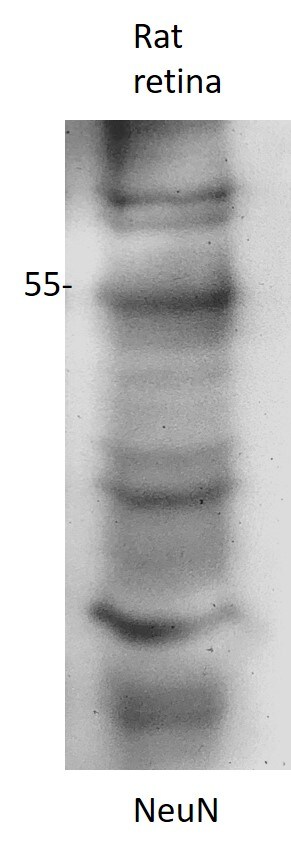
Application: Western BlotSample Tested: retinaSpecies: RatVerified Customer | Posted 07/28/2020good reaction. Little non-specific.

-
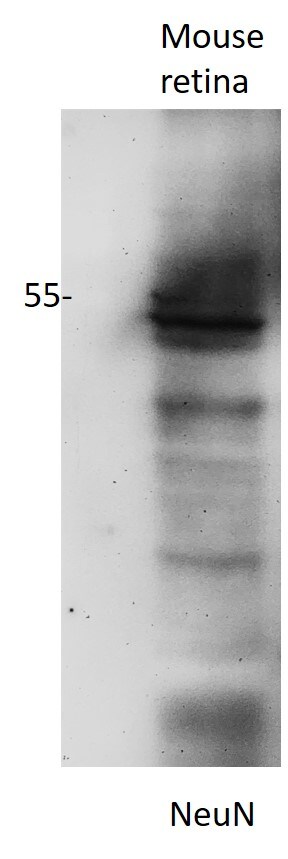
Application: Western BlotSample Tested: retinaSpecies: MouseVerified Customer | Posted 07/28/2020good reaction
-
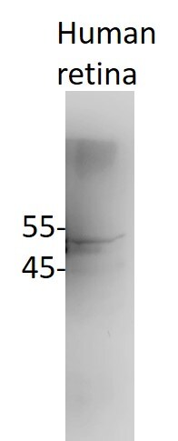
Application: Simple WesternSample Tested: Adult eye and muscle tissue (labeled E and MSpecies: HumanVerified Customer | Posted 07/21/2020Human retinal lysates
-
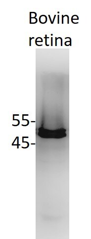
Application: Western BlotSample Tested: retinaSpecies: BovineVerified Customer | Posted 07/21/2020Works excellent
-
Application: Western BlotSample Tested: retinaSpecies: PigVerified Customer | Posted 07/21/2020Pig retina lysates-NeuN
-
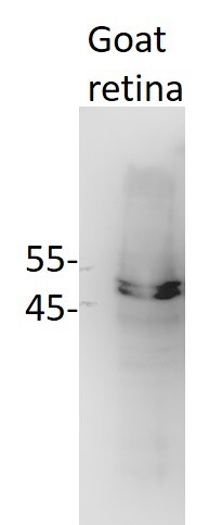
Application: Western BlotSample Tested: retinaSpecies: GoatVerified Customer | Posted 07/21/2020Goat retina-NeuN
-
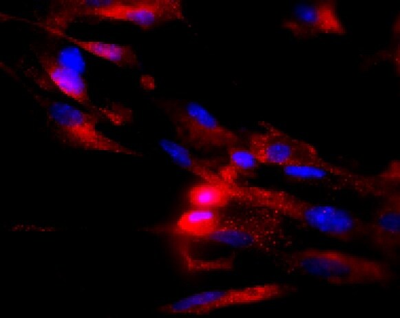
Application: ImmunocytochemistrySample Tested: mixed primary neuronal cultureSpecies: HumanVerified Customer | Posted 06/23/2020
There are no reviews that match your criteria.
Protocols
View specific protocols for RBFOX3/NeuN Antibody - BSA Free (NBP1-77686):
Immunocytochemistry Protocol
Culture cells to appropriate density in 35 mm culture dishes or 6-well plates.
1. Remove culture medium and add 10% formalin to the dish. Fix at room temperature for 30 minutes.
2. Remove the formalin and add ice cold methanol. Incubate for 5-10 minutes.
3. Remove methanol and add washing solution (i.e. PBS). Be sure to not let the specimen dry out. Wash three times for 10 minutes.
4. To block nonspecific antibody binding incubate in 10% normal goat serum from 1 hour to overnight at room temperature.
5. Add primary antibody at appropriate dilution and incubate at room temperature from 2 hours to overnight at room temperature.
6. Remove primary antibody and replace with washing solution. Wash three times for 10 minutes.
7. Add secondary antibody at appropriate dilution. Incubate for 1 hour at room temperature.
8. Remove antibody and replace with wash solution, then wash for 10 minutes. Add Hoechst 33258 to wash solution at 1:25,0000 and incubate for 10 minutes. Wash a third time for 10 minutes.
9. Cells can be viewed directly after washing. The plates can also be stored in PBS containing Azide covered in Parafilm (TM). Cells can also be cover-slipped using Fluoromount, with appropriate sealing.
*The above information is only intended as a guide. The researcher should determine what protocol best meets their needs. Please follow safe laboratory procedures.
Immunohistochemistry-Paraffin Embedded Sections
Antigen Unmasking:
Bring slides to a boil in 10 mM sodium citrate buffer (pH 6.0) then maintain at a sub-boiling temperature for 10 minutes. Cool slides on bench-top for 30 minutes.
Staining:
1. Wash sections in deionized water three times for 5 minutes each.
2. Wash sections in wash buffer for 5 minutes.
3. Block each section with 100-400 ul blocking solution for 1 hour at room temperature.
4. Remove blocking solution and add 100-400 ul diluted primary antibody. Incubate overnight at 4C.
5. Remove antibody solution and wash sections in wash buffer three times for 5 minutes each.
6. Add 100-400 ul biotinylated diluted secondary antibody. Incubate 30 minutes at room temperature.
7. Remove secondary antibody solution and wash sections three times with wash buffer for 5 minutes each.
8. Add 100-400 ul Streptavidin-HRP reagent to each section and incubate for 30 minutes at room temperature.
9. Wash sections three times in wash buffer for 5 minutes each.
10. Add 100-400 ul DAB substrate to each section and monitor staining closely.
11. As soon as the sections develop, immerse slides in deionized water.
12. Counterstain sections in hematoxylin.
13. Wash sections in deionized water two times for 5 minutes each.
14. Dehydrate sections.
15. Mount coverslips.
*The above information is only intended as a guide. The researcher should determine what protocol best meets their needs. Please follow safe laboratory procedures.
Find general support by application which include: protocols, troubleshooting, illustrated assays, videos and webinars.
- 7-Amino Actinomycin D (7-AAD) Cell Viability Flow Cytometry Protocol
- Antigen Retrieval Protocol (PIER)
- Antigen Retrieval for Frozen Sections Protocol
- Appropriate Fixation of IHC/ICC Samples
- Cellular Response to Hypoxia Protocols
- Chromogenic IHC Staining of Formalin-Fixed Paraffin-Embedded (FFPE) Tissue Protocol
- Chromogenic Immunohistochemistry Staining of Frozen Tissue
- ClariTSA™ Fluorophore Kits
- Detection & Visualization of Antibody Binding
- Extracellular Membrane Flow Cytometry Protocol
- Flow Cytometry Protocol for Cell Surface Markers
- Flow Cytometry Protocol for Staining Membrane Associated Proteins
- Flow Cytometry Staining Protocols
- Flow Cytometry Troubleshooting Guide
- Fluorescent IHC Staining of Frozen Tissue Protocol
- Graphic Protocol for Heat-induced Epitope Retrieval
- Graphic Protocol for the Preparation and Fluorescent IHC Staining of Frozen Tissue Sections
- Graphic Protocol for the Preparation and Fluorescent IHC Staining of Paraffin-embedded Tissue Sections
- Graphic Protocol for the Preparation of Gelatin-coated Slides for Histological Tissue Sections
- ICC Cell Smear Protocol for Suspension Cells
- ICC Immunocytochemistry Protocol Videos
- ICC for Adherent Cells
- IHC Sample Preparation (Frozen sections vs Paraffin)
- Immunocytochemistry (ICC) Protocol
- Immunocytochemistry Troubleshooting
- Immunofluorescence of Organoids Embedded in Cultrex Basement Membrane Extract
- Immunofluorescent IHC Staining of Formalin-Fixed Paraffin-Embedded (FFPE) Tissue Protocol
- Immunohistochemistry (IHC) and Immunocytochemistry (ICC) Protocols
- Immunohistochemistry Frozen Troubleshooting
- Immunohistochemistry Paraffin Troubleshooting
- Intracellular Flow Cytometry Protocol Using Alcohol (Methanol)
- Intracellular Flow Cytometry Protocol Using Detergents
- Intracellular Nuclear Staining Flow Cytometry Protocol Using Detergents
- Intracellular Staining Flow Cytometry Protocol Using Alcohol Permeabilization
- Intracellular Staining Flow Cytometry Protocol Using Detergents to Permeabilize Cells
- Preparing Samples for IHC/ICC Experiments
- Preventing Non-Specific Staining (Non-Specific Binding)
- Primary Antibody Selection & Optimization
- Propidium Iodide Cell Viability Flow Cytometry Protocol
- Protocol for Heat-Induced Epitope Retrieval (HIER)
- Protocol for Liperfluo
- Protocol for Making a 4% Formaldehyde Solution in PBS
- Protocol for VisUCyte™ HRP Polymer Detection Reagent
- Protocol for the Characterization of Human Th22 Cells
- Protocol for the Characterization of Human Th9 Cells
- Protocol for the Fluorescent ICC Staining of Cell Smears - Graphic
- Protocol for the Fluorescent ICC Staining of Cultured Cells on Coverslips - Graphic
- Protocol for the Preparation & Fixation of Cells on Coverslips
- Protocol for the Preparation and Chromogenic IHC Staining of Frozen Tissue Sections
- Protocol for the Preparation and Chromogenic IHC Staining of Frozen Tissue Sections - Graphic
- Protocol for the Preparation and Chromogenic IHC Staining of Paraffin-embedded Tissue Sections
- Protocol for the Preparation and Chromogenic IHC Staining of Paraffin-embedded Tissue Sections - Graphic
- Protocol for the Preparation and Fluorescent ICC Staining of Cells on Coverslips
- Protocol for the Preparation and Fluorescent ICC Staining of Non-adherent Cells
- Protocol for the Preparation and Fluorescent ICC Staining of Stem Cells on Coverslips
- Protocol for the Preparation and Fluorescent IHC Staining of Frozen Tissue Sections
- Protocol for the Preparation and Fluorescent IHC Staining of Paraffin-embedded Tissue Sections
- Protocol for the Preparation of Gelatin-coated Slides for Histological Tissue Sections
- Protocol for the Preparation of a Cell Smear for Non-adherent Cell ICC - Graphic
- Protocol: Annexin V and PI Staining by Flow Cytometry
- Protocol: Annexin V and PI Staining for Apoptosis by Flow Cytometry
- R&D Systems Quality Control Western Blot Protocol
- TUNEL and Active Caspase-3 Detection by IHC/ICC Protocol
- The Importance of IHC/ICC Controls
- Troubleshooting Guide: Fluorokine Flow Cytometry Kits
- Troubleshooting Guide: Immunohistochemistry
- Troubleshooting Guide: Western Blot Figures
- Western Blot Conditions
- Western Blot Protocol
- Western Blot Protocol for Cell Lysates
- Western Blot Troubleshooting
- Western Blot Troubleshooting Guide
- View all Protocols, Troubleshooting, Illustrated assays and Webinars
Loading...