RECQ1 Antibody - BSA Free
Novus Biologicals | Catalog # NB100-619
![Western Blot: RECQ1 Antibody [NB100-619]](https://resources.rndsystems.com/images/products/RECQ1-Antibody-Western-Blot-NB100-619-img0007.jpg "Western Blot: RECQ1 Antibody [NB100-619]")
Key Product Details
Validated by
Independent Antibodies, Biological Validation
Species Reactivity
Validated:
Human
Cited:
Human
Predicted:
Orangutan (100%). Backed by our 100% Guarantee.
Applications
Validated:
Immunohistochemistry, Immunohistochemistry-Paraffin, Western Blot, Immunoprecipitation
Cited:
Western Blot
Label
Unconjugated
Antibody Source
Polyclonal Rabbit IgG
Format
BSA Free
Loading...
Product Specifications
Immunogen
The immunogen recognized by this antibody maps to a region between residue 600 and the C-terminus (residue 649) of human RecQ protein-like (DNA helicase Q1-like) 1 using the numbering given in Swiss-Prot entry P46063 (GeneID 5965).
Clonality
Polyclonal
Host
Rabbit
Isotype
IgG
Theoretical MW
73 kDa.
Disclaimer note: The observed molecular weight of the protein may vary from the listed predicted molecular weight due to post translational modifications, post translation cleavages, relative charges, and other experimental factors.
Disclaimer note: The observed molecular weight of the protein may vary from the listed predicted molecular weight due to post translational modifications, post translation cleavages, relative charges, and other experimental factors.
Scientific Data Images for RECQ1 Antibody - BSA Free
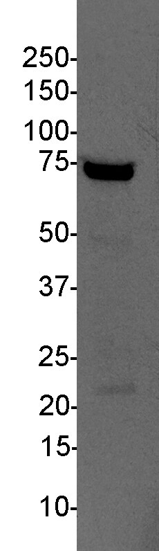
Western Blot: RECQ1 Antibody [NB100-619]
Western Blot: RECQ1 Antibody [NB100-619] - Whole cell lysate (50 ug) from HeLa and 293T cells prepared using NETN lysis buffer. Affinity purified rabbit anti-RecQ1 antibody NB100-619 used for WB at 0.1 ug/mL. Detection: Chemiluminescence with an exposure time of 1 second.![Immunohistochemistry: RECQ1 Antibody [NB100-619]](https://resources.rndsystems.com/images/products/RECQ1-Antibody-Immunohistochemistry-NB100-619-img0005.jpg "Immunohistochemistry: RECQ1 Antibody [NB100-619]")
Immunohistochemistry: RECQ1 Antibody [NB100-619]
Immunohistochemistry: RECQ1 Antibody [NB100-619] - Sample: FFPE section of human ovarian carcinoma. Antibody: Affinity purified rabbit anti- RecQ1 at 1:5000 (0.2 ug/mL). Detection: DAB![Immunoprecipitation: RECQ1 Antibody [NB100-619]](https://resources.rndsystems.com/images/products/RECQ1-Antibody-Immunoprecipitation-NB100-619-img0009.jpg "Immunoprecipitation: RECQ1 Antibody [NB100-619]")
Immunoprecipitation: RECQ1 Antibody [NB100-619]
Immunoprecipitation: RECQ1 Antibody [NB100-619] - Detection of human RecQ1 by western blot of immunoprecipitates. Samples: Whole cell lysate (0.5 or 1.0 mg per IP reaction; 20% of IP loaded) from HeLa cells prepared using NETN lysis buffer. Antibodies: Affinity purified rabbit anti-RecQ1 antibody NB100-619 used for IP at 6 ug per reaction. RecQ1 was also immunoprecipitated by rabbit anti-RecQ1 antibody NB100-618. For blotting immunoprecipitated RecQ1, NB100-619 was used at 1 ug/ml. Detection: Chemiluminescence with an exposure time of 1 second.Applications for RECQ1 Antibody - BSA Free
Application
Recommended Usage
Immunohistochemistry
1:2000-1:10000
Immunohistochemistry-Paraffin
1:2000-1:10000
Immunoprecipitation
2-10 ug/mg lysate
Western Blot
1:2000-1:10000
Reviewed Applications
Read 3 reviews rated 4.3 using NB100-619 in the following applications:
Formulation, Preparation, and Storage
Purification
Immunogen affinity purified
Formulation
Tris-Citrate/Phosphate (pH 7.0 - 8.0)
Format
BSA Free
Preservative
0.09% Sodium Azide
Concentration
1.0 mg/ml
Shipping
The product is shipped with polar packs. Upon receipt, store it immediately at the temperature recommended below.
Stability & Storage
Store at 4C. Do not freeze.
Background: RECQ1
Alternate Names
DNA helicase, RecQ-like type 1, DNA-dependent ATPase Q1, EC 3.6.1, EC 3.6.4.12, RecQ protein-like (DNA helicase Q1-like), RecQ protein-like 1, RECQ1, RecQ1ATP-dependent DNA helicase Q1, RecQL1
Entrez Gene IDs
5965 (Human)
Gene Symbol
RECQL
UniProt
Additional RECQ1 Products
Product Documents for RECQ1 Antibody - BSA Free
Certificate of Analysis
To download a Certificate of Analysis, please enter a lot or batch number in the search box below.
Product Specific Notices for RECQ1 Antibody - BSA Free
This product is for research use only and is not approved for use in humans or in clinical diagnosis. Primary Antibodies are guaranteed for 1 year from date of receipt.
Citations for RECQ1 Antibody - BSA Free
 Powered by Bioz
Customer Reviews for RECQ1 Antibody - BSA Free (3)
4.3 out of 5
3 Customer Ratings
Have you used RECQ1 Antibody - BSA Free?
Submit a review and receive an Amazon gift card!
$25/€18/£15/$25CAN/¥2500 Yen for a review with an image
$10/€7/£6/$10CAN/¥1110 Yen for a review without an image
Submit a review
Customer Images
Showing
1
-
3 of
3 reviews
Showing All
Filter By:
-
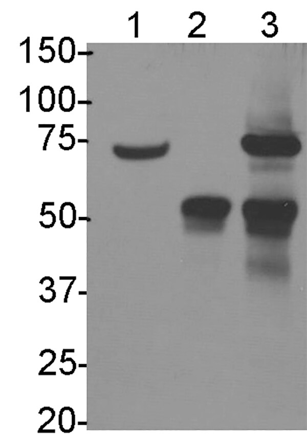
Application: ImmunoprecipitationSample Tested: b-cellSpecies: HumanVerified Customer | Posted 12/04/2018RECQ1 immunoprecipitation in human b-cells. L1: 40ug input, L2: IgG, L3: IP10ug/mg lysate

-
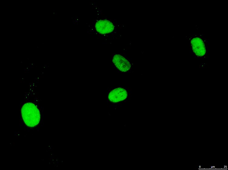
Application: ImmunocytochemistrySample Tested: fibroblastsSpecies: HumanVerified Customer | Posted 03/13/2018BIOSCARF 1:250 in 1% BSA/0.3% Triton X-100 / PBS
-
Application: Western BlotSample Tested: human lymphocytes, whole cell lysateSpecies: HumanVerified Customer | Posted 12/12/201725ug protein, 10 second exposure. 1:2000 dilution in 5% milk/TBS-T. Longer exposure (1 min had several bands).25ug protein, 10 second exposure. 1:2000 dilution in 5% milk/TBS-T. Longer exposure (1 min had several bands).
There are no reviews that match your criteria.
Protocols
Find general support by application which include: protocols, troubleshooting, illustrated assays, videos and webinars.
- Antigen Retrieval Protocol (PIER)
- Antigen Retrieval for Frozen Sections Protocol
- Appropriate Fixation of IHC/ICC Samples
- Cellular Response to Hypoxia Protocols
- Chromogenic IHC Staining of Formalin-Fixed Paraffin-Embedded (FFPE) Tissue Protocol
- Chromogenic Immunohistochemistry Staining of Frozen Tissue
- ClariTSA™ Fluorophore Kits
- Detection & Visualization of Antibody Binding
- Fluorescent IHC Staining of Frozen Tissue Protocol
- Graphic Protocol for Heat-induced Epitope Retrieval
- Graphic Protocol for the Preparation and Fluorescent IHC Staining of Frozen Tissue Sections
- Graphic Protocol for the Preparation and Fluorescent IHC Staining of Paraffin-embedded Tissue Sections
- Graphic Protocol for the Preparation of Gelatin-coated Slides for Histological Tissue Sections
- IHC Sample Preparation (Frozen sections vs Paraffin)
- Immunofluorescent IHC Staining of Formalin-Fixed Paraffin-Embedded (FFPE) Tissue Protocol
- Immunohistochemistry (IHC) and Immunocytochemistry (ICC) Protocols
- Immunohistochemistry Frozen Troubleshooting
- Immunohistochemistry Paraffin Troubleshooting
- Immunoprecipitation Protocol
- Preparing Samples for IHC/ICC Experiments
- Preventing Non-Specific Staining (Non-Specific Binding)
- Primary Antibody Selection & Optimization
- Protocol for Heat-Induced Epitope Retrieval (HIER)
- Protocol for Making a 4% Formaldehyde Solution in PBS
- Protocol for VisUCyte™ HRP Polymer Detection Reagent
- Protocol for the Preparation & Fixation of Cells on Coverslips
- Protocol for the Preparation and Chromogenic IHC Staining of Frozen Tissue Sections
- Protocol for the Preparation and Chromogenic IHC Staining of Frozen Tissue Sections - Graphic
- Protocol for the Preparation and Chromogenic IHC Staining of Paraffin-embedded Tissue Sections
- Protocol for the Preparation and Chromogenic IHC Staining of Paraffin-embedded Tissue Sections - Graphic
- Protocol for the Preparation and Fluorescent IHC Staining of Frozen Tissue Sections
- Protocol for the Preparation and Fluorescent IHC Staining of Paraffin-embedded Tissue Sections
- Protocol for the Preparation of Gelatin-coated Slides for Histological Tissue Sections
- R&D Systems Quality Control Western Blot Protocol
- TUNEL and Active Caspase-3 Detection by IHC/ICC Protocol
- The Importance of IHC/ICC Controls
- Troubleshooting Guide: Immunohistochemistry
- Troubleshooting Guide: Western Blot Figures
- Western Blot Conditions
- Western Blot Protocol
- Western Blot Protocol for Cell Lysates
- Western Blot Troubleshooting
- Western Blot Troubleshooting Guide
- View all Protocols, Troubleshooting, Illustrated assays and Webinars
Loading...