Ribosomal Protein S6/RPS6 Antibody
Novus Biologicals | Catalog # NB100-1595
![Western Blot: Ribosomal Protein S6/RPS6 Antibody [NB100-1595]](https://resources.rndsystems.com/images/products/Ribosomal-Protein-S6-RPS6-Antibody-Western-Blot-NB100-1595-img0007.jpg "Western Blot: Ribosomal Protein S6/RPS6 Antibody [NB100-1595]")
Loading...
Key Product Details
Species Reactivity
Validated:
Human, Mouse
Cited:
Human, Mouse
Predicted:
Bovine (100%), Chicken (100%), Rat (100%), Xenopus (100%). Backed by our 100% Guarantee.
Applications
Validated:
Immunohistochemistry, Immunohistochemistry-Paraffin, Western Blot, Immunoprecipitation
Cited:
Western Blot, Immunofluorescence
Label
Unconjugated
Antibody Source
Polyclonal Rabbit IgG
Loading...
Product Specifications
Immunogen
The immunogen recognized by this antibody maps to a region between residue 200 and the C-terminus (residue 249) of human Ribosomal Protein S6 using the numbering given in entry NP_001001.2 (GeneID 6194).
Reactivity Notes
X. laevis (100%).
Clonality
Polyclonal
Host
Rabbit
Isotype
IgG
Theoretical MW
29 kDa.
Disclaimer note: The observed molecular weight of the protein may vary from the listed predicted molecular weight due to post translational modifications, post translation cleavages, relative charges, and other experimental factors.
Disclaimer note: The observed molecular weight of the protein may vary from the listed predicted molecular weight due to post translational modifications, post translation cleavages, relative charges, and other experimental factors.
Scientific Data Images for Ribosomal Protein S6/RPS6 Antibody
Western Blot: Ribosomal Protein S6/RPS6 Antibody [NB100-1595]
Western Blot: Ribosomal Protein S6/RPS6 Antibody [NB100-1595] - Detection of human and mouse RPS6 by western blot. Samples: Whole cell lysate (50 ug) from HeLa, HEK293T, Jurkat, mouse TCMK-1, and mouse NIH 3T3 cells prepared using NETN lysis buffer. Antibody: Affinity purified rabbit anti-RPS6 antibody NB100-1595 used for WB at 0.04 ug/ml. Detection: Chemiluminescence with an exposure time of 1 second.Previous![Immunohistochemistry-Paraffin: Ribosomal Protein S6/RPS6 Antibody [NB100-1595]](https://resources.rndsystems.com/images/products/Ribosomal-Protein-S6-RPS6-Antibody-Immunohistochemistry-NB100-1595-img0009.jpg "Immunohistochemistry-Paraffin: Ribosomal Protein S6/RPS6 Antibody [NB100-1595]")
Immunohistochemistry-Paraffin: Ribosomal Protein S6/RPS6 Antibody [NB100-1595]
Immunohistochemistry-Paraffin: Ribosomal Protein S6/RPS6 Antibody [NB100-1595] - Section of human lung carcinoma. Antibody: Affinity purified rabbit anti-RPS6 (NB100-1595). Detection: DAB![Immunoprecipitation: Ribosomal Protein S6/RPS6 Antibody [NB100-1595]](https://resources.rndsystems.com/images/products/Ribosomal-Protein-S6-RPS6-Antibody-Immunoprecipitation-NB100-1595-img0008.jpg "Immunoprecipitation: Ribosomal Protein S6/RPS6 Antibody [NB100-1595]")
Immunoprecipitation: Ribosomal Protein S6/RPS6 Antibody [NB100-1595]
Immunoprecipitation: Ribosomal Protein S6/RPS6 Antibody [NB100-1595] - Detection of human RPS6 by western blot of immunoprecipitates. Samples: Whole cell lysate (1.0 mg per IP reaction; 20% of IP loaded) from HeLa cells prepared using NETN lysis buffer. Antibodies: Affinity purified rabbit anti-RPS6 antibody NB100-1595 (lot NB100-1595-2) used for IP at 6 ug per reaction. RPS6 was also immunoprecipitated by a previous lot of this antibody (lot NB100-1595-1). For blotting immunoprecipitated RPS6, NB100-1595 was used at 0.1 ug/ml. Detection: Chemiluminescence with an exposure time of 1 second.
Western Blot: Ribosomal Protein S6/RPS6 Antibody [NB100-1595] -
Western Blot: Ribosomal Protein S6/RPS6 Antibody [NB100-1595] - Loss of SPAG5 & CLUH lead to hyperactivation of mTORC1 signaling.(A, B) MSigDB Hallmark pathways of downregulated (E) or upregulated (F) proteins (with a cutoff of p ≤ 0.05; q ≤ 0.15) detected in proteomics analysis of SPAG5 ind-KO cells (Figure 4A, Supplementary file 3) using the EnrichR webtool. (C) Western blots of WT & SPAG5 ind-KO HeLa cells transfected with siRNA against CLUH or untargeted control siRNA. Cells were induced for 4 days with doxycycline, additionally downregulated for the last 3 days & grown for the last 8 hr in basal or HBSS media without doxycycline. Pan-actin was used as loading control. (D) Quantification of experiments as shown in C (n = 4 independent experiments). Antibody signal was normalized to pan-actin signal, & signal of phospho-protein was normalized to signal of the total protein. Bars show mean ± standard error of the mean (SEM) & dots represent values of individual replicates. One-way analysis of variance (ANOVA) with post hoc Tukey’s multiple comparison tests were performed with *p ≤ 0.05; **p ≤ 0.01; ***p ≤ 0.001.Figure 5—figure supplement 1—source data 1.Uncropped blots for Figure 5—figure supplement 1C.Figure 5—figure supplement 1—source data 2.Unedited blots for Figure 5—figure supplement 1C.Uncropped blots for Figure 5—figure supplement 1C.Unedited blots for Figure 5—figure supplement 1C. Image collected & cropped by CiteAb from the following publication (https://pubmed.ncbi.nlm.nih.gov/35559794), licensed under a CC-BY license. Not internally tested by Novus Biologicals.
Immunocytochemistry/ Immunofluorescence: Ribosomal Protein S6/RPS6 Antibody [NB100-1595] -
L1 bodies (LBs) are large cytoplasmic compartments enriched in ORF1p, L1 mRNA, and ribosomes.(A–E) Electron micrographs exemplifying the formation of LBs in Mael-/- spermatocytes. Boxed areas in A and B identify small cytoplasmic aggregates magnified in A’ and B’, respectively. Boxed area in C identifies an intermediate size LB magnified in C’. D and E show more prominent LBs, partially surrounded by a bilayer membrane (red arrows). Nuc: nucleus. Cyto: cytoplasm. NE: nuclear envelope. (F) Immunofluorescence staining of L1 ORF1p on Mael-/- spermatocytes shows ORF1p accumulation in LBs (left panel, magenta). 3D reconstruction of LBs (magenta) and nuclei (blue) (middle and right panels); numbers [1–5] are used to label corresponding cells. Scale bars: 5 μm. (G) HCR RNA-FISH of L1 RNA (green) and immunofluorescence staining of L1 ORF1p (magenta) on Mael+/- and Mael-/- testis sections. Scale bars: 5 μm. (H) Double immunofluorescence staining of ribosomal protein S6 (RPS6, top two rows, green) or L28 (RPL28, bottom two rows, green) and L1 ORF1p (magenta) showing their colocalization in LBs. Dotted white lines mark seminiferous tubule boundaries. Scale bars: 15 μm. See also S1 Fig. Image collected and cropped by CiteAb from the following open publication (https://pubmed.ncbi.nlm.nih.gov/37307272), licensed under a CC-BY license. Not internally tested by Novus Biologicals.Applications for Ribosomal Protein S6/RPS6 Antibody
Application
Recommended Usage
Immunohistochemistry-Paraffin
1:200-1:1000
Immunoprecipitation
2-10 ug/mg lysate
Western Blot
1:2000-1:10000
Application Notes
Epitope retrieval with citrate buffer pH6.0 is recommended for FFPE tissue sections.
Reviewed Applications
Read 1 review rated 4 using NB100-1595 in the following applications:
Formulation, Preparation, and Storage
Purification
Immunogen affinity purified
Formulation
TBS and 0.1% BSA
Preservative
0.09% Sodium Azide
Concentration
0.2 mg/ml
Shipping
The product is shipped with polar packs. Upon receipt, store it immediately at the temperature recommended below.
Stability & Storage
Store at 4C. Do not freeze.
Background: Ribosomal Protein S6/RPS6
Additional Ribosomal Protein S6/RPS6 Products
Product Documents for Ribosomal Protein S6/RPS6 Antibody
Certificate of Analysis
To download a Certificate of Analysis, please enter a lot or batch number in the search box below.
Product Specific Notices for Ribosomal Protein S6/RPS6 Antibody
This product is for research use only and is not approved for use in humans or in clinical diagnosis. Primary Antibodies are guaranteed for 1 year from date of receipt.
Related Research Areas
Citations for Ribosomal Protein S6/RPS6 Antibody
 Powered by Bioz
Customer Reviews for Ribosomal Protein S6/RPS6 Antibody (1)
4 out of 5
1 Customer Rating
Have you used Ribosomal Protein S6/RPS6 Antibody?
Submit a review and receive an Amazon gift card!
$25/€18/£15/$25CAN/¥2500 Yen for a review with an image
$10/€7/£6/$10CAN/¥1110 Yen for a review without an image
Submit a review
Customer Images
Showing
1
-
1 of
1 review
Showing All
Filter By:
-
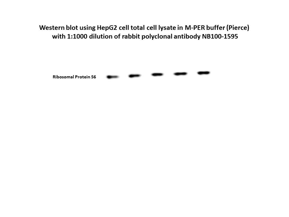
Application: Western BlotSample Tested: HepG2Species: HumanVerified Customer | Posted 09/18/2015RPS6 in HepG2 total cell lysate

There are no reviews that match your criteria.
Protocols
Find general support by application which include: protocols, troubleshooting, illustrated assays, videos and webinars.
- Antigen Retrieval Protocol (PIER)
- Antigen Retrieval for Frozen Sections Protocol
- Appropriate Fixation of IHC/ICC Samples
- Cellular Response to Hypoxia Protocols
- Chromogenic IHC Staining of Formalin-Fixed Paraffin-Embedded (FFPE) Tissue Protocol
- Chromogenic Immunohistochemistry Staining of Frozen Tissue
- ClariTSA™ Fluorophore Kits
- Detection & Visualization of Antibody Binding
- Fluorescent IHC Staining of Frozen Tissue Protocol
- Graphic Protocol for Heat-induced Epitope Retrieval
- Graphic Protocol for the Preparation and Fluorescent IHC Staining of Frozen Tissue Sections
- Graphic Protocol for the Preparation and Fluorescent IHC Staining of Paraffin-embedded Tissue Sections
- Graphic Protocol for the Preparation of Gelatin-coated Slides for Histological Tissue Sections
- IHC Sample Preparation (Frozen sections vs Paraffin)
- Immunofluorescent IHC Staining of Formalin-Fixed Paraffin-Embedded (FFPE) Tissue Protocol
- Immunohistochemistry (IHC) and Immunocytochemistry (ICC) Protocols
- Immunohistochemistry Frozen Troubleshooting
- Immunohistochemistry Paraffin Troubleshooting
- Immunoprecipitation Protocol
- Preparing Samples for IHC/ICC Experiments
- Preventing Non-Specific Staining (Non-Specific Binding)
- Primary Antibody Selection & Optimization
- Protocol for Heat-Induced Epitope Retrieval (HIER)
- Protocol for Making a 4% Formaldehyde Solution in PBS
- Protocol for VisUCyte™ HRP Polymer Detection Reagent
- Protocol for the Preparation & Fixation of Cells on Coverslips
- Protocol for the Preparation and Chromogenic IHC Staining of Frozen Tissue Sections
- Protocol for the Preparation and Chromogenic IHC Staining of Frozen Tissue Sections - Graphic
- Protocol for the Preparation and Chromogenic IHC Staining of Paraffin-embedded Tissue Sections
- Protocol for the Preparation and Chromogenic IHC Staining of Paraffin-embedded Tissue Sections - Graphic
- Protocol for the Preparation and Fluorescent IHC Staining of Frozen Tissue Sections
- Protocol for the Preparation and Fluorescent IHC Staining of Paraffin-embedded Tissue Sections
- Protocol for the Preparation of Gelatin-coated Slides for Histological Tissue Sections
- R&D Systems Quality Control Western Blot Protocol
- TUNEL and Active Caspase-3 Detection by IHC/ICC Protocol
- The Importance of IHC/ICC Controls
- Troubleshooting Guide: Immunohistochemistry
- Troubleshooting Guide: Western Blot Figures
- Western Blot Conditions
- Western Blot Protocol
- Western Blot Protocol for Cell Lysates
- Western Blot Troubleshooting
- Western Blot Troubleshooting Guide
- View all Protocols, Troubleshooting, Illustrated assays and Webinars
Loading...
Associated Pathways

