Rictor Antibody - BSA Free
Novus Biologicals | Catalog # NB100-612
![Knockdown Validated: Rictor Antibody [NB100-612]](https://resources.rndsystems.com/images/products/Rictor-Antibody-Western-Blot-NB100-612-img0025.jpg "Western Blot: Rictor Antibody [NB100-612]")
![Western Blot: Rictor Antibody [NB100-612]](https://resources.rndsystems.com/images/products/Rictor-Antibody-Western-Blot-NB100-612-img0024.jpg "Western Blot: Rictor Antibody [NB100-612]")
![Western Blot: Rictor Antibody [NB100-612]](https://resources.rndsystems.com/images/products/Rictor-Antibody-Western-Blot-NB100-612-img0020.jpg "Western Blot: Rictor Antibody [NB100-612]")
Key Product Details
Validated by
Knockout/Knockdown, Independent Antibodies, Biological Validation
Species Reactivity
Validated:
Human, Mouse
Cited:
Human, Mouse
Applications
Validated:
Immunohistochemistry, Immunohistochemistry-Paraffin, Western Blot, Immunocytochemistry/ Immunofluorescence, Immunoprecipitation, Knockdown Validated
Cited:
Western Blot, Immunocytochemistry/ Immunofluorescence, Immunoprecipitation
Label
Unconjugated
Antibody Source
Polyclonal Rabbit IgG
Format
BSA Free
Loading...
Product Specifications
Immunogen
The immunogen recognized by this antibody maps to a region between residue 1650 and the C-terminus (residue 1708) of human Rapamycin-Insensitive Companion of mTOR using the numbering given in TrEMBL entry Q6R327 (GeneID 253260).
Clonality
Polyclonal
Host
Rabbit
Isotype
IgG
Theoretical MW
192 kDa.
Disclaimer note: The observed molecular weight of the protein may vary from the listed predicted molecular weight due to post translational modifications, post translation cleavages, relative charges, and other experimental factors.
Disclaimer note: The observed molecular weight of the protein may vary from the listed predicted molecular weight due to post translational modifications, post translation cleavages, relative charges, and other experimental factors.
Scientific Data Images for Rictor Antibody - BSA Free
Western Blot: Rictor Antibody [NB100-612]
Western Blot: Rictor Antibody [NB100-612] - Whole cell lysate (50 ug) from HeLa, HEK293T, and Jurkat cells prepared using NETN lysis buffer. Antibody: Affinity purified rabbit anti-Rictor antibody used for WB at 0.1 ug/ml. Detection: Chemiluminescence with an exposure time of 30 seconds.![Immunoprecipitation: Rictor Antibody [NB100-612]](https://resources.rndsystems.com/images/products/Rictor-Antibody-Immunoprecipitation-NB100-612-img0022.jpg "Immunoprecipitation: Rictor Antibody [NB100-612]")
Immunoprecipitation: Rictor Antibody [NB100-612]
Immunoprecipitation: Rictor Antibody [NB100-612] - Detection of human Rictor by western blot of immunoprecipitates. Samples: Whole cell lysate (1.0 mg per IP reaction; 20% of IP loaded) from HeLa cells prepared using NETN lysis buffer. Antibodies: Affinity purified rabbit anti-Rictor antibody NB100-612 (lot NB100-612-4) used for IP at 3 ug per reaction. Rictor was also immunoprecipitated by a previous lot of this antibody (lot NB100-612-3) and rabbit anti-Rictor antibody NB100-611. For blotting immunoprecipitated Rictor, NB100-612 was used at 1 ug/ml. Detection: Chemiluminescence with an exposure time of 10 seconds.![Immunohistochemistry-Paraffin: Rictor Antibody [NB100-612]](https://resources.rndsystems.com/images/products/Rictor-Antibody-Immunohistochemistry-Paraffin-NB100-612-img0019.jpg "Immunohistochemistry-Paraffin: Rictor Antibody [NB100-612]")
Immunohistochemistry-Paraffin: Rictor Antibody [NB100-612]
Immunohistochemistry-Paraffin: Rictor Antibody [NB100-612] - Human lung carcinoma. Antibody: Affinity purified rabbit anti-Rictor used at a dilution of 1:1,000 (1ug/ml). Detection: DAB![Western Blot: Rictor Antibody [NB100-612]](https://resources.rndsystems.com/images/products/Rictor-Antibody-Western-Blot-NB100-612-img0008.jpg "Western Blot: Rictor Antibody [NB100-612]")
![Western Blot: Rictor Antibody [NB100-612]](https://resources.rndsystems.com/images/products/Rictor-Antibody-Western-Blot-NB100-612-img0021.jpg "Western Blot: Rictor Antibody [NB100-612]")
Western Blot: Rictor Antibody [NB100-612]
Western Blot: Rictor Antibody [NB100-612] - Whole cell lysate (50 ug) from TCMK-1 and NIH 3T3 cells prepared using NETN lysis buffer. Antibody: Affinity purified rabbit anti-Rictor antibody used for WB at 0.1 ug/ml. Detection: Chemiluminescence with an exposure time of 3 minutes.![Western Blot: Rictor Antibody [NB100-612]](https://resources.rndsystems.com/images/products/Rictor-Antibody-Western-Blot-NB100-612-img0023.jpg "Western Blot: Rictor Antibody [NB100-612]")

Western Blot: Rictor Antibody [NB100-612] -
Western Blot: Rictor Antibody [NB100-612] - Truncated Sin1 displaces endogenous Sin1 from mTORC2 in DLD1 colon cancer cellsA. Schematic indicating the domain structure of Sin1 & the constructs used to displace endogenous Sin1 from mTORC2. B. Expression of myc tagged Sin1 constructs can be detected only after induction with Doxycycline (Dox). Cells were treated with 100nM of doxycycline (+) for 72 hours & expressed proteins were detected by immunoblot of whole cell lysates with anti-myc (9E10) antibodies. C. & D. Sin1 constructs incorporate into mTORC2 & displace endogenous Sin1. Constructs were induced for 72 hours prior to immune precipitation. (C) mTORC2 subunits, mTOR & Rictor, only appear in myc immunoprecipitates after induction with doxycycline (Left panels); myc-∆Sin1 cannot be directly detected in precipitates due to secondary antibody cross reaction with precipitating IgG. Right panels indicate unchanging expression levels of Rictor & mTOR in immune precipitation input lysates, which is further quantified from 3 independent experiments E. Endogenous Sin1 & Rictor immunoprecipitates demonstrate displacement of endogenous Sin1 from mTORC2. Following induction, band shifted myc-tagged FL Sin1 can be detected in Sin1 & Rictor precipitates (Left panels). Truncated ∆Sin1 can be detected in Rictor, but not Sin1, immunoprecipitates as the Sin1 antibody epitope is deleted from ∆Sin1. F. Quantification of Sin1 levels detected in Rictor immunoprecipitates indicates the level of endogenous mTORC2 disruption following Sin1 construct induction (data are mean +/- S.D; n = 3). Myc-∆Sin1 displaces >80% of endogenous Sin1 while levels of myc-FL Sin1 associated with Rictor are comparable with endogenous Sin1 levels. Image collected & cropped by CiteAb from the following publication (https://www.oncotarget.com/lookup/doi/10.18632/oncotarget.20086), licensed under a CC-BY license. Not internally tested by Novus Biologicals.
Western Blot: Rictor Antibody [NB100-612] -
Western Blot: Rictor Antibody [NB100-612] - Truncated Sin1 displaces endogenous Sin1 from mTORC2 in DLD1 colon cancer cellsA. Schematic indicating the domain structure of Sin1 & the constructs used to displace endogenous Sin1 from mTORC2. B. Expression of myc tagged Sin1 constructs can be detected only after induction with Doxycycline (Dox). Cells were treated with 100nM of doxycycline (+) for 72 hours & expressed proteins were detected by immunoblot of whole cell lysates with anti-myc (9E10) antibodies. C. & D. Sin1 constructs incorporate into mTORC2 & displace endogenous Sin1. Constructs were induced for 72 hours prior to immune precipitation. (C) mTORC2 subunits, mTOR & Rictor, only appear in myc immunoprecipitates after induction with doxycycline (Left panels); myc-∆Sin1 cannot be directly detected in precipitates due to secondary antibody cross reaction with precipitating IgG. Right panels indicate unchanging expression levels of Rictor & mTOR in immune precipitation input lysates, which is further quantified from 3 independent experiments E. Endogenous Sin1 & Rictor immunoprecipitates demonstrate displacement of endogenous Sin1 from mTORC2. Following induction, band shifted myc-tagged FL Sin1 can be detected in Sin1 & Rictor precipitates (Left panels). Truncated ∆Sin1 can be detected in Rictor, but not Sin1, immunoprecipitates as the Sin1 antibody epitope is deleted from ∆Sin1. F. Quantification of Sin1 levels detected in Rictor immunoprecipitates indicates the level of endogenous mTORC2 disruption following Sin1 construct induction (data are mean +/- S.D; n = 3). Myc-∆Sin1 displaces >80% of endogenous Sin1 while levels of myc-FL Sin1 associated with Rictor are comparable with endogenous Sin1 levels. Image collected & cropped by CiteAb from the following publication (https://www.oncotarget.com/lookup/doi/10.18632/oncotarget.20086), licensed under a CC-BY license. Not internally tested by Novus Biologicals.Applications for Rictor Antibody - BSA Free
Application
Recommended Usage
Immunocytochemistry/ Immunofluorescence
1:50 - 1:250
Immunohistochemistry
1:500-1:2000
Immunohistochemistry-Paraffin
1:500-1:2000
Immunoprecipitation
2-5 ug/mg lysate
Western Blot
1:2000-1:10000
Application Notes
Use in ICC/IF reported in scientific literature (PMID 25378594). Epitope retrieval with citrate buffer pH6.0 is recommended for FFPE tissue sections.
Reviewed Applications
Read 2 reviews rated 5 using NB100-612 in the following applications:
Formulation, Preparation, and Storage
Purification
Immunogen affinity purified
Formulation
Tris-Citrate/Phosphate (pH 7.0 - 8.0)
Format
BSA Free
Preservative
0.09% Sodium Azide
Concentration
1.0 mg/ml
Shipping
The product is shipped with polar packs. Upon receipt, store it immediately at the temperature recommended below.
Stability & Storage
Store at 4C. Do not freeze.
Background: Rictor
Long Name
Rapamycin-insensitive Companion of mTOR
Alternate Names
mAVO3, Pianissimo
Gene Symbol
RICTOR
UniProt
Additional Rictor Products
Product Documents for Rictor Antibody - BSA Free
Certificate of Analysis
To download a Certificate of Analysis, please enter a lot or batch number in the search box below.
Product Specific Notices for Rictor Antibody - BSA Free
This product is for research use only and is not approved for use in humans or in clinical diagnosis. Primary Antibodies are guaranteed for 1 year from date of receipt.
Related Research Areas
Citations for Rictor Antibody - BSA Free
 Powered by Bioz
Customer Reviews for Rictor Antibody - BSA Free (2)
5 out of 5
2 Customer Ratings
Have you used Rictor Antibody - BSA Free?
Submit a review and receive an Amazon gift card!
$25/€18/£15/$25CAN/¥2500 Yen for a review with an image
$10/€7/£6/$10CAN/¥1110 Yen for a review without an image
Submit a review
Customer Images
Showing
1
-
2 of
2 reviews
Showing All
Filter By:
-
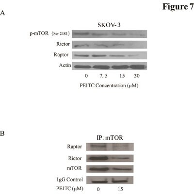
Application: Western BlotSample Tested: SKOV3 whole cell lysateSpecies: HumanVerified Customer | Posted 08/26/2012

-
Application: Western BlotSample Tested: HumanSpecies: HumanVerified Customer | Posted 12/09/2011
There are no reviews that match your criteria.
Protocols
Find general support by application which include: protocols, troubleshooting, illustrated assays, videos and webinars.
- Antigen Retrieval Protocol (PIER)
- Antigen Retrieval for Frozen Sections Protocol
- Appropriate Fixation of IHC/ICC Samples
- Cellular Response to Hypoxia Protocols
- Chromogenic IHC Staining of Formalin-Fixed Paraffin-Embedded (FFPE) Tissue Protocol
- Chromogenic Immunohistochemistry Staining of Frozen Tissue
- ClariTSA™ Fluorophore Kits
- Detection & Visualization of Antibody Binding
- Fluorescent IHC Staining of Frozen Tissue Protocol
- Graphic Protocol for Heat-induced Epitope Retrieval
- Graphic Protocol for the Preparation and Fluorescent IHC Staining of Frozen Tissue Sections
- Graphic Protocol for the Preparation and Fluorescent IHC Staining of Paraffin-embedded Tissue Sections
- Graphic Protocol for the Preparation of Gelatin-coated Slides for Histological Tissue Sections
- ICC Cell Smear Protocol for Suspension Cells
- ICC Immunocytochemistry Protocol Videos
- ICC for Adherent Cells
- IHC Sample Preparation (Frozen sections vs Paraffin)
- Immunocytochemistry (ICC) Protocol
- Immunocytochemistry Troubleshooting
- Immunofluorescence of Organoids Embedded in Cultrex Basement Membrane Extract
- Immunofluorescent IHC Staining of Formalin-Fixed Paraffin-Embedded (FFPE) Tissue Protocol
- Immunohistochemistry (IHC) and Immunocytochemistry (ICC) Protocols
- Immunohistochemistry Frozen Troubleshooting
- Immunohistochemistry Paraffin Troubleshooting
- Immunoprecipitation Protocol
- Preparing Samples for IHC/ICC Experiments
- Preventing Non-Specific Staining (Non-Specific Binding)
- Primary Antibody Selection & Optimization
- Protocol for Heat-Induced Epitope Retrieval (HIER)
- Protocol for Making a 4% Formaldehyde Solution in PBS
- Protocol for VisUCyte™ HRP Polymer Detection Reagent
- Protocol for the Fluorescent ICC Staining of Cell Smears - Graphic
- Protocol for the Fluorescent ICC Staining of Cultured Cells on Coverslips - Graphic
- Protocol for the Preparation & Fixation of Cells on Coverslips
- Protocol for the Preparation and Chromogenic IHC Staining of Frozen Tissue Sections
- Protocol for the Preparation and Chromogenic IHC Staining of Frozen Tissue Sections - Graphic
- Protocol for the Preparation and Chromogenic IHC Staining of Paraffin-embedded Tissue Sections
- Protocol for the Preparation and Chromogenic IHC Staining of Paraffin-embedded Tissue Sections - Graphic
- Protocol for the Preparation and Fluorescent ICC Staining of Cells on Coverslips
- Protocol for the Preparation and Fluorescent ICC Staining of Non-adherent Cells
- Protocol for the Preparation and Fluorescent ICC Staining of Stem Cells on Coverslips
- Protocol for the Preparation and Fluorescent IHC Staining of Frozen Tissue Sections
- Protocol for the Preparation and Fluorescent IHC Staining of Paraffin-embedded Tissue Sections
- Protocol for the Preparation of Gelatin-coated Slides for Histological Tissue Sections
- Protocol for the Preparation of a Cell Smear for Non-adherent Cell ICC - Graphic
- R&D Systems Quality Control Western Blot Protocol
- TUNEL and Active Caspase-3 Detection by IHC/ICC Protocol
- The Importance of IHC/ICC Controls
- Troubleshooting Guide: Immunohistochemistry
- Troubleshooting Guide: Western Blot Figures
- Western Blot Conditions
- Western Blot Protocol
- Western Blot Protocol for Cell Lysates
- Western Blot Troubleshooting
- Western Blot Troubleshooting Guide
- View all Protocols, Troubleshooting, Illustrated assays and Webinars
Loading...
Associated Pathways

