RNA Polymerase II/POLR2A [p Ser2] Antibody
Novus Biologicals | Catalog # NB100-1805
![Western Blot: RNA Polymerase II/POLR2A [p Ser2] Antibody [NB100-1805]](https://resources.rndsystems.com/images/products/RNA-Polymerase-II-POLR2A-[p-Ser2]-Antibody-Western-Blot-NB100-1805-img0027.jpg "Western Blot: RNA Polymerase II/POLR2A [p Ser2] Antibody [NB100-1805]")
Loading...
Key Product Details
Validated by
Biological Validation
Species Reactivity
Validated:
Human, Mouse, Yeast
Cited:
Human, Yeast
Predicted:
Drosophila (100%). Backed by our 100% Guarantee.
Applications
Validated:
Immunohistochemistry, Immunohistochemistry-Paraffin, Western Blot, Immunoprecipitation, Single Cell Western
Cited:
Western Blot, Proximity Ligation Assay
Label
Unconjugated
Antibody Source
Polyclonal Rabbit IgG
Loading...
Product Specifications
Immunogen
Immunogen for this antibody was a phosphorylated synthetic peptide, which represented a portion of human RNA Polymerase II (GeneID 5430) around serine 2 of the C-terminal repeat YSPTSPS.
Reactivity Notes
Yeast reactivity reported in scientific literature (PMID: 25416796).
Modification
p Ser2
Specificity
RNA Polymerase 2 Phosphospecific [Ser2]
Clonality
Polyclonal
Host
Rabbit
Isotype
IgG
Scientific Data Images for RNA Polymerase II/POLR2A [p Ser2] Antibody
Western Blot: RNA Polymerase II/POLR2A [p Ser2] Antibody [NB100-1805]
Western Blot: RNA Polymerase II/POLR2A [p Ser2] Antibody [NB100-1805] - Detection of Human Phospho RNA Polymerase II (S2) by Western Blot. Samples: Whole cell lysate (50 and 15 ug) from HeLa prepared using NETN buffer. Lysate was mock treated (-) or treated with phosphatases (+). Antibodies: Affinity purified rabbit anti-Phospho RNA Polymerase II (S2) antibody NB100-1805 was used for WB at 0.04 ug/ml (upper panel). To examine total RNA Polymerase II, rabbit anti-RNA Polymerase II antibody was used at 1 ug/ml (lower panel). Detection: Chemiluminescence with exposure time of 3 seconds.![Immunohistochemistry-Paraffin: RNA Polymerase II/POLR2A [p Ser2] Antibody [NB100-1805]](https://resources.rndsystems.com/images/products/RNA-Polymerase-II-POLR2A-[p-Ser2]-Antibody-Immunohistochemistry-Paraffin-NB100-1805-img0029.jpg "Immunohistochemistry-Paraffin: RNA Polymerase II/POLR2A [p Ser2] Antibody [NB100-1805]")
Immunohistochemistry-Paraffin: RNA Polymerase II/POLR2A [p Ser2] Antibody [NB100-1805]
Immunohistochemistry-Paraffin: RNA Polymerase II/POLR2A [p Ser2] Antibody [NB100-1805] - Section of mouse teratoma. Antibody: Affinity purified rabbit anti-PhosphoRNA Polymerase II (S2) used at a dilution of 1:200 (1ug/ml). Detection: DAB![Western Blot: RNA Polymerase II/POLR2A [p Ser2] Antibody [NB100-1805]](https://resources.rndsystems.com/images/products/RNA-Polymerase-II-POLR2A-[p-Ser2]-Antibody-Western-Blot-NB100-1805-img0021.jpg "Western Blot: RNA Polymerase II/POLR2A [p Ser2] Antibody [NB100-1805]")
Western Blot: RNA Polymerase II/POLR2A [p Ser2] Antibody [NB100-1805]
Western Blot: RNA Polymerase II/POLR2A [p Ser2] Antibody [NB100-1805] - Hela whole cell lysate (50 ug each lane) loaded and probed by anti-RNA polymerase II Antibody. NB100-1805 was pre-incubated without peptide (-), with non-phosphorylated peptide (NP), with phosphorylated S2 peptide, or with phosphorylated S5 peptide.![Western Blot: RNA Polymerase II/POLR2A [p Ser2] Antibody [NB100-1805]](https://resources.rndsystems.com/images/products/RNA-Polymerase-II-POLR2A-[p-Ser2]-Antibody-Western-Blot-NB100-1805-img0022.jpg "Western Blot: RNA Polymerase II/POLR2A [p Ser2] Antibody [NB100-1805]")
Western Blot: RNA Polymerase II/POLR2A [p Ser2] Antibody [NB100-1805]
Western Blot: RNA Polymerase II/POLR2A [p Ser2] Antibody [NB100-1805] - Whole cell lysate (15 and 50 ug) from NIH3T3 cells. Antibody used at 0.08 ug/ml.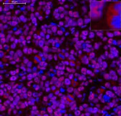
Immunohistochemistry-Paraffin: RNA Polymerase II/POLR2A [p Ser2] Antibody [NB100-1805] - Analysis of POLR2A in MCF-7 xenograft using anti-POLR2A [p Ser2] antibody. Image from verified customer review.
![Immunohistochemistry-Paraffin: RNA Polymerase II/POLR2A [p Ser2] Antibody [NB100-1805]](https://resources.rndsystems.com/images/products/RNA-Polymerase-II-POLR2A-[p-Ser2]-Antibody-Immunohistochemistry-Paraffin-NB100-1805-img0028.jpg "Immunohistochemistry-Paraffin: RNA Polymerase II/POLR2A [p Ser2] Antibody [NB100-1805]")
Immunohistochemistry-Paraffin: RNA Polymerase II/POLR2A [p Ser2] Antibody [NB100-1805]
Immunohistochemistry-Paraffin: RNA Polymerase II/POLR2A [p Ser2] Antibody [NB100-1805] - Human ovarian carcinoma. Antibody: Affinity purified rabbit antiPhospho-RNA Polymerase II (S2) used at a dilution of 1:1000 (0.2ug/ml). Detection: DAB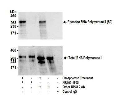
RNA Polymerase II/POLR2A [p Ser2] Antibody [NB100-1805] - Detection of Phospho RNA Polymerase II (S2) by western blot of immunoprecipitates. Samples: Whole cell lysate (0.5 or 1 mg per IP reaction, 20% of IP loaded) from HeLa cells prepared using NETN lysis buffer. immunoprecipitates were either phosphatase treated (+) or mock treated (-). Antibody: Affinity purified rabbit anti-Phospho RNA Polymerase II (S2) antibody NB100-1805 used for IP at 5 ug per reaction and another Affinity purified rabbit anti-RNA Polymerase II antibody used for IP at 5 ug per reaction. For blotting immunoprecipitated Phospho RNA Polymerase II (S2) and total RNA Polymerase II, NB100-1805 was used at 0.1 ug/ml (upper panel) and the other antibody was used at 1 ug/ml (lower panel). Detection: Chemiluminescence with an exposure time of 10 seconds (upper) and 75 seconds (lower).
Applications for RNA Polymerase II/POLR2A [p Ser2] Antibody
Application
Recommended Usage
Immunohistochemistry
1:200 - 1:1000
Immunohistochemistry-Paraffin
1:200 - 1:1000
Immunoprecipitation
2-10 mg/mg
Single Cell Western
100 ug/mL
Western Blot
1:2000-1:10000
Application Notes
Epitope retrieval with citrate buffer pH6.0 is recommended for FFPE tissue sections.
Reviewed Applications
Read 2 reviews rated 5 using NB100-1805 in the following applications:
Formulation, Preparation, and Storage
Purification
Immunogen affinity purified
Formulation
TBS and 0.1% BSA
Preservative
0.09% Sodium Azide
Concentration
0.2 mg/ml
Shipping
The product is shipped with polar packs. Upon receipt, store it immediately at the temperature recommended below.
Stability & Storage
Store at 4C. Do not freeze.
Background: RNA Polymerase II/POLR2A
Long Name
DNA-directed RNA Polymerase II Subunit RPB1
Alternate Names
POLR2, POLR2A, POLRA, RPB1, RPO2, RPOL2
Entrez Gene IDs
5430 (Human)
Gene Symbol
POLR2A
UniProt
Additional RNA Polymerase II/POLR2A Products
Product Documents for RNA Polymerase II/POLR2A [p Ser2] Antibody
Certificate of Analysis
To download a Certificate of Analysis, please enter a lot or batch number in the search box below.
Product Specific Notices for RNA Polymerase II/POLR2A [p Ser2] Antibody
This product is for research use only and is not approved for use in humans or in clinical diagnosis. Primary Antibodies are guaranteed for 1 year from date of receipt.
Related Research Areas
Citations for RNA Polymerase II/POLR2A [p Ser2] Antibody
 Powered by Bioz
Customer Reviews for RNA Polymerase II/POLR2A [p Ser2] Antibody (2)
5 out of 5
2 Customer Ratings
Have you used RNA Polymerase II/POLR2A [p Ser2] Antibody?
Submit a review and receive an Amazon gift card!
$25/€18/£15/$25CAN/¥2500 Yen for a review with an image
$10/€7/£6/$10CAN/¥1110 Yen for a review without an image
Submit a review
Customer Images
![RNA Polymerase II/POLR2A [p Ser2] Antibody NB100-1805](https://resources.rndsystems.com/images/reviews/Immunohistochemisty-Paraffin__NB100-1805_22181.png)
![RNA Polymerase II/POLR2A [p Ser2] Antibody NB100-1805](https://resources.rndsystems.com/images/reviews/Western-Blot_RNA-polymerase-II-Antibody-(NB100-1805)-(01-ml)_NB100-1805_9526.jpg)
Showing
1
-
2 of
2 reviews
Showing All
Filter By:
-
Application: Immunohistochemistry-ParaffinSample Tested: MCF7Species: HumanVerified Customer | Posted 10/29/20151:250 POLR2A staining on MCF7 xenograft
![RNA Polymerase II/POLR2A [p Ser2] Antibody NB100-1805](data:image/png;base64,R0lGODlhAQABAAD/ACwAAAAAAQABAAACADs=)
-
Application: Western BlotSample Tested: 293T cellsSpecies: HumanVerified Customer | Posted 08/26/2014WB for RNA Pol II pSer 2 in 293T cells
There are no reviews that match your criteria.
Protocols
Find general support by application which include: protocols, troubleshooting, illustrated assays, videos and webinars.
- Antigen Retrieval Protocol (PIER)
- Antigen Retrieval for Frozen Sections Protocol
- Appropriate Fixation of IHC/ICC Samples
- Cellular Response to Hypoxia Protocols
- Chromogenic IHC Staining of Formalin-Fixed Paraffin-Embedded (FFPE) Tissue Protocol
- Chromogenic Immunohistochemistry Staining of Frozen Tissue
- ClariTSA™ Fluorophore Kits
- Detection & Visualization of Antibody Binding
- Fluorescent IHC Staining of Frozen Tissue Protocol
- Graphic Protocol for Heat-induced Epitope Retrieval
- Graphic Protocol for the Preparation and Fluorescent IHC Staining of Frozen Tissue Sections
- Graphic Protocol for the Preparation and Fluorescent IHC Staining of Paraffin-embedded Tissue Sections
- Graphic Protocol for the Preparation of Gelatin-coated Slides for Histological Tissue Sections
- IHC Sample Preparation (Frozen sections vs Paraffin)
- Immunofluorescent IHC Staining of Formalin-Fixed Paraffin-Embedded (FFPE) Tissue Protocol
- Immunohistochemistry (IHC) and Immunocytochemistry (ICC) Protocols
- Immunohistochemistry Frozen Troubleshooting
- Immunohistochemistry Paraffin Troubleshooting
- Immunoprecipitation Protocol
- Preparing Samples for IHC/ICC Experiments
- Preventing Non-Specific Staining (Non-Specific Binding)
- Primary Antibody Selection & Optimization
- Protocol for Heat-Induced Epitope Retrieval (HIER)
- Protocol for Making a 4% Formaldehyde Solution in PBS
- Protocol for VisUCyte™ HRP Polymer Detection Reagent
- Protocol for the Preparation & Fixation of Cells on Coverslips
- Protocol for the Preparation and Chromogenic IHC Staining of Frozen Tissue Sections
- Protocol for the Preparation and Chromogenic IHC Staining of Frozen Tissue Sections - Graphic
- Protocol for the Preparation and Chromogenic IHC Staining of Paraffin-embedded Tissue Sections
- Protocol for the Preparation and Chromogenic IHC Staining of Paraffin-embedded Tissue Sections - Graphic
- Protocol for the Preparation and Fluorescent IHC Staining of Frozen Tissue Sections
- Protocol for the Preparation and Fluorescent IHC Staining of Paraffin-embedded Tissue Sections
- Protocol for the Preparation of Gelatin-coated Slides for Histological Tissue Sections
- R&D Systems Quality Control Western Blot Protocol
- TUNEL and Active Caspase-3 Detection by IHC/ICC Protocol
- The Importance of IHC/ICC Controls
- Troubleshooting Guide: Immunohistochemistry
- Troubleshooting Guide: Western Blot Figures
- Western Blot Conditions
- Western Blot Protocol
- Western Blot Protocol for Cell Lysates
- Western Blot Troubleshooting
- Western Blot Troubleshooting Guide
- View all Protocols, Troubleshooting, Illustrated assays and Webinars
Loading...